|
Frontal Sinuses
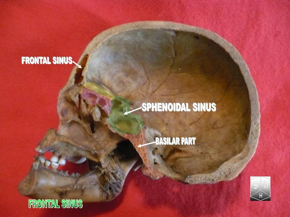
The frontal sinuses are one of the four pairs of paranasal sinuses that are situated behind the brow ridges. Sinuses are mucosa-lined airspaces within the bones of the face and skull. Each opens into the anterior part of the corresponding middle nasal meatus of the nose through the frontonasal duct which traverses the anterior part of the labyrinth of the ethmoid. These structures then open into the semilunar hiatus in the middle meatus. Structure Each frontal sinus is situated between the external and internal plates of the frontal bone. Their average measurements are as follows: height 28 mm, breadth 24 mm, depth 20 mm, creating a space of 6-7 ml. Each frontal sinus extends into the squamous part of the frontal bone superiorly, and into the orbital part of frontal bone posteriorly to come to occupy the medial part of the roof of the orbit. Each sinus drains through an opening in its inferomedial part into the frontonasal duct. Vasculature The mucous m ... [...More Info...] [...Related Items...] OR: [Wikipedia] [Google] [Baidu] |
Supra-orbital Artery
The supraorbital artery is a branch of the ophthalmic artery. It passes anteriorly within the orbit to exit the orbit through the supraorbital foramen or notch alongside the supraorbital nerve, splitting into two terminal branches which go on to form anastomoses with arteries of the head. Structure Origin The supraorbital artery arises from the ophthalmic artery. Course and relations It travels anteriorly in the orbit by passing superior to the eye and medial to the superior rectus and levator palpebrae superioris. It then joins the supraorbital nerve to jointly pass between the periosteum of the roof of the orbit and the levator palpebrae superioris towards the supraorbital foramen or notch. After passing through the supraorbital foramen or notch, it often splits into a superficial branch and a deep branch. Distribution The supraorbital artery contributes arterial supply to: the superior rectus muscle, superior oblique muscle, levator palpebrae muscles, periorbita, the ... [...More Info...] [...Related Items...] OR: [Wikipedia] [Google] [Baidu] |
Submandibular Lymph Nodes
The submandibular lymph nodes (submaxillary glands in older texts), are some 3-6 lymph nodes situated at the inferior border of the ramus of mandible. Anatomy They are situated just superficial to the submandibular salivary gland, and posterolateral to the anterior belly of either digastric muscle. One gland, the ''middle gland of Stahr'', which lies on the facial artery as it turns over the mandible, is the most constant of the series; small lymph glands are sometimes found on the deep surface of the submandibular gland. Afferents They drain the upper lip, body of tongue, cheeks, anterior portion of the hard palate, and most teeth with their associated periodontium and gingiva (except for the mandibular incisor teeth and third molar teeth). The facial and submental lymph nodes may also drain into the submandibular glands. Efferents They drain to the superior deep cervical lymph nodes. Clinical significance The most common causes of enlargement of the submandibular l ... [...More Info...] [...Related Items...] OR: [Wikipedia] [Google] [Baidu] |
Choanae
The choanae (: choana), posterior nasal apertures or internal nostrils are two openings found at the back of the nasal passage between the nasal cavity and the pharynx, in humans and other mammals (as well as crocodilians and most skinks). They are considered one of the most important synapomorphies of tetrapodomorphs, that allowed the passage from water to land. In animals with secondary palates, they allow breathing when the mouth is closed. Janvier, Philippe (2004) "Wandering nostrils". ''Nature'', 432 (7013): 23–24. In tetrapods without secondary palates their function relates primarily to olfaction (sense of smell). The choanae are separated in two by the vomer. Boundaries A choana is the opening between the nasal cavity and the nasopharynx. It is therefore not a structure but a space bounded as follows: * anteriorly and inferiorly by the horizontal plate of palatine bone, * superiorly and posteriorly by the sphenoid bone * laterally by the medial pterygoid pla ... [...More Info...] [...Related Items...] OR: [Wikipedia] [Google] [Baidu] |
Ciliated
The cilium (: cilia; ; in Medieval Latin and in anatomy, ''cilium'') is a short hair-like membrane protrusion from many types of eukaryotic cell. (Cilia are absent in bacteria and archaea.) The cilium has the shape of a slender threadlike projection that extends from the surface of the much larger cell body. Eukaryotic flagella found on sperm cells and many protozoans have a similar structure to motile cilia that enables swimming through liquids; they are longer than cilia and have a different undulating motion. There are two major classes of cilia: ''motile'' and ''non-motile'' cilia, each with two subtypes, giving four types in all. A cell will typically have one primary cilium or many motile cilia. The structure of the cilium core, called the axoneme, determines the cilium class. Most motile cilia have a central pair of single microtubules surrounded by nine pairs of double microtubules called a 9+2 axoneme. Most non-motile cilia have a 9+0 axoneme that lacks the centra ... [...More Info...] [...Related Items...] OR: [Wikipedia] [Google] [Baidu] |
Puberty
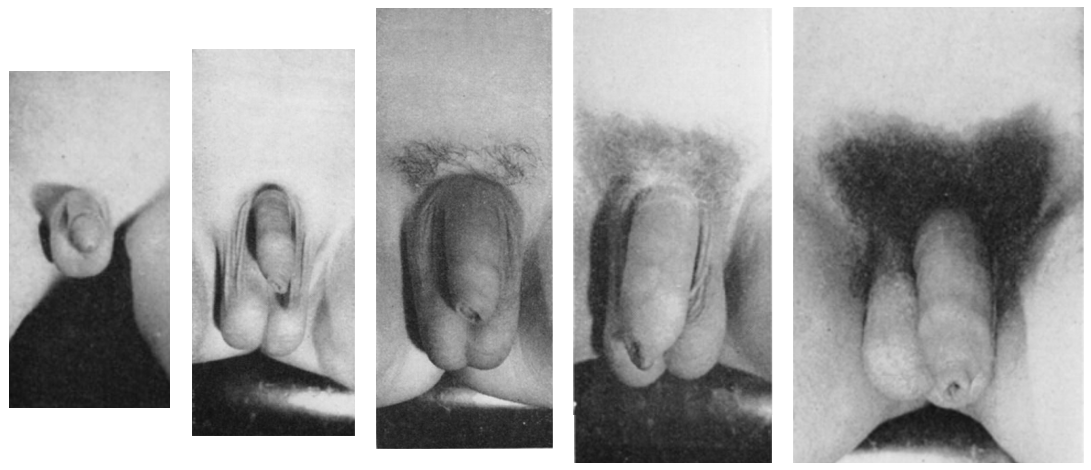
Puberty is the process of physical changes through which a child's body matures into an adult body capable of sexual reproduction. It is initiated by hormonal signals from the brain to the gonads: the ovaries in a female, the testicles in a male. In response to the signals, the gonads produce hormones that stimulate libido and the growth, function, and transformation of the brain, bones, muscle, blood, skin, hair, breasts, and sex organs. Physical growth—height and weight—accelerates in the first half of puberty and is completed when an adult body has been developed. Before puberty, the external sex organs, known as primary sexual characteristics, are sex characteristics that distinguish males and females. Puberty leads to sexual dimorphism through the development of the secondary sex characteristics, which further distinguish the sexes. On average, females begin puberty at age 10½ and complete puberty at ages 15-17; males begin at ages 11½-12 and complete pube ... [...More Info...] [...Related Items...] OR: [Wikipedia] [Google] [Baidu] |
Diploë
Diploë ( or ) is the spongy cancellous bone separating the inner and outer layers of the cortical bone of the skull. It is a subclass of trabecular bone. In the cranial bones, the layers of compact cortical tissue are familiarly known as the tables of the skull; the outer one is thick and tough; the inner is thin, dense, and brittle, and hence is termed the ''vitreous table''. The intervening cancellous tissue is called the diploë. In certain regions of the skull The skull, or cranium, is typically a bony enclosure around the brain of a vertebrate. In some fish, and amphibians, the skull is of cartilage. The skull is at the head end of the vertebrate. In the human, the skull comprises two prominent ..., this becomes absorbed so as to leave spaces filled with liquid between the two tables. Etymology From Ancient Greek διπλόη (''diplóē'', “literally, a fold”), noun use of feminine of διπλόος (''diplóos'', “double”) References External ... [...More Info...] [...Related Items...] OR: [Wikipedia] [Google] [Baidu] |
Sexual Characteristics
Sexual characteristics are physical traits of an organism (typically of a sexually dimorphic organism) which are indicative of or resultant from biological sexual factors. These include both primary sex characteristics, such as gonads, and secondary sex characteristics. Humans In humans, sex organs or primary sexual characteristics, which are those a person is born with, can be distinguished from secondary sex characteristics, which develop later in life, usually during puberty. The development of both is controlled by sex hormones produced by the body after the initial fetal stage where the presence or absence of the Y-chromosome and/or the SRY gene determine development. Male primary sex characteristics are the penis, the scrotum and the ability to ejaculate when matured. Female primary sex characteristics are the vulva, vagina, uterus, fallopian tubes, cervix, and the ability to give birth and menstruate when matured. Hormones that express sexual differen ... [...More Info...] [...Related Items...] OR: [Wikipedia] [Google] [Baidu] |
Sexual Dimorphism
Sexual dimorphism is the condition where sexes of the same species exhibit different Morphology (biology), morphological characteristics, including characteristics not directly involved in reproduction. The condition occurs in most dioecy, dioecious species, which consist of most animals and some plants. Differences may include secondary sex characteristics, size, weight, color, markings, or behavioral or cognitive traits. Male-male reproductive competition has evolved a diverse array of sexually dimorphic traits. Aggressive utility traits such as "battle" teeth and blunt heads reinforced as battering rams are used as weapons in aggressive interactions between rivals. Passive displays such as ornamental feathering or song-calling have also evolved mainly through sexual selection. These differences may be subtle or exaggerated and may be subjected to sexual selection and natural selection. The opposite of dimorphism is ''monomorphism'', when both biological sexes are phenotype, ... [...More Info...] [...Related Items...] OR: [Wikipedia] [Google] [Baidu] |
Septum
In biology, a septum (Latin language, Latin for ''something that encloses''; septa) is a wall, dividing a Body cavity, cavity or structure into smaller ones. A cavity or structure divided in this way may be referred to as septate. Examples Human anatomy * Interatrial septum, the wall of tissue that is a sectional part of the left and right atria of the heart * Interventricular septum, the wall separating the left and right ventricles of the heart * Lingual septum, a vertical layer of fibrous tissue that separates the halves of the tongue *Nasal septum: the cartilage wall separating the nostrils of the nose * Alveolar septum: the thin wall which separates the Pulmonary alveolus, alveoli from each other in the lungs * Orbital septum, a palpebral ligament in the upper and lower eyelids * Septum pellucidum or septum lucidum, a thin structure separating two fluid pockets in the brain * Uterine septum, a malformation of the uterus * Septum of the penis, Penile septum, a fibrous w ... [...More Info...] [...Related Items...] OR: [Wikipedia] [Google] [Baidu] |
Ophthalmic Nerve
The ophthalmic nerve (CN V1) is a sensory nerve of the head. It is one of three divisions of the trigeminal nerve (CN V), a cranial nerve. It has three major branches which provide sensory innervation to the eye, and the skin of the upper face and anterior scalp, as well as other structures of the head. Structure Origin The ophthalmic nerve is the first branch of the trigeminal nerve (CN V), the first and smallest of its three divisions. It arises from the superior part of the trigeminal ganglion. Course It passes anterior-ward along the lateral wall of the cavernous sinus inferior to the oculomotor nerve (CN III) and trochlear nerve (N IV). It exits the skull into the orbit through the superior orbital fissure. Branches Within the skull, the ophthalmic nerve produces: * meningeal branch (tentorial nerve) The ophthalmic nerve divides into three major branches which pass through the superior orbital fissure: * frontal nerve ** supraorbital nerve ** supratrochlea ... [...More Info...] [...Related Items...] OR: [Wikipedia] [Google] [Baidu] |
Secretion
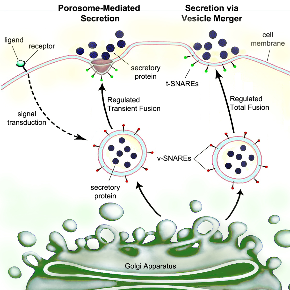
Secretion is the movement of material from one point to another, such as a secreted chemical substance from a cell or gland. In contrast, excretion is the removal of certain substances or waste products from a cell or organism. The classical mechanism of cell secretion is via secretory portals at the plasma membrane called porosomes. Porosomes are permanent cup-shaped lipoprotein structures embedded in the cell membrane, where secretory vesicles transiently dock and fuse to release intra-vesicular contents from the cell. Secretion in bacterial species means the transport or translocation of effector molecules. For example: proteins, enzymes or toxins (such as cholera toxin in pathogenic bacteria e.g. '' Vibrio cholerae'') from across the interior (cytoplasm or cytosol) of a bacterial cell to its exterior. Secretion is a very important mechanism in bacterial functioning and operation in their natural surrounding environment for adaptation and survival. In eukaryotic cells ... [...More Info...] [...Related Items...] OR: [Wikipedia] [Google] [Baidu] |
Mucus
Mucus (, ) is a slippery aqueous secretion produced by, and covering, mucous membranes. It is typically produced from cells found in mucous glands, although it may also originate from mixed glands, which contain both Serous fluid, serous and mucous cells. It is a viscous colloid containing inorganic ions, inorganic salts, antimicrobial enzymes (such as lysozymes), Antibody, immunoglobulins (especially Immunoglobulin A, IgA), and glycoproteins such as lactoferrin and mucins, which are produced by goblet cells in the mucous membranes and submucosal glands. Mucus covers the Epithelium, epithelial cells that interact with outside environment, serves to protect the linings of the respiratory system, respiratory, Digestion#Digestive system, digestive, and Genitourinary system, urogenital systems, and structures in the Visual system, visual and auditory systems from pathogenic Fungus, fungi, bacteria and viruses. Most of the mucus in the body is produced in the gastrointestinal tract. ... [...More Info...] [...Related Items...] OR: [Wikipedia] [Google] [Baidu] |


