|
Foramen Ovale (skull)
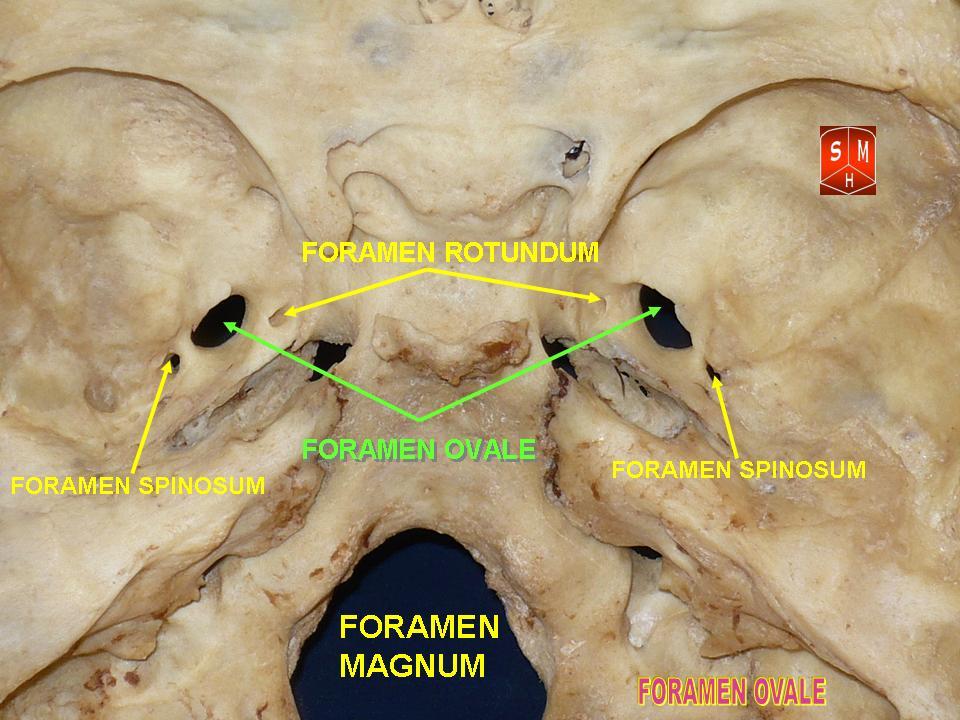
The foramen ovale (Latin: oval window) is a hole in the posterior part of the sphenoid bone, posterolateral to the foramen rotundum. It is one of the larger of the several holes (the foramina) in the skull. It transmits the mandibular nerve, a branch of the trigeminal nerve. Structure The foramen ovale is an opening in the greater wing of the sphenoid bone. The foramen ovale is one of two cranial foramina in the greater wing, the other being the foramen spinosum. The foramen ovale is posterolateral to the foramen rotundum and anteromedial to the foramen spinosum. Posterior and medial to the foramen is the opening for the carotid canal. Variation Similar to other foramina, the foramen ovale differs in shape and size throughout the natural life. The earliest perfect ring-shaped formation of the foramen ovale was observed in the 7th fetal month and the latest in 3 years after birth, in a study using over 350 skulls.In a study conducted on 100 skulls, the foramen ovale ... [...More Info...] [...Related Items...] OR: [Wikipedia] [Google] [Baidu] |
Sphenoid Bone
The sphenoid bone is an unpaired bone of the neurocranium. It is situated in the middle of the skull towards the front, in front of the basilar part of the occipital bone. The sphenoid bone is one of the seven bones that articulate to form the orbit. Its shape somewhat resembles that of a butterfly or bat with its wings extended. Structure It is divided into the following parts: * a median portion, known as the body of sphenoid bone, containing the sella turcica, which houses the pituitary gland as well as the paired paranasal sinuses, the sphenoidal sinuses * two greater wings on the lateral side of the body and two lesser wings from the anterior side. * Pterygoid processes of the sphenoides, directed downwards from the junction of the body and the greater wings. Two sphenoidal conchae are situated at the anterior and inferior part of the body. Intrinsic ligaments of the sphenoid The more important of these are: * the pterygospinous, stretching between the spin ... [...More Info...] [...Related Items...] OR: [Wikipedia] [Google] [Baidu] |
Accessory Meningeal Artery
The accessory meningeal artery (also accessory branch of middle meningeal artery, pterygomeningeal artery, small meningeal or parvidural branch) is a branch of the maxillary artery, sometimes derived from the middle meningeal artery. Course It enters the skull through the foramen ovale, and supplies the trigeminal ganglion and dura mater. Nomenclature Only about 10% of the blood flowing through this artery reaches intracranial structures. The remaining blood flow is dispersed to extracranial structures around the infratemporal fossa. Reflecting this fact, Terminologia Anatomica ''Terminologia Anatomica'' is the international standard for human anatomical terminology. It is developed by the Federative International Programme on Anatomical Terminology, a program of the International Federation of Associations of Anatomis ... lists entries for both "accessory branch of middle meningeal artery" and "pterygomeningeal artery". References External links * * () Arteries ... [...More Info...] [...Related Items...] OR: [Wikipedia] [Google] [Baidu] |
Temporal Lobe
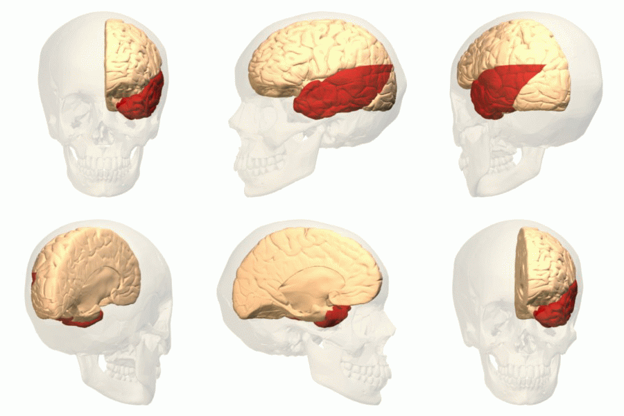
The temporal lobe is one of the four major lobes of the cerebral cortex in the brain of mammals. The temporal lobe is located beneath the lateral fissure on both cerebral hemispheres of the mammalian brain. The temporal lobe is involved in processing sensory input into derived meanings for the appropriate retention of visual memory, language comprehension, and emotion association. ''Temporal'' refers to the head's temples. Structure The temporal lobe consists of structures that are vital for declarative or long-term memory. Declarative (denotative) or explicit memory is conscious memory divided into semantic memory (facts) and episodic memory (events). Medial temporal lobe structures that are critical for long-term memory include the hippocampus, along with the surrounding hippocampal region consisting of the perirhinal, parahippocampal, and entorhinal neocortical regions. The hippocampus is critical for memory formation, and the surrounding medial temporal cortex is curre ... [...More Info...] [...Related Items...] OR: [Wikipedia] [Google] [Baidu] |
Trigeminal Neuralgia
Trigeminal neuralgia (TN or TGN), also called Fothergill disease, tic douloureux, or trifacial neuralgia is a long-term pain disorder that affects the trigeminal nerve, the nerve responsible for sensation in the face and motor functions such as biting and chewing. It is a form of neuropathic pain. There are two main types: typical and atypical trigeminal neuralgia. The typical form results in episodes of severe, sudden, shock-like pain in one side of the face that lasts for seconds to a few minutes. Groups of these episodes can occur over a few hours. The atypical form results in a constant burning pain that is less severe. Episodes may be triggered by any touch to the face. Both forms may occur in the same person. It is regarded as one of the most painful disorders known to medicine, and often results in depression. The exact cause is unknown, but believed to involve loss of the myelin of the trigeminal nerve. This might occur due to compression from a blood vessel as the ner ... [...More Info...] [...Related Items...] OR: [Wikipedia] [Google] [Baidu] |
Pterygoid Plexus
The pterygoid plexus (; in Merriam-Webster Online Dictionary '. from ''pteryx'', "wing" and ''eidos'', "shape") is a venous plexus of considerable size, and is situated between the temporalis muscle and lateral pterygoid muscle, and partly between the two pterygoid mus ... [...More Info...] [...Related Items...] OR: [Wikipedia] [Google] [Baidu] |
Cavernous Sinus
The cavernous sinus within the human head is one of the dural venous sinuses creating a cavity called the lateral sellar compartment bordered by the temporal bone of the skull and the sphenoid bone, lateral to the sella turcica. Structure The cavernous sinus is one of the dural venous sinuses of the head. It is a network of veins that sit in a cavity. It sits on both sides of the sphenoidal bone and pituitary gland, approximately 1 × 2 cm in size in an adult. The carotid siphon of the internal carotid artery, and cranial nerves III, IV, V (branches V1 and V2) and VI all pass through this blood filled space. Both sides of cavernous sinus is connected to each other via intercavernous sinuses. The cavernous sinus lies in between the inner and outer layers of dura mater. Nearby structures * Above: optic tract, optic chiasma, internal carotid artery. * Inferiorly: foramen lacerum, and the junction of the body and greater wing of sphenoid bone. * Medially: pi ... [...More Info...] [...Related Items...] OR: [Wikipedia] [Google] [Baidu] |
Emissary Vein
The emissary veins connect the extracranial venous system with the intracranial venous sinuses. They connect the veins outside the cranium to the venous sinuses inside the cranium. They drain from the scalp, through the skull, into the larger meningeal veins and dural venous sinuses. Emissary veins have an important role in selective cooling of the head. They also serve as routes where infections are carried into the cranial cavity from the extracranial veins to the intracranial veins. There are several types of emissary veins including posterior condyloid, mastoid, occipital and parietal emissary vein. Structure There are also emissary veins passing through the foramen ovale, jugular foramen, foramen lacerum, and hypoglossal canal. Function Because the emissary veins are valveless, they are an important part in selective brain cooling through bidirectional flow of cooler blood from the evaporating surface of the head. In general, blood flow is from external to internal but ... [...More Info...] [...Related Items...] OR: [Wikipedia] [Google] [Baidu] |
Foramen Petrosum
The lesser petrosal nerve (also known as the small superficial petrosal nerve) is the general visceral efferent (GVE) component of the glossopharyngeal nerve (CN IX), carrying parasympathetic preganglionic fibers from the tympanic plexus to the parotid gland. It synapses in the otic ganglion, from where the postganglionic fibers emerge. Structure After arising in the tympanic plexus, the lesser petrosal nerve passes forward and then through the hiatus for lesser petrosal nerve on the anterior surface of the petrous part of the temporal bone into the middle cranial fossa. It travels across the floor of the middle cranial fossa, then exits the skull via canaliculus innominatus to reach the infratemporal fossa. The fibres synapse in the otic ganglion, and post-ganglionic fibres then travel briefly with the auriculotemporal nerve (a branch of V3) before entering the body of the parotid gland. The lesser petrosal nerve will distribute its parasympathetic post-ganglionic (GVE) fiber ... [...More Info...] [...Related Items...] OR: [Wikipedia] [Google] [Baidu] |
Glossopharyngeal Nerve
The glossopharyngeal nerve (), also known as the ninth cranial nerve, cranial nerve IX, or simply CN IX, is a cranial nerve that exits the brainstem from the sides of the upper medulla, just anterior (closer to the nose) to the vagus nerve. Being a mixed nerve (sensorimotor), it carries afferent sensory and efferent motor information. The motor division of the glossopharyngeal nerve is derived from the basal plate of the embryonic medulla oblongata, whereas the sensory division originates from the cranial neural crest. Structure From the anterior portion of the medulla oblongata, the glossopharyngeal nerve passes laterally across or below the flocculus, and leaves the skull through the central part of the jugular foramen. From the superior and inferior ganglia in jugular foramen, it has its own sheath of dura mater. The inferior ganglion on the inferior surface of petrous part of temporal is related with a triangular depression into which the aqueduct of cochlea opens. On the ... [...More Info...] [...Related Items...] OR: [Wikipedia] [Google] [Baidu] |
Lesser Petrosal Nerve
The lesser petrosal nerve (also known as the small superficial petrosal nerve) is the general visceral efferent (GVE) component of the glossopharyngeal nerve (CN IX), carrying parasympathetic preganglionic fibers from the tympanic plexus to the parotid gland. It synapses in the otic ganglion, from where the postganglionic fibers emerge. Structure After arising in the tympanic plexus, the lesser petrosal nerve passes forward and then through the hiatus for lesser petrosal nerve on the anterior surface of the petrous part of the temporal bone into the middle cranial fossa. It travels across the floor of the middle cranial fossa, then exits the skull via canaliculus innominatus to reach the infratemporal fossa. The fibres synapse in the otic ganglion, and post-ganglionic fibres then travel briefly with the auriculotemporal nerve (a branch of V3) before entering the body of the parotid gland. The lesser petrosal nerve will distribute its parasympathetic post-ganglionic (GVE) fibe ... [...More Info...] [...Related Items...] OR: [Wikipedia] [Google] [Baidu] |
Carotid Canal
The carotid canal is a passageway in the temporal bone of the skull through which the internal carotid artery enters the middle cranial fossa from the neck. Structure The carotid canal is located within the middle cranial fossa, at the petrous part of the temporal bone. Anteriorly, it is limited by posterior margin of the greater wing of sphenoid bone. Posteromedially, it is limited by basilar part of occipital bone. It is divided in three parts, namely, ascending petrous, transverse petrous, and ascending cavernous parts. The carotid canal has two openings, namely internal and external openings. The internal opening is situated laterally to foramen lacerum. The external opening of the carotid canal is located posterolaterally to the foramen lacerum. Both internal and external openings of the carotid canal lies anterior to the jugular foramen, where the latter is located inside the posterior cranial fossa. The carotid canal is separated from middle ear and inner ear by a thi ... [...More Info...] [...Related Items...] OR: [Wikipedia] [Google] [Baidu] |
Sphenoid Bone
The sphenoid bone is an unpaired bone of the neurocranium. It is situated in the middle of the skull towards the front, in front of the basilar part of the occipital bone. The sphenoid bone is one of the seven bones that articulate to form the orbit. Its shape somewhat resembles that of a butterfly or bat with its wings extended. Structure It is divided into the following parts: * a median portion, known as the body of sphenoid bone, containing the sella turcica, which houses the pituitary gland as well as the paired paranasal sinuses, the sphenoidal sinuses * two greater wings on the lateral side of the body and two lesser wings from the anterior side. * Pterygoid processes of the sphenoides, directed downwards from the junction of the body and the greater wings. Two sphenoidal conchae are situated at the anterior and inferior part of the body. Intrinsic ligaments of the sphenoid The more important of these are: * the pterygospinous, stretching between the spin ... [...More Info...] [...Related Items...] OR: [Wikipedia] [Google] [Baidu] |


