|
Flocculonodular Lobe
The flocculonodular lobe (vestibulocerebellum) is a lobe of the cerebellum consisting of the nodule and the flocculus. The two flocculi are connected to the midline structure called the nodulus by thin pedicles. It is placed on the anteroinferior surface of cerebellum. This region of the cerebellum has important connections to the vestibular nuclei and uses information about head movement to influence eye movement. Lesions to this area can result in multiple deficits in visual tracking and oculomotor control (such as nystagmus and vertigo), integration of vestibular information for eye and head control, as well as control of axial muscles for balance. This lobe is also involved in the maintenance of balance equilibrium and muscle tone. The most common cause of damage to the flocculonodular lobe is medulloblastoma in childhood. References External links NIF Search - Flocculonodular lobevia the Neuroscience Information Framework The Neuroscience Information Framework is a rep ... [...More Info...] [...Related Items...] OR: [Wikipedia] [Google] [Baidu] |
Anatomy Of The Cerebellum
The anatomy of the cerebellum can be viewed at three levels. At the level of gross anatomy, the cerebellum consists of a tightly folded and crumpled layer of cortex, with white matter underneath, several deep nuclei embedded in the white matter, and a fluid-filled ventricle in the middle. At the intermediate level, the cerebellum and its auxiliary structures can be broken down into several hundred or thousand independently functioning modules or compartments known as microzones. At the microscopic level, each module consists of the same small set of neuronal elements, laid out with a highly stereotyped geometry. Gross anatomy The cerebellum is located at the base of the brain, with the large mass of the cerebral cortex above it and the portion of the brainstem called the pons in front of it. It is separated from the overlying cerebrum by a layer of tough dura mater; all of its connections with other parts of the brain travel through the pons. Anatomists classify the cerebel ... [...More Info...] [...Related Items...] OR: [Wikipedia] [Google] [Baidu] |
Cerebellum
The cerebellum (Latin for "little brain") is a major feature of the hindbrain of all vertebrates. Although usually smaller than the cerebrum, in some animals such as the mormyrid fishes it may be as large as or even larger. In humans, the cerebellum plays an important role in motor control. It may also be involved in some cognition, cognitive functions such as attention and language as well as emotion, emotional control such as regulating fear and pleasure responses, but its movement-related functions are the most solidly established. The human cerebellum does not initiate movement, but contributes to Motor coordination, coordination, precision, and accurate timing: it receives input from sensory systems of the spinal cord and from other parts of the brain, and integrates these inputs to fine-tune motor activity. Cerebellar damage produces disorders in Fine motor skill, fine movement, Equilibrioception, equilibrium, Human positions, posture, and motor learning in humans. Anatomica ... [...More Info...] [...Related Items...] OR: [Wikipedia] [Google] [Baidu] |
Nodule Of Vermis
The nodule (nodular lobe), or anterior end of the inferior vermis, abuts against the roof of the fourth ventricle, and can only be distinctly seen after the cerebellum has been separated from the medulla oblongata and pons The pons (from Latin , "bridge") is part of the brainstem that in humans and other bipeds lies inferior to the midbrain, superior to the medulla oblongata and anterior to the cerebellum. The pons is also called the pons Varolii ("bridge of Va .... On either side of the nodule is a thin layer of white substance, named the posterior medullary velum. It is semilunar in form, its convex border being continuous with the white substance of the cerebellum; it extends on either side as far as the flocculus. Additional Images File:Slide2SEER.JPG, Cerebellum. Inferior surface. File:Slide3EER.JPG, Cerebellum. Inferior surface. File:Slide4SER.JPG, Cerebellum. Inferior surface. External links * * https://web.archive.org/web/20010514005529/http://www.ib.amwaw.edu ... [...More Info...] [...Related Items...] OR: [Wikipedia] [Google] [Baidu] |
Flocculus (cerebellar)
The flocculus (Latin: ''tuft of wool'', diminutive) is a small lobe of the cerebellum at the posterior border of the middle cerebellar peduncle anterior to the biventer lobule. Like other parts of the cerebellum, the flocculus is involved in motor control. It is an essential part of the vestibulo-ocular reflex, and aids in the learning of basic motor skills in the brain. It is associated with the nodulus of the vermis; together, these two structures compose the vestibular part of the cerebellum. At its base, the flocculus receives input from the inner ear's vestibular system and regulates balance. Many floccular projections connect to the motor nuclei involved in control of eye movement. Structure The flocculus is contained within the flocculonodular lobe which is connected to the cerebellum. The cerebellum is the section of the brain that is essential for motor control. As a part of the cerebellum, the flocculus plays a part of the vestibulo-ocular reflex system, a system th ... [...More Info...] [...Related Items...] OR: [Wikipedia] [Google] [Baidu] |
Pedicel
Pedicle or pedicel may refer to: Human anatomy *Pedicle of vertebral arch, the segment between the transverse process and the vertebral body, and is often used as a radiographic marker and entry point in vertebroplasty and kyphoplasty procedures *Pedicle of a skin flap (medicine) *Hilum of kidney, also called the renal pedicle *Pedicel, a foot process of a renal podocyte Animal anatomy * Pedicle in brachiopods, a fleshy line used to attach and anchor brachiopods and some bivalve mollusks to a substrate *Pedicle (cervidae), the attachment point for antlers in cervids * Pedicel (antenna), the second segment of the antenna in the class Insecta, where the Johnston's organ is found * Pedicel or petiole (insect), the stem formed by a restricted abdominal segment which connects the thorax with the gaster (the remaining abdominal segments) in the suborder Apocrita * Pedicel (spider), the narrow segment connecting the cephalothorax with the abdomen Other *Pedicel (botany), the stalk of ... [...More Info...] [...Related Items...] OR: [Wikipedia] [Google] [Baidu] |
Vestibular Nuclei
The vestibular nuclei (VN) are the cranial nuclei for the vestibular nerve located in the brainstem. In Terminologia Anatomica they are grouped in both the pons and the medulla in the brainstem. Structure Path The fibers of the vestibular nerve enter the medulla oblongata on the medial side of those of the cochlear, and pass between the inferior peduncle and the spinal tract of the trigeminal nerve. They then divide into ascending and descending fibers. The latter end by arborizing around the cells of the medial nucleus, which is situated in the area acustica of the rhomboid fossa. The ascending fibers either end in the same manner or in the lateral nucleus, which is situated lateral to the area acustica and farther from the ventricular floor. Some of the axons of the cells of the lateral nucleus, and possibly also of the medial nucleus, are continued upward through the inferior peduncle to the roof nuclei of the opposite side of the cerebellum, to which also other fibers of th ... [...More Info...] [...Related Items...] OR: [Wikipedia] [Google] [Baidu] |
Nystagmus
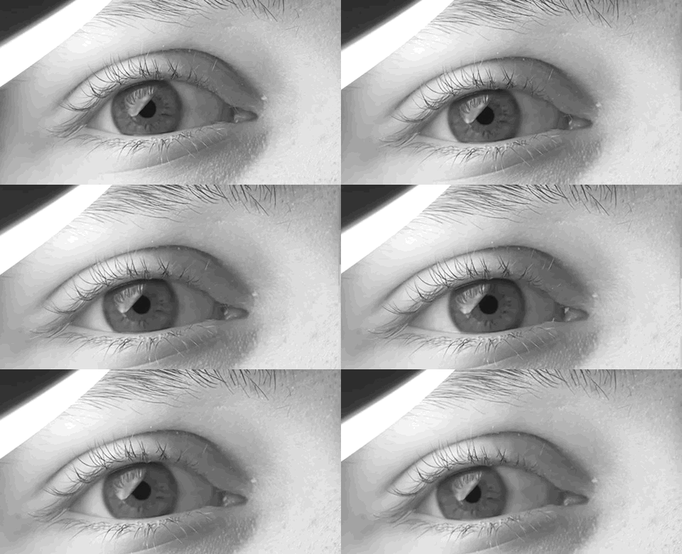
Nystagmus is a condition of involuntary (or voluntary, in some cases) eye movement. Infants can be born with it but more commonly acquire it in infancy or later in life. In many cases it may result in reduced or limited vision. Due to the involuntary movement of the eye, it has been called "dancing eyes". In normal eyesight, while the head rotates about an axis, distant visual images are sustained by rotating eyes in the opposite direction of the respective axis. The semicircular canals in the vestibule of the ear sense angular acceleration, and send signals to the nuclei for eye movement in the brain. From here, a signal is relayed to the extraocular muscles to allow one's gaze to fix on an object as the head moves. Nystagmus occurs when the semicircular canals are stimulated (e.g., by means of the caloric test, or by disease) while the head is stationary. The direction of ocular movement is related to the semicircular canal that is being stimulated. There are two key form ... [...More Info...] [...Related Items...] OR: [Wikipedia] [Google] [Baidu] |
Vertigo
Vertigo is a condition where a person has the sensation of movement or of surrounding objects moving when they are not. Often it feels like a spinning or swaying movement. This may be associated with nausea, vomiting, sweating, or difficulties walking. It is typically worse when the head is moved. Vertigo is the most common type of dizziness. The most common disorders that result in vertigo are benign paroxysmal positional vertigo (BPPV), Ménière's disease, and labyrinthitis. Less common causes include stroke, brain tumors, brain injury, multiple sclerosis, migraines, trauma, and uneven pressures between the middle ears. Physiologic vertigo may occur following being exposed to motion for a prolonged period such as when on a ship or simply following spinning with the eyes closed. Other causes may include toxin exposures such as to carbon monoxide, alcohol, or aspirin. Vertigo typically indicates a problem in a part of the vestibular system. Other causes of dizziness incl ... [...More Info...] [...Related Items...] OR: [Wikipedia] [Google] [Baidu] |
Neuroscience Information Framework
The Neuroscience Information Framework is a repository of global neuroscience web resources, including experimental, clinical, and translational neuroscience databases, knowledge bases, atlases, and genetic/ genomic resources and provides many authoritative links throughout the neuroscience portal of Wikipedia. Description The Neuroscience Information Framework (NIF) is an initiative of the NIH Blueprint for Neuroscience Research, which was established in 2004 by the National Institutes of Health. Development of the NIF started in 2008, when the University of California, San Diego School of Medicine obtained an NIH contract to create and maintain "a dynamic inventory of web-based neurosciences data, resources, and tools that scientists and students can access via any computer connected to the Internet". The project is headed by Maryann Martone, co-director of the National Center for Microscopy and Imaging Research (NCMIR), part of the multi-disciplinary Center for Research in Bio ... [...More Info...] [...Related Items...] OR: [Wikipedia] [Google] [Baidu] |


