|
Femoral Triangle
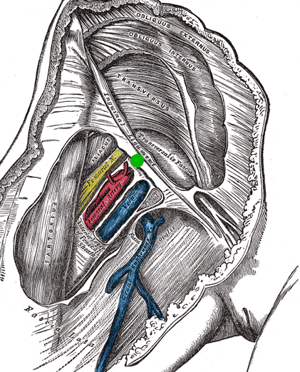
The femoral triangle (or Scarpa's triangle) is an anatomical region of the upper third of the thigh. It is a subfascial space which appears as a triangular depression below the inguinal ligament when the thigh is flexed, abducted and laterally rotated. Structure The femoral triangle is bounded: * superiorly (also known as the base) by the inguinal ligament. * medially by the medial border of the adductor longus muscle. (Some people consider the femoral triangle to be smaller hence the medial border being at the lateral border of the adductor longus muscle.) * laterally by the medial border of the sartorius muscle. The apex of the triangle is continuous with the adductor canal. The roof is formed by the skin, superficial fascia, and deep fascia (fascia lata). The superficial fascia contains the superficial inguinal lymph nodes, femoral branch of the genitofemoral nerve, branches of the ilioinguinal nerve, superficial branches of the femoral artery with accompanying veins, and u ... [...More Info...] [...Related Items...] OR: [Wikipedia] [Google] [Baidu] |
Femoral Vein
In the human body, the femoral vein is a blood vessel that accompanies the femoral artery in the femoral sheath. It begins at the adductor hiatus (an opening in the adductor magnus muscle) as the continuation of the popliteal vein. It ends at the inferior margin of the inguinal ligament where it becomes the external iliac vein. The femoral vein bears valves which are mostly bicuspid and whose number is variable between individuals and often between left and right leg. Structure Segments *The common femoral vein is the segment of the femoral vein between the branching point of the deep femoral vein and the inferior margin of the inguinal ligament.Page 590 in: *The subsartorial vein or superficial femoral vein are designat ... [...More Info...] [...Related Items...] OR: [Wikipedia] [Google] [Baidu] |
Mid-inguinal Point
The mid-inguinal point (MIP) is located on the inguinal ligament, halfway between the anterior superior iliac spine (ASIS) and the pubic symphysis. It is not to be confused with the midpoint of the inguinal ligament itself, which is located halfway between the anterior superior iliac spine and the pubic tubercle. Significance The external iliac artery becomes the femoral artery The femoral artery is a large artery in the thigh and the main arterial supply to the thigh and leg. The femoral artery gives off the deep femoral artery or profunda femoris artery and descends along the anteromedial part of the thigh in the f ... when it passes deep to the inguinal ligament, at the mid-inguinal point. As such, the point is along the superior boundary of the femoral triangle. References {{Authority control Abdomen Ligaments ... [...More Info...] [...Related Items...] OR: [Wikipedia] [Google] [Baidu] |
Venipuncture
In medicine, venipuncture or venepuncture is the process of obtaining intravenous access for the purpose of venous blood sampling (also called '' phlebotomy'') or intravenous therapy. In healthcare, this procedure is performed by medical laboratory scientists, medical practitioners, some EMTs, paramedics, phlebotomists, dialysis technicians, and other nursing staff. In veterinary medicine, the procedure is performed by veterinarians and veterinary technicians. It is essential to follow a standard procedure for the collection of blood specimens to get accurate laboratory results. Any error in collecting the blood or filling the test tubes may lead to erroneous laboratory results. Venipuncture is one of the most routinely performed invasive procedures and is carried out for any of five reasons: # to obtain blood for diagnostic purposes; # to monitor levels of blood components; # to administer therapeutic treatments including medications, nutrition, or chemotherapy; # to rem ... [...More Info...] [...Related Items...] OR: [Wikipedia] [Google] [Baidu] |
Angioplasty
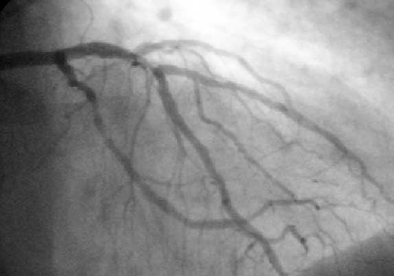
Angioplasty, is also known as balloon angioplasty and percutaneous transluminal angioplasty (PTA), is a minimally invasive endovascular procedure used to widen narrowed or obstructed arteries or veins, typically to treat arterial atherosclerosis. A deflated balloon attached to a catheter (a balloon catheter) is passed over a guide-wire into the narrowed vessel and then inflated to a fixed size. The balloon forces expansion of the blood vessel and the surrounding muscular wall, allowing an improved blood flow. A stent may be inserted at the time of ballooning to ensure the vessel remains open, and the balloon is then deflated and withdrawn. Angioplasty has come to include all manner of vascular interventions that are typically performed percutaneously. The word is composed of the combining forms of the Greek words ἀνγεῖον ' "vessel" or "cavity" (of the human body) and πλάσσω ' "form" or "mould". Uses and indications Coronary angioplasty A coronary an ... [...More Info...] [...Related Items...] OR: [Wikipedia] [Google] [Baidu] |
Clitoris
The clitoris ( or ) is a female sex organ present in mammals, ostriches and a limited number of other animals. In humans, the visible portion – the glans – is at the front junction of the labia minora (inner lips), above the opening of the urethra. Unlike the penis, the male homologue (equivalent) to the clitoris, it usually does not contain the distal portion (or opening) of the urethra and is therefore not used for urination. In most species, the clitoris lacks any reproductive function. While few animals urinate through the clitoris or use it reproductively, the spotted hyena, which has an especially large clitoris, urinates, mates, and gives birth via the organ. Some other mammals, such as lemurs and spider monkeys, also have a large clitoris. The clitoris is the human female's most sensitive erogenous zone and generally the primary anatomical source of human female sexual pleasure. In humans and other mammals, it develops from an outgrowth in the em ... [...More Info...] [...Related Items...] OR: [Wikipedia] [Google] [Baidu] |
Glans Penis
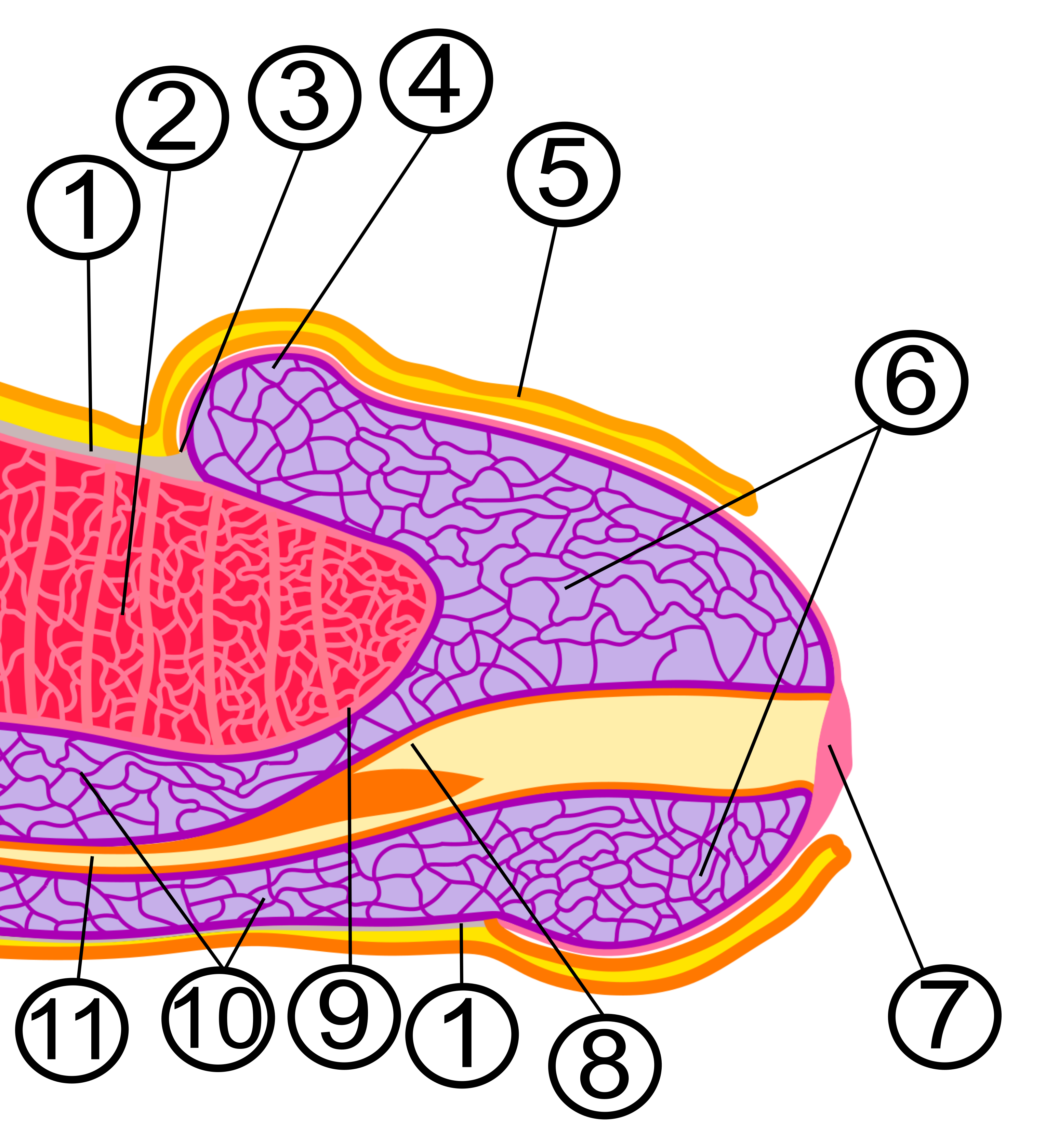
In male human anatomy, the glans penis, commonly referred to as the glans, is the bulbous structure at the distal end of the human penis that is the human male's most sensitive erogenous zone and their primary anatomical source of sexual pleasure. It is anatomically homologous to the clitoral glans. The glans penis is part of the male reproductive organs in humans and other mammals where it may appear smooth, spiny, elongated or divided. It is externally lined with mucosal tissue, which creates a smooth texture and glossy appearance. In humans, the glans is a continuation of the corpus spongiosum of the penis. At the summit appears the urinary meatus and at the base forms the corona glandis. An elastic band of tissue, known as the frenulum, runs on its ventral surface. In men who are not circumcised, it is completely or partially covered by the foreskin. In adults, the foreskin can generally be retracted over and past the glans manually or sometimes automatically dur ... [...More Info...] [...Related Items...] OR: [Wikipedia] [Google] [Baidu] |
Femoral Canal
The femoral canal is the medial (and smallest) compartment of the three compartments of the femoral sheath. It is conical in shape. The femoral canal contains lymphatic vessels, and adipose and loose connective tissue, as well as - sometimes - a deep inguinal lymph node. The function of the femoral canal is to accommodate the distension of the femoral vein when venous return from the leg is increased or temporarily restricted (e.g. during a Valsalva maneuver). The proximal, abdominal end of the femoral canal forms the femoral ring. The femoral canal should not be confused with the nearby adductor canal. Anatomy The femoral canal is bordered: * anterosuperiorly by the inguinal ligament * posteriorly by the pectineal ligament lying anterior to the superior pubic ramus * Medially by the lacunar ligament * Laterally by the femoral vein Physiological significance The position of the femoral canal medially to the femoral vein is of physiologic importance. The space of the canal a ... [...More Info...] [...Related Items...] OR: [Wikipedia] [Google] [Baidu] |
Deep Inguinal Lymph Nodes
Inguinal lymph nodes are lymph nodes in the human groin. Located in the femoral triangle of the inguinal region, they are grouped into superficial and deep lymph nodes. The superficial have three divisions: the superomedial, superolateral, and inferior superficial. Superficial inguinal lymph nodes * The superficial inguinal lymph nodes are the inguinal lymph nodes that form a chain immediately below the inguinal ligament. They lie deep to the fascia of Camper that overlies the femoral vessels at the medial aspect of the thigh. They are bounded superiorly by the inguinal ligament in the femoral triangle; laterally by the border of the sartorius muscle, and medially by the adductor longus muscle. They are divided into three groups: * inferior – inferior of the saphenous opening of the leg, receive drainage from lower legs * superolateral – on the side of the saphenous opening, receive drainage from the side buttocks and the lower abdominal wall. * superomedial – located at ... [...More Info...] [...Related Items...] OR: [Wikipedia] [Google] [Baidu] |
Great Saphenous Vein
The great saphenous vein (GSV, alternately "long saphenous vein"; ) is a large, subcutaneous, superficial vein of the leg. It is the longest vein in the body, running along the length of the lower limb, returning blood from the foot, leg and thigh to the deep femoral vein at the femoral triangle. Structure The great saphenous vein originates from where the dorsal vein of the big toe (the hallux) merges with the dorsal venous arch of the foot. After passing in front of the medial malleolus (where it often can be visualized and palpated), it runs up the medial side of the leg. At the knee, it runs over the posterior border of the medial epicondyle of the femur bone. In the proximal anterior thigh inferolateral to the pubic tubercle, the great saphenous vein dives down deep through the cribriform fascia of the saphenous opening to join the femoral vein. It forms an arch, the saphenous arch, to join the common femoral vein in the region of the femoral triangle at the saphe ... [...More Info...] [...Related Items...] OR: [Wikipedia] [Google] [Baidu] |
Femoral Vein
In the human body, the femoral vein is a blood vessel that accompanies the femoral artery in the femoral sheath. It begins at the adductor hiatus (an opening in the adductor magnus muscle) as the continuation of the popliteal vein. It ends at the inferior margin of the inguinal ligament where it becomes the external iliac vein. The femoral vein bears valves which are mostly bicuspid and whose number is variable between individuals and often between left and right leg. Structure Segments *The common femoral vein is the segment of the femoral vein between the branching point of the deep femoral vein and the inferior margin of the inguinal ligament.Page 590 in: *The subsartorial vein or superficial femoral vein are designat ... [...More Info...] [...Related Items...] OR: [Wikipedia] [Google] [Baidu] |
Pelvic Bone
The hip bone (os coxae, innominate bone, pelvic bone or coxal bone) is a large flat bone, constricted in the center and expanded above and below. In some vertebrates (including humans before puberty) it is composed of three parts: the ilium, ischium, and the pubis. The two hip bones join at the pubic symphysis and together with the sacrum and coccyx (the pelvic part of the spine) comprise the skeletal component of the pelvis – the pelvic girdle which surrounds the pelvic cavity. They are connected to the sacrum, which is part of the axial skeleton, at the sacroiliac joint. Each hip bone is connected to the corresponding femur (thigh bone) (forming the primary connection between the bones of the lower limb and the axial skeleton) through the large ball and socket joint of the hip. Structure The hip bone is formed by three parts: the ilium, ischium, and pubis. At birth, these three components are separated by hyaline cartilage. They join each other in a Y-shaped portion of ... [...More Info...] [...Related Items...] OR: [Wikipedia] [Google] [Baidu] |


.png)


