|
Fascia Lata
The fascia lata is the deep fascia of the thigh. It encloses the thigh muscles and forms the outer limit of the fascial compartments of thigh, which are internally separated by the medial intermuscular septum and the lateral intermuscular septum. The fascia lata is thickened at its lateral side where it forms the iliotibial tract, a structure that runs to the tibia and serves as a site of muscle attachment. Structure The fascia lata is an investment for the whole of the thigh, but varies in thickness in different parts. It is thicker in the upper and lateral part of the thigh, where it receives a fibrous expansion from the gluteus maximus, and where the tensor fasciae latae is inserted between its layers; it is very thin behind and at the upper and medial part, where it covers the adductor muscles, and again becomes stronger around the knee, receiving fibrous expansions from the tendon of the biceps femoris laterally, from the sartorius medially, and from the quadriceps f ... [...More Info...] [...Related Items...] OR: [Wikipedia] [Google] [Baidu] |
Deep Fascia
Deep fascia (or investing fascia) is a fascia, a layer of dense connective tissue that can surround individual muscles and groups of muscles to separate into fascial compartments. This fibrous connective tissue interpenetrates and surrounds the muscles, bones, nerves, and blood vessels of the body. It provides connection and communication in the form of aponeuroses, ligaments, tendons, retinacula, joint capsules, and septa. The deep fasciae envelop all bone (periosteum and endosteum); cartilage ( perichondrium), and blood vessels (tunica externa) and become specialized in muscles (epimysium, perimysium, and endomysium) and nerves ( epineurium, perineurium, and endoneurium). The high density of collagen fibers gives the deep fascia its strength and integrity. The amount of elastin fiber determines how much extensibility and resilience it will have. Examples Examples include: * Fascia lata * Deep fascia of leg * Brachial fascia * Buck's fascia Fascial dynamics Deep f ... [...More Info...] [...Related Items...] OR: [Wikipedia] [Google] [Baidu] |
Inguinal Ligament
The inguinal ligament (), also known as Poupart's ligament or groin ligament, is a band running from the pubic tubercle to the anterior superior iliac spine. It forms the base of the inguinal canal through which an indirect inguinal hernia may develop. Structure The inguinal ligament runs from the anterior superior iliac crest of the ilium to the pubic tubercle of the pubic bone. It is formed by the external abdominal oblique aponeurosis and is continuous with the fascia lata of the thigh. There is some dispute over the attachments. Structures that pass deep to the inguinal ligament include: *Psoas major, iliacus, pectineus * Femoral nerve, artery, and vein * Lateral cutaneous nerve of thigh *Lymphatics Function The ligament serves to contain soft tissues as they course anteriorly from the trunk to the lower extremity. This structure demarcates the superior border of the femoral triangle. It demarcates the inferior border of the inguinal triangle. The midpoint of ... [...More Info...] [...Related Items...] OR: [Wikipedia] [Google] [Baidu] |
Quadriceps Femoris Muscle
The quadriceps femoris muscle (, also called the quadriceps extensor, quadriceps or quads) is a large muscle group that includes the four prevailing muscles on the front of the thigh. It is the sole extensor muscle of the knee, forming a large fleshy mass which covers the front and sides of the femur. The name derives . Structure Parts The quadriceps femoris muscle is subdivided into four separate muscles (the 'heads'), with the first superficial to the other three over the femur (from the trochanters to the condyles): *The rectus femoris muscle occupies the middle of the thigh, covering most of the other three quadriceps muscles. It originates on the ilium. It is named for its straight course. *The vastus lateralis muscle is on the ''lateral side'' of the femur (i.e. on the outer side of the thigh). *The vastus medialis muscle is on the ''medial side'' of the femur (i.e. on the inner part thigh). *The vastus intermedius muscle lies between vastus lateralis and vastus medi ... [...More Info...] [...Related Items...] OR: [Wikipedia] [Google] [Baidu] |
Patella
The patella, also known as the kneecap, is a flat, rounded triangular bone which articulates with the femur (thigh bone) and covers and protects the anterior articular surface of the knee joint. The patella is found in many tetrapods, such as mice, cats, birds and dogs, but not in whales, or most reptiles. In humans, the patella is the largest sesamoid bone (i.e., embedded within a tendon or a muscle) in the body. Babies are born with a patella of soft cartilage which begins to ossify into bone at about four years of age. Structure The patella is a sesamoid bone roughly triangular in shape, with the apex of the patella facing downwards. The apex is the most inferior (lowest) part of the patella. It is pointed in shape, and gives attachment to the patellar ligament. The front and back surfaces are joined by a thin margin and towards centre by a thicker margin. The tendon of the quadriceps femoris muscle attaches to the base of the patella., with the vastus intermediu ... [...More Info...] [...Related Items...] OR: [Wikipedia] [Google] [Baidu] |
Head Of The Fibula
The fibula or calf bone is a leg bone on the lateral side of the tibia, to which it is connected above and below. It is the smaller of the two bones and, in proportion to its length, the most slender of all the long bones. Its upper extremity is small, placed toward the back of the head of the tibia, below the knee joint and excluded from the formation of this joint. Its lower extremity inclines a little forward, so as to be on a plane anterior to that of the upper end; it projects below the tibia and forms the lateral part of the ankle joint. Structure The bone has the following components: * Lateral malleolus * Interosseous membrane connecting the fibula to the tibia, forming a syndesmosis joint * The superior tibiofibular articulation is an arthrodial joint between the lateral condyle of the tibia and the head of the fibula. * The inferior tibiofibular articulation (tibiofibular syndesmosis) is formed by the rough, convex surface of the medial side of the lower end of the ... [...More Info...] [...Related Items...] OR: [Wikipedia] [Google] [Baidu] |
Condyle (anatomy)
A condyle (; in Merriam-Webster Online Dictionary '. la, condylus, from el, kondylos; κόνδυλος knuckle) is the round prominence at the end of a , most often part of a joint – an articulation with another bone. It is one of the markings or features of bones, and can refer to: * On the , in the knee joi ... [...More Info...] [...Related Items...] OR: [Wikipedia] [Google] [Baidu] |
Knee Joint
In humans and other primates, the knee joins the thigh with the human leg, leg and consists of two joints: one between the femur and tibia (tibiofemoral joint), and one between the femur and patella (patellofemoral joint). It is the largest joint in the human body. The knee is a modified hinge joint, which permits flexion and extension (kinesiology), extension as well as slight internal and external rotation. The knee is vulnerable to injury and to the development of osteoarthritis. It is often termed a ''compound joint'' having tibiofemoral and patellofemoral components. (The fibular collateral ligament is often considered with tibiofemoral components.) Structure The knee is a modified hinge joint, a type of synovial joint, which is composed of three functional compartments: the patellofemoral articulation, consisting of the patella, or "kneecap", and the patellar groove on the front of the femur through which it slides; and the medial and lateral tibiofemoral articulation ... [...More Info...] [...Related Items...] OR: [Wikipedia] [Google] [Baidu] |
Hip Joint
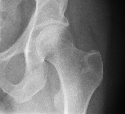
In vertebrate anatomy, hip (or "coxa"Latin ''coxa'' was used by Celsus in the sense "hip", but by Pliny the Elder in the sense "hip bone" (Diab, p 77) in medical terminology) refers to either an anatomical region or a joint. The hip region is located lateral and anterior to the gluteal region, inferior to the iliac crest, and overlying the greater trochanter of the femur, or "thigh bone". In adults, three of the bones of the pelvis have fused into the hip bone or acetabulum which forms part of the hip region. The hip joint, scientifically referred to as the acetabulofemoral joint (''art. coxae''), is the joint between the head of the femur and acetabulum of the pelvis and its primary function is to support the weight of the body in both static (e.g., standing) and dynamic (e.g., walking or running) postures. The hip joints have very important roles in retaining balance, and for maintaining the pelvic inclination angle. Pain of the hip may be the result of numerous cause ... [...More Info...] [...Related Items...] OR: [Wikipedia] [Google] [Baidu] |
Iliotibial Band
The iliotibial tract or iliotibial band (ITB; also known as Maissiat's band or the IT band) is a longitudinal fibrous reinforcement of the fascia lata. The action of the muscles associated with the ITB (tensor fasciae latae and some fibers of gluteus maximus) flex, extend, abduct, and laterally and medially rotate the hip. The ITB contributes to lateral knee stabilization. During knee extension the ITB moves anterior to the lateral condyle of the femur, while ~30 degrees knee flexion, the ITB moves posterior to the lateral condyle. However, it has been suggested that this is only an illusion due to the changing tension in the anterior and posterior fibers during movement. It originates at the anterolateral iliac tubercle portion of the external lip of the iliac crest and inserts at the lateral condyle of the tibia at Gerdy's tubercle. The figure shows only the proximal part of the iliotibial tract. The part of the iliotibial band which lies beneath the tensor fasciae latae is p ... [...More Info...] [...Related Items...] OR: [Wikipedia] [Google] [Baidu] |
Gluteus Medius
The gluteus medius, one of the three gluteal muscles, is a broad, thick, radiating muscle. It is situated on the outer surface of the pelvis. Its posterior third is covered by the gluteus maximus, its anterior two-thirds by the gluteal aponeurosis, which separates it from the superficial fascia and integument. Structure The gluteus medius muscle starts, or "originates", on the outer surface of the ilium between the iliac crest and the posterior gluteal line above, and the anterior gluteal line below; the gluteus medius also originates from the gluteal aponeurosis that covers its outer surface. The fibers of the muscle converge into a strong flattened tendon that inserts on the lateral surface of the greater trochanter. More specifically, the muscle's tendon inserts into an oblique ridge that runs downward and forward on the lateral surface of the greater trochanter. Relations A bursa separates the tendon of the muscle from the surface of the trochanter over which it glides. V ... [...More Info...] [...Related Items...] OR: [Wikipedia] [Google] [Baidu] |
Sacrotuberous Ligament
The sacrotuberous ligament (great or posterior sacrosciatic ligament) is situated at the lower and back part of the pelvis. It is flat, and triangular in form; narrower in the middle than at the ends. Structure It runs from the sacrum (the lower transverse sacral tubercles, the inferior margins sacrum and the upper coccyx) to the tuberosity of the ischium. It is a remnant of part of Biceps femoris muscle. The sacrotuberous ligament is attached by its broad base to the posterior superior iliac spine, the posterior sacroiliac ligaments (with which it is partly blended), to the lower transverse sacral tubercles and the lateral margins of the lower sacrum and upper coccyx. Its oblique fibres descend laterally, converging to form a thick, narrow band that widens again below and is attached to the medial margin of the ischial tuberosity. It then spreads along the ischial ramus as the falciform process, whose concave edge blends with the fascial sheath of the internal pudendal vessels ... [...More Info...] [...Related Items...] OR: [Wikipedia] [Google] [Baidu] |
Ischium
The ischium () forms the lower and back region of the hip bone (''os coxae''). Situated below the ilium and behind the pubis, it is one of three regions whose fusion creates the coxal bone. The superior portion of this region forms approximately one-third of the . Structure The i ...[...More Info...] [...Related Items...] OR: [Wikipedia] [Google] [Baidu] |


