|
Facial Vein
The facial vein (or anterior facial vein) is a relatively large vein in the human face. It commences at the side of the root of the nose and is a direct continuation of the angular vein where it also receives a small nasal branch. It lies behind the facial artery and follows a less tortuous course. It receives blood from the external palatine vein before it either joins the anterior branch of the retromandibular vein to form the common facial vein, or drains directly into the internal jugular vein. A common misconception states that the facial vein has no valves, but this has been contradicted by recent studies. Its walls are not so flaccid as most superficial veins. Path From its origin it runs obliquely downward and backward, beneath the zygomaticus major muscle and zygomatic head of the quadratus labii superioris, descends along the anterior border and then on the superficial surface of the masseter, crosses over the body of the mandible, and passes obliquely backward, benea ... [...More Info...] [...Related Items...] OR: [Wikipedia] [Google] [Baidu] |
Masseter
In human anatomy, the masseter is one of the muscles of mastication. Found only in mammals, it is particularly powerful in herbivores to facilitate chewing of plant matter. The most obvious muscle of mastication is the masseter muscle, since it is the most superficial and one of the strongest. Structure The masseter is a thick, somewhat quadrilateral muscle, consisting of three heads, superficial, deep and coronoid. The fibers of superficial and deep heads are continuous at their insertion. Superficial head The superficial head, the larger, arises by a thick, tendinous aponeurosis from the temporal process of the zygomatic bone, and from the anterior two-thirds of the inferior border of the zygomatic arch. Its fibers pass inferior and posterior, to be inserted into the angle of the mandible and inferior half of the lateral surface of the ramus of the mandible. Deep head The deep head is much smaller, and more muscular in texture. It arises from the posterior third of the lower ... [...More Info...] [...Related Items...] OR: [Wikipedia] [Google] [Baidu] |
Superficial Veins
Superficial veins are veins that are close to the surface of the body, as opposed to deep veins, which are far from the surface. Superficial veins are not paired with an artery, unlike the deep veins, which are typically associated with an artery of the same name. Superficial veins are important physiologically for cooling of the body. When the body is too hot, the body shunts blood from the deep veins to the superficial veins to facilitate heat transfer to the body's surroundings. Superficial veins are often visible underneath the skin. Those below the level of the heart tend to bulge out, which can be readily witnessed in the hand, where the veins bulge significantly less after the arm has been raised above the head for a short time. Veins become more visually prominent when lifting heavy weight, especially after a period of proper strength training. Physiologically, the superficial veins are not as important as the deep veins (as they carry less blood) and are sometimes r ... [...More Info...] [...Related Items...] OR: [Wikipedia] [Google] [Baidu] |
Dissection
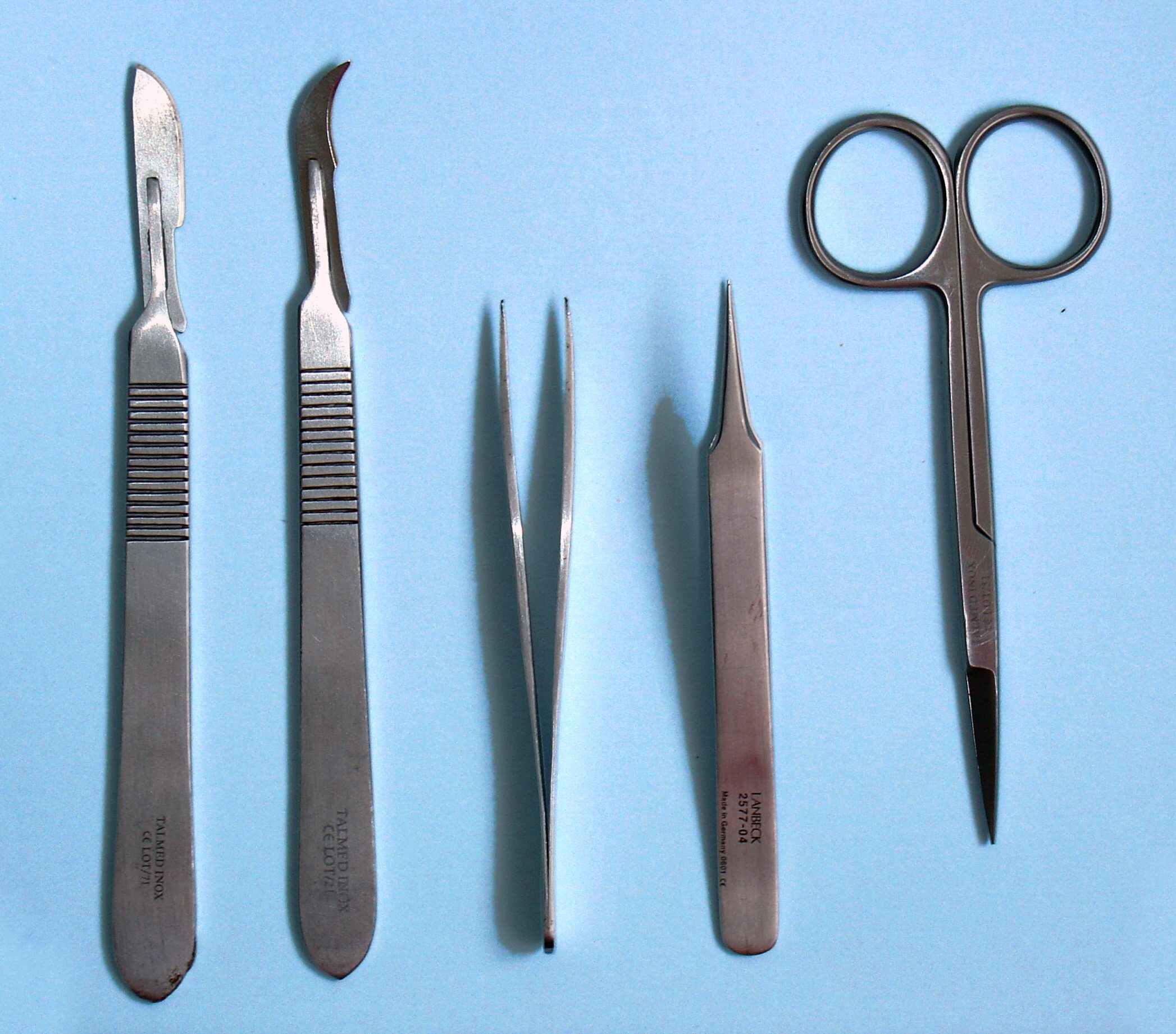
Dissection (from Latin ' "to cut to pieces"; also called anatomization) is the dismembering of the body of a deceased animal or plant to study its anatomical structure. Autopsy is used in pathology and forensic medicine to determine the cause of death in humans. Less extensive dissection of plants and smaller animals preserved in a formaldehyde solution is typically carried out or demonstrated in biology and natural science classes in middle school and high school, while extensive dissections of cadavers of adults and children, both fresh and preserved are carried out by medical students in medical schools as a part of the teaching in subjects such as anatomy, pathology and forensic medicine. Consequently, dissection is typically conducted in a morgue or in an anatomy lab. Dissection has been used for centuries to explore anatomy. Objections to the use of cadavers have led to the use of alternatives including virtual dissection of computer models. Overview Plant and animal b ... [...More Info...] [...Related Items...] OR: [Wikipedia] [Google] [Baidu] |
Dural Venous Sinuses
The dural venous sinuses (also called dural sinuses, cerebral sinuses, or cranial sinuses) are venous channels found between the endosteal and meningeal layers of dura mater in the brain. They receive blood from the cerebral veins, receive cerebrospinal fluid (CSF) from the subarachnoid space via arachnoid granulations, and mainly empty into the internal jugular vein. Venous sinuses Structure The walls of the dural venous sinuses are composed of dura mater lined with endothelium, a specialized layer of flattened cells found in blood vessels. They differ from other blood vessels in that they lack a full set of vessel layers (e.g. tunica media) characteristic of arteries and veins. It also lacks valves (in veins; with exception of materno-fetal blood circulation i.e. placental artery and pulmonary arteries both of which carry deoxygenated blood). Clinical relevance The sinuses can be injured by trauma in which damage to the dura mater, may result in blood clot formation (throm ... [...More Info...] [...Related Items...] OR: [Wikipedia] [Google] [Baidu] |
Cavernous Sinus
The cavernous sinus within the human head is one of the dural venous sinuses creating a cavity called the lateral sellar compartment bordered by the temporal bone of the skull and the sphenoid bone, lateral to the sella turcica. Structure The cavernous sinus is one of the dural venous sinuses of the head. It is a network of veins that sit in a cavity. It sits on both sides of the sphenoidal bone and pituitary gland, approximately 1 × 2 cm in size in an adult. The carotid siphon of the internal carotid artery, and cranial nerves III, IV, V (branches V1 and V2) and VI all pass through this blood filled space. Both sides of cavernous sinus is connected to each other via intercavernous sinuses. The cavernous sinus lies in between the inner and outer layers of dura mater. Nearby structures * Above: optic tract, optic chiasma, internal carotid artery. * Inferiorly: foramen lacerum, and the junction of the body and greater wing of sphenoid bone. * Medially: pituitary gla ... [...More Info...] [...Related Items...] OR: [Wikipedia] [Google] [Baidu] |
Thrombophlebitis
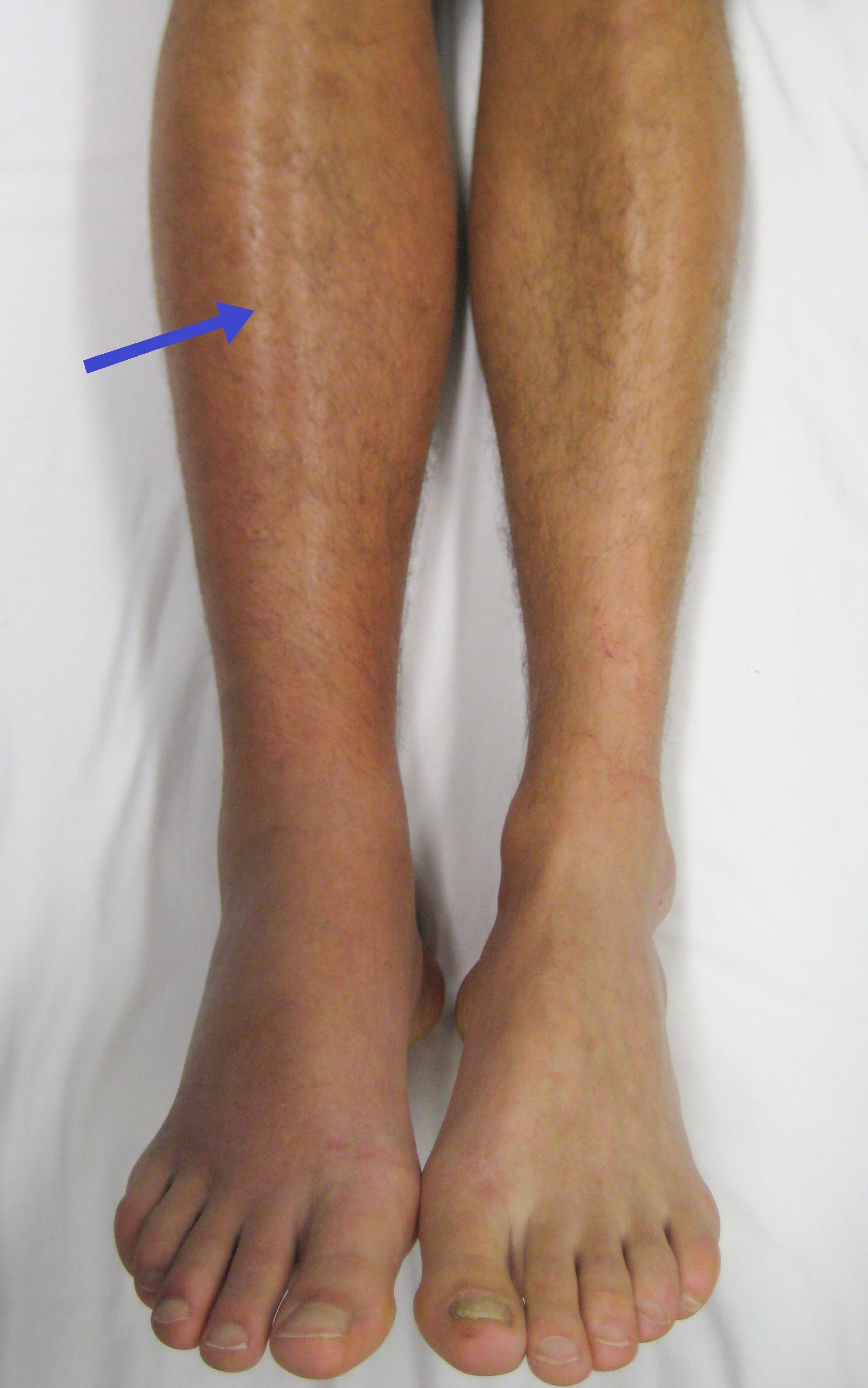
Thrombophlebitis is a phlebitis (inflammation of a vein) related to a thrombus (blood clot). When it occurs repeatedly in different locations, it is known as thrombophlebitis migrans (migratory thrombophlebitis). Signs and symptoms The following symptoms or signs are often associated with thrombophlebitis, although thrombophlebitis is not restricted to the veins of the legs. * Pain (area affected) * Skin redness/inflammation * Edema * Veins hard and cord-like * Tenderness Complications In terms of complications, one of the most serious occurs when the superficial blood clot is associated with a deep vein thrombosis; this can then dislodge, traveling through the heart and occluding the dense capillary network of the lungs This is a pulmonary embolism which can be life-threatening. Causes Thrombophlebitis causes include disorders related to increased tendency for blood clotting and reduced speed of blood in the veins such as prolonged immobility; prolonged traveling (sitting) may ... [...More Info...] [...Related Items...] OR: [Wikipedia] [Google] [Baidu] |
Stylohyoideus
The stylohyoid muscle is a Gracility, slender muscle, lying anterior and Anatomical terms of location#Superior and inferior, superior of the posterior belly of the digastric muscle. It is one of the suprahyoid muscles. It shares this muscle's innervation by the facial nerve, and functions to draw the lingual bone, hyoid bone backwards and elevate the tongue. Its origin is the styloid process of the temporal bone. It inserts on the body of the hyoid. Structure The stylohyoid muscle originates from the posterior and lateral surface of the styloid process (temporal), styloid process of the temporal bone, near the base. Passing inferior and anterior, it inserts into the body of the hyoid bone, at its junction with the greater cornu, and just superior to the omohyoid muscle. It belongs to the group of suprahyoid muscles. It is perforated, near its insertion, by the intermediate tendon of the digastric muscle. The stylohyoid muscle has vascular supply from the lingual artery, a branch ... [...More Info...] [...Related Items...] OR: [Wikipedia] [Google] [Baidu] |
Digastricus
The digastric muscle (also digastricus) (named ''digastric'' as it has two 'bellies') is a small muscle located under the jaw. The term "digastric muscle" refers to this specific muscle. However, other muscles that have two separate muscle bellies include the suspensory muscle of duodenum, omohyoid, occipitofrontalis. It lies below the body of the mandible, and extends, in a curved form, from the mastoid notch to the mandibular symphysis. It belongs to the suprahyoid muscles group. A broad aponeurotic layer is given off from the tendon of the digastric muscle on either side, to be attached to the body and greater cornu of the hyoid bone; this is termed the suprahyoid aponeurosis. Structure The digastricus (digastric muscle) consists of two muscular bellies united by an intermediate rounded tendon. The two bellies of the digastric muscle have different embryological origins, and are supplied by different cranial nerves. Each person has a right and left digastric muscle. In mo ... [...More Info...] [...Related Items...] OR: [Wikipedia] [Google] [Baidu] |
Submandibular Gland
The paired submandibular glands (historically known as submaxillary glands) are major salivary glands located beneath the floor of the mouth. They each weigh about 15 grams and contribute some 60–67% of unstimulated saliva secretion; on stimulation their contribution decreases in proportion as the parotid secretion rises to 50%. The average length of the normal human submandibular salivary gland is approximately 27mm, while the average width is approximately 14.3mm. Structure Lying superior to the digastric muscles, each submandibular gland is divided into superficial and deep lobes, which are separated by the mylohyoid muscle: * The superficial lobe comprises most of the gland, with the mylohyoid muscle runs under it * The deep lobe is the smaller part Secretions are delivered into the submandibular duct on the deep portion after which they hook around the posterior edge of the mylohyoid muscle and proceed on the superior surface laterally. The excretory ducts are then cross ... [...More Info...] [...Related Items...] OR: [Wikipedia] [Google] [Baidu] |
Cervical Fascia
The cervical fascia is fascia found in the region of the neck. It usually refers to the deep cervical fascia. However, there is also a superficial cervical fascia Superficial cervical fascia is a thin layer of subcutaneous connective tissue that lies between the dermis of the skin and the deep cervical fascia. It contains the platysma, cutaneous nerves, blood, and lymphatic vessels. It also contains a varyi .... Fascia {{musculoskeletal-stub ... [...More Info...] [...Related Items...] OR: [Wikipedia] [Google] [Baidu] |
Platysma
The platysma muscle is a superficial muscle of the human neck that overlaps the sternocleidomastoid. It covers the anterior surface of the neck superficially. When it contracts, it produces a slight wrinkling of the neck, and a "bowstring" effect on either side of the neck. Structure The platysma muscle is a broad sheet of muscle arising from the fascia covering the upper parts of the pectoralis major muscle and deltoid muscle. Its fibers cross the clavicle, and proceed obliquely upward and medially along the side of the neck. This leaves the inferior part of the neck in the midline deficient of significant muscle cover. Fibres at the front of the muscle from the left and right sides intermingle together below and behind the mandibular symphysis, the junction where the two lateral halves of the mandible are fused at an early period of life (although not a true symphysis). Fibres at the back of the muscle cross the mandible, some being inserted into the bone below the oblique li ... [...More Info...] [...Related Items...] OR: [Wikipedia] [Google] [Baidu] |
Quadratus Labii Superioris
The levator labii superioris (pl. ''levatores labii superioris'', also called quadratus labii superioris, pl. ''quadrati labii superioris'') is a muscle of the human body used in facial expression. It is a broad sheet, the origin of which extends from the side of the nose to the zygomatic bone. Structure Its medial fibers form the ''angular head'' (also known as the levator labii superioris alaeque nasi muscle,) which arises by a pointed extremity from the upper part of the frontal process of the maxilla and passing obliquely downward and lateralward divides into two slips. One of these is inserted into the greater alar cartilage and skin of the nose; the other is prolonged into the lateral part of the upper lip, blending with the infraorbital head and with the orbicularis oris. The intermediate portion or ''infraorbital head'' arises from the lower margin of the orbit immediately above the infraorbital foramen, some of its fibers being attached to the maxilla, others to the ... [...More Info...] [...Related Items...] OR: [Wikipedia] [Google] [Baidu] |


