|
Epiboly
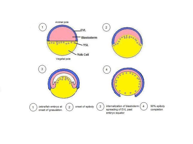
Epiboly describes one of the five major types of cell movements that occur in the gastrulation stage of embryonic development of some organisms. Epiboly is the spreading and thinning of the ectoderm while the endoderm and mesoderm layers move to the inside of the embryo. When undergoing epiboly, a monolayer of cells must undergo a physical change in shape in order to spread. Alternatively, multiple layers of cells can also undergo epiboly as the position of cells is changed or the cell layers undergo intercalation. While human embryos do not experience epiboly, this movement can be studied in sea urchins, tunicates, amphibians, and most commonly zebrafish. Zebrafish General movements Epiboly in zebrafish is the first coordinated cell movement, and begins once the embryo has completed the blastula stage. At this point the zebrafish embryo contains three portions: an epithelial monolayer known as the enveloping layer (EVL), a yolk syncytial layer (YSL) which is a membrane-encl ... [...More Info...] [...Related Items...] OR: [Wikipedia] [Google] [Baidu] |
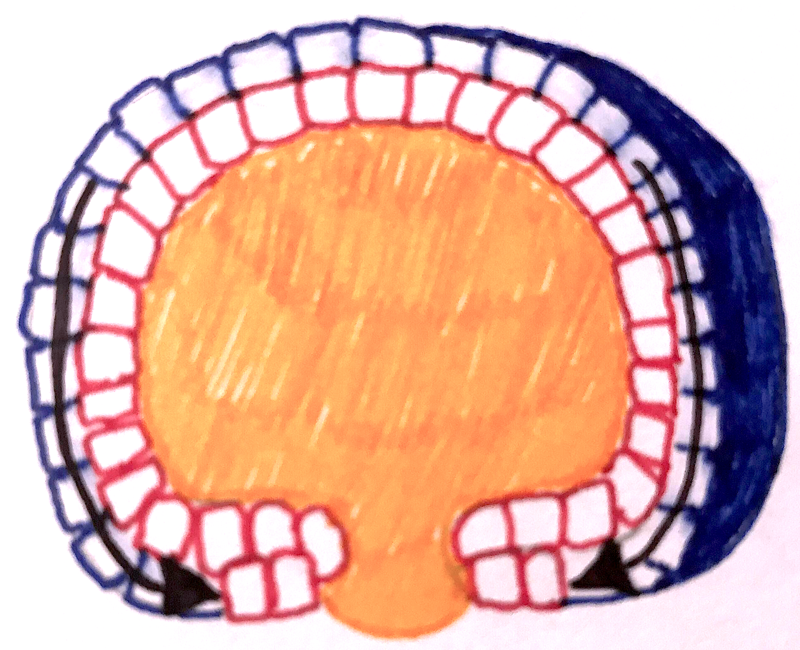
Epiboly Drawing
Epiboly describes one of the five major types of cell movements that occur in the gastrulation stage of embryonic development of some organisms. Epiboly is the spreading and thinning of the ectoderm while the endoderm and mesoderm layers move to the inside of the embryo. When undergoing epiboly, a monolayer of cells must undergo a physical change in shape in order to spread. Alternatively, multiple layers of cells can also undergo epiboly as the position of cells is changed or the cell layers undergo intercalation. While human embryos do not experience epiboly, this movement can be studied in sea urchins, tunicates, amphibians, and most commonly zebrafish. Zebrafish General movements Epiboly in zebrafish is the first coordinated cell movement, and begins once the embryo has completed the blastula stage. At this point the zebrafish embryo contains three portions: an epithelial monolayer known as the enveloping layer (EVL), a yolk syncytial layer (YSL) which is a membrane-enclosed ... [...More Info...] [...Related Items...] OR: [Wikipedia] [Google] [Baidu] |
Zebrafish
The zebrafish (''Danio rerio'') is a freshwater fish belonging to the minnow family ( Cyprinidae) of the order Cypriniformes. Native to South Asia, it is a popular aquarium fish, frequently sold under the trade name zebra danio (and thus often called a "tropical fish" although both tropical and subtropical). It is also found in private ponds. The zebrafish is an important and widely used vertebrate model organism in scientific research, for example in drug development, in particular pre-clinical development. It is also notable for its regenerative abilities, and has been modified by researchers to produce many transgenic strains. Taxonomy The zebrafish is a derived member of the genus '' Brachydanio'', of the family Cyprinidae. It has a sister-group relationship with ''Danio aesculapii''. Zebrafish are also closely related to the genus ''Devario'', as demonstrated by a phylogenetic tree of close species. Distribution Range The zebrafish is native to fresh water h ... [...More Info...] [...Related Items...] OR: [Wikipedia] [Google] [Baidu] |
Ectoderm
The ectoderm is one of the three primary germ layers formed in early embryonic development. It is the outermost layer, and is superficial to the mesoderm (the middle layer) and endoderm (the innermost layer). It emerges and originates from the outer layer of germ cells. The word ectoderm comes from the Greek ''ektos'' meaning "outside", and ''derma'' meaning "skin".Gilbert, Scott F. Developmental Biology. 9th ed. Sunderland, MA: Sinauer Associates, 2010: 333-370. Print. Generally speaking, the ectoderm differentiates to form epithelial and neural tissues (spinal cord, peripheral nerves and brain). This includes the skin, linings of the mouth, anus, nostrils, sweat glands, hair and nails, and tooth enamel. Other types of epithelium are derived from the endoderm. In vertebrate embryos, the ectoderm can be divided into two parts: the dorsal surface ectoderm also known as the external ectoderm, and the neural plate, which invaginates to form the neural tube and neural crest. Th ... [...More Info...] [...Related Items...] OR: [Wikipedia] [Google] [Baidu] |
Gastrulation
Gastrulation is the stage in the early embryonic development of most animals, during which the blastula (a single-layered hollow sphere of cells), or in mammals the blastocyst is reorganized into a multilayered structure known as the gastrula. Before gastrulation, the embryo is a continuous epithelial sheet of cells; by the end of gastrulation, the embryo has begun differentiation to establish distinct cell lineages, set up the basic axes of the body (e.g. dorsal-ventral, anterior-posterior), and internalized one or more cell types including the prospective gut. In triploblastic organisms, the gastrula is trilaminar (three-layered). These three germ layers are the ectoderm (outer layer), mesoderm (middle layer), and endoderm (inner layer).Mundlos 2009p. 422/ref>McGeady, 2004: p. 34 In diploblastic organisms, such as Cnidaria and Ctenophora, the gastrula has only ectoderm and endoderm. The two layers are also sometimes referred to as the ''hypoblast'' and ''epiblast''. Sponges ... [...More Info...] [...Related Items...] OR: [Wikipedia] [Google] [Baidu] |
Amniotes
Amniotes are a clade of tetrapod vertebrates that comprises sauropsids (including all reptiles and birds, and extinct parareptiles and non-avian dinosaurs) and synapsids (including pelycosaurs and therapsids such as mammals). They are distinguished from the other tetrapod clade — the amphibians — by the development of three extraembryonic membranes ( amnion for embryoic protection, chorion for gas exchange, and allantois for metabolic waste disposal or storage), thicker and more keratinized skin, and costal respiration (breathing by expanding/constricting the rib cage). All three main features listed above, namely the presence of an amniotic buffer, water-impermeable cutes and a robust respiratory system, are very important for amniotes to live on land as true terrestrial animals – the ability to reproduce in locations away from water bodies, better homeostasis in drier environments, and more efficient air respiration to power terrestrial locomotions, although they migh ... [...More Info...] [...Related Items...] OR: [Wikipedia] [Google] [Baidu] |
Xenopus Laevis
The African clawed frog (''Xenopus laevis'', also known as the xenopus, African clawed toad, African claw-toed frog or the ''platanna'') is a species of African aquatic frog of the family Pipidae. Its name is derived from the three short claws on each hind foot, which it uses to tear apart its food. The word ''Xenopus'' means 'strange foot' and ''laevis'' means 'smooth'. The species is found throughout much of Sub-Saharan Africa (Nigeria and Sudan to South Africa),Weldon; du Preez; Hyatt; Muller; and Speare (2004). Origin of the Amphibian Chytrid Fungus.' Emerging Infectious Diseases 10(12). and in isolated, introduced populations in North America, South America, Europe, and Asia. All species of the family Pipidae are tongueless, toothless and completely aquatic. They use their hands to shove food in their mouths and down their throats and a hyobranchial pump to draw or suck things in their mouth. Pipidae have powerful legs for swimming and lunging after food. They also use the ... [...More Info...] [...Related Items...] OR: [Wikipedia] [Google] [Baidu] |
Cadherin
Cadherins (named for "calcium-dependent adhesion") are a type of cell adhesion molecule (CAM) that is important in the formation of adherens junctions to allow cells to adhere to each other . Cadherins are a class of type-1 transmembrane proteins, and they are dependent on calcium (Ca2+) ions to function, hence their name. Cell-cell adhesion is mediated by extracellular cadherin domains, whereas the intracellular cytoplasmic tail associates with numerous adaptors and signaling proteins, collectively referred to as the cadherin adhesome. The cadherin family is essential in maintaining the cell-cell contact and regulating cytoskeletal complexes. The cadherin superfamily includes cadherins, protocadherins, desmogleins, desmocollins, and more. In structure, they share ''cadherin repeats'', which are the extracellular Ca2+-binding domains. There are multiple classes of cadherin molecules, each designated with a prefix (in general, noting the types of tissue with which it is associated). ... [...More Info...] [...Related Items...] OR: [Wikipedia] [Google] [Baidu] |
Claudin
Claudins are a family of proteins which, along with occludin, are the most important components of the tight junctions ( zonulae occludentes). Tight junctions establish the paracellular barrier that controls the flow of molecules in the intercellular space between the cells of an epithelium. They have four transmembrane domains, with the N-terminus and the C-terminus in the cytoplasm. Structure Claudins are small (20–24/27 kilodalton (kDa)) transmembrane proteins which are found in many organisms, ranging from nematodes to human beings. They all have a very similar structure. Claudins span the cellular membrane 4 times, with the N-terminal end and the C-terminal end both located in the cytoplasm, and two extracellular loops which show the highest degree of conservation. Claudins have both cis and trans interactions between cell membranes. Cis-interactions is when claudins on the same membrane interact, one way they interact is by transmembrane domain having molecular intera ... [...More Info...] [...Related Items...] OR: [Wikipedia] [Google] [Baidu] |
Tight Junctions
Tight junctions, also known as occluding junctions or ''zonulae occludentes'' (singular, ''zonula occludens''), are multiprotein junctional complexes whose canonical function is to prevent leakage of solutes and water and seals between the epithelial cells. They also play a critical role maintaining the structure and permeability of endothelial cells. Tight junctions may also serve as leaky pathways by forming selective channels for small cations, anions, or water. The corresponding junctions that occur in invertebrates are septate junctions. Structure Tight junctions are composed of a branching network of sealing strands, each strand acting independently from the others. Therefore, the efficiency of the junction in preventing ion passage increases exponentially with the number of strands. Each strand is formed from a row of transmembrane proteins embedded in both plasma membranes, with extracellular domains joining one another directly. There are at least 40 different protei ... [...More Info...] [...Related Items...] OR: [Wikipedia] [Google] [Baidu] |
Cytochalasin B
Cytochalasin B, the name of which comes from the Greek ''cytos'' (cell) and ''chalasis'' (relaxation), is a cell-permeable mycotoxin. It was found that substoichimetric concentrations of cytochalasin B (CB) strongly inhibit network formation by actin filaments. Due to this, it is often used in cytological research. It inhibits cytoplasmic division by blocking the formation of contractile microfilaments. It inhibits cell movement and induces nuclear extrusion. Cytochalasin B shortens actin filaments by blocking monomer addition at the fast-growing end of polymers. Cytochalasin B inhibits glucose transport and platelet aggregation. It blocks adenosine-induced apoptotic body formation without affecting activation of endogenous ADP-ribosylation in leukemia HL-60 cells. It is also used in cloning through nuclear transfer. Here enucleated recipient cells are treated with cytochalasin B. Cytochalasin B makes the cytoplasm of the oocytes more fluid and makes it possible to aspirate the nuc ... [...More Info...] [...Related Items...] OR: [Wikipedia] [Google] [Baidu] |
Nocodazole
Nocodazole is an antineoplastic agent which exerts its effect in cells by interfering with the polymerization of microtubules. Microtubules are one type of fibre which constitutes the cytoskeleton, and the dynamic microtubule network has several important roles in the cell, including vesicular transport, forming the mitotic spindle and in cytokinesis. Several drugs including vincristine and colcemid are similar to nocodazole in that they interfere with microtubule polymerization. Nocodazole has been shown to decrease the oncogenic potential of cancer cells via another microtubules-independent mechanisms. Nocodazole stimulates the expression of LATS2 which potently inhibits the Wnt signaling pathway by abrogating the interaction between the Wnt-dependent transcriptional co-factors beta-catenin and BCL9. It is related to mebendazole by replacement of the left most benzene ring by thiophene. Use in cell biology research As nocodazole affects the cytoskeleton, it is often used in ... [...More Info...] [...Related Items...] OR: [Wikipedia] [Google] [Baidu] |


.jpg)


