|
Ependymoma
An ependymoma is a tumor that arises from the ependyma, a tissue of the central nervous system. Usually, in pediatric cases the location is intracranial, while in adults it is spinal. The common location of intracranial ependymomas is the fourth ventricle. Rarely, ependymomas can occur in the pelvic cavity. Syringomyelia can be caused by an ependymoma. Ependymomas are also seen with neurofibromatosis type II. Signs and symptoms Source: * severe headache * visual loss (due to papilledema) * vomiting * bilateral Babinski sign * drowsiness (after several hours of the above symptoms) * gait change (rotation of feet when walking) * impaction/constipation * back flexibility Morphology Ependymomas are composed of cells with regular, round to oval nuclei. There is a variably dense fibrillary background. Tumor cells may form gland-like round or elongated structures that resemble the embryologic ependymal canal, with long, delicate processes extending into the lumen; more frequently pre ... [...More Info...] [...Related Items...] OR: [Wikipedia] [Google] [Baidu] |
Neurofibromatosis Type II
Neurofibromatosis type II (also known as MISME syndrome – multiple inherited schwannomas, meningiomas, and ependymomas) is a genetic condition that may be inherited or may arise spontaneously, and causes benign tumors of the brain, spinal cord, and peripheral nerves. The types of tumors frequently associated with NF2 include vestibular schwannomas, meningiomas, and ependymomas. The main manifestation of the condition is the development of bilateral benign brain tumors in the nerve sheath of the cranial nerve VIII, which is the "auditory-vestibular nerve" that transmits sensory information from the inner ear to the brain. Besides, other benign brain and spinal tumors occur. Symptoms depend on the presence, localisation and growth of the tumor(s), in which multiple cranial nerves can be involved. Many people with this condition also experience vision problems. Neurofibromatosis type II (NF2 ''or'' NF II) is caused by mutations of the "Merlin" gene, which seems to influence the for ... [...More Info...] [...Related Items...] OR: [Wikipedia] [Google] [Baidu] |
Pseudorosette
In histopathology, a palisade is a single layer of relatively long cells, arranged loosely perpendicular to a surface and parallel to each other. A rosette is a palisade in a halo or spoke-and-wheel arrangement, surrounding a central core or hub. A pseudorosette is a perivascular radial arrangement of neoplastic cells around a small blood vessel. Rosette A ''rosette'' is a cell formation in a halo or spoke-and-wheel arrangement, surrounding a central core or hub. The central hub may consist of an empty-appearing lumen or a space filled with cytoplasmic processes. The cytoplasm of each of the cells in the rosette is often wedge-shaped with the apex directed toward the central core: the nuclei of the cells participating in the rosette are peripherally positioned and form a ring or halo around the hub. Pathogenesis Rosettes may be considered primary or secondary manifestations of tumor architecture. Primary rosettes form as a characteristic growth pattern of a given tumor type wh ... [...More Info...] [...Related Items...] OR: [Wikipedia] [Google] [Baidu] |
Brain Tumor
A brain tumor occurs when abnormal cells form within the brain. There are two main types of tumors: malignant tumors and benign (non-cancerous) tumors. These can be further classified as primary tumors, which start within the brain, and secondary tumors, which most commonly have spread from tumors located outside the brain, known as brain metastasis tumors. All types of brain tumors may produce symptoms that vary depending on the size of the tumor and the part of the brain that is involved. Where symptoms exist, they may include headaches, seizures, problems with vision, vomiting and mental changes. Other symptoms may include difficulty walking, speaking, with sensations, or unconsciousness. The cause of most brain tumors is unknown. Uncommon risk factors include exposure to vinyl chloride, Epstein–Barr virus, ionizing radiation, and inherited syndromes such as neurofibromatosis, tuberous sclerosis, and von Hippel-Lindau Disease. Studies on mobile phone exposure hav ... [...More Info...] [...Related Items...] OR: [Wikipedia] [Google] [Baidu] |
Sacrococcygeal Teratoma
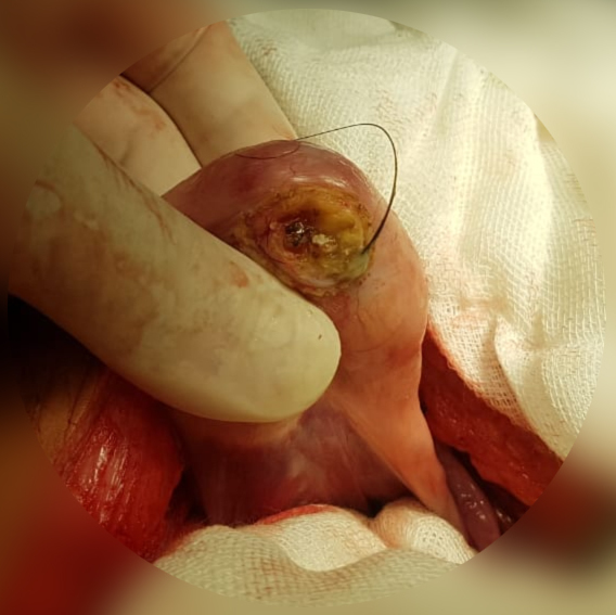
Sacrococcygeal teratoma (SCT) is a type of tumor known as a teratoma that develops at the base of the coccyx (tailbone) and is thought to be primarily derived from remnants of the primitive streak. Sacrococcygeal teratomas are benign 75% of the time, malignant 12% of the time, and the remainder are considered "immature teratomas" that share benign and malignant features. Benign sacrococcygeal teratomas are more likely to develop in younger children who are less than 5 months old, and older children are more likely to develop malignant sacrococcygeal teratomas. The Currarino syndrome, due to an autosomal dominant mutation in the MNX1 gene, consists of a presacral mass (usually a mature teratoma or anterior meningocele), anorectal malformation and sacral dysgenesis. Presentation Complications Maternal complications of pregnancy may include mirror syndrome. Maternal complications of delivery may include a Cesarean section or, alternatively, a vaginal delivery with mechanical dyst ... [...More Info...] [...Related Items...] OR: [Wikipedia] [Google] [Baidu] |
Teratoma
A teratoma is a tumor made up of several different types of tissue, such as hair, muscle, teeth, or bone. Teratomata typically form in the ovary, testicle, or coccyx. Symptoms Symptoms may be minimal if the tumor is small. A testicular teratoma may present as a painless lump. Complications may include ovarian torsion, testicular torsion, or hydrops fetalis. They are a type of germ cell tumor (a tumor that begins in the cells that give rise to sperm or eggs). They are divided into two types: mature and immature. Mature teratomas include dermoid cysts and are generally benign. Immature teratomas may be cancerous. Most ovarian teratomas are mature. In adults, testicular teratomas are generally cancerous. Definitive diagnosis is based on a tissue biopsy. Treatment of coccyx, testicular, and ovarian teratomas is generally by surgery. Testicular and immature ovarian teratomas are also frequently treated with chemotherapy. Teratomas occur in the coccyx in about one in 30,000 n ... [...More Info...] [...Related Items...] OR: [Wikipedia] [Google] [Baidu] |
Neuro-oncology
Neuro-oncology is the study of brain neoplasms, brain and Spinal neoplasms, spinal cord neoplasms, many of which are (at least eventually) very dangerous and life-threatening (astrocytoma, glioma, glioblastoma multiforme, ependymoma, pontine glioma, and brain stem tumors are among the many examples of these). Among the malignant brain cancers, gliomas of the brainstem and pons, glioblastoma multiforme, and Grading (tumors), high-grade (highly anaplastic) astrocytoma/oligodendroglioma are among the worst. In these cases, untreated survival usually amounts to only a few months, and survival with current radiation and chemotherapy treatments may extend that time from around a year to a year and a half, possibly two or more, depending on the patient's condition, immune function, treatments used, and the specific type of malignant brain neoplasm. Surgery may in some cases be curative, but, as a general rule, malignant brain cancers tend to regenerate and emerge from remission (medicine) ... [...More Info...] [...Related Items...] OR: [Wikipedia] [Google] [Baidu] |
Fourth Ventricle
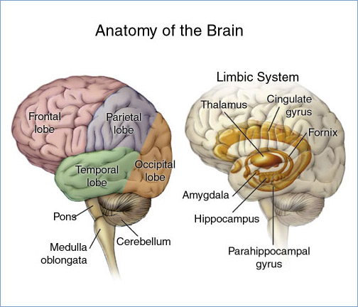
The fourth ventricle is one of the four connected fluid-filled cavities within the human brain. These cavities, known collectively as the ventricular system, consist of the left and right lateral ventricles, the third ventricle, and the fourth ventricle. The fourth ventricle extends from the cerebral aqueduct (''aqueduct of Sylvius'') to the obex, and is filled with cerebrospinal fluid (CSF). The fourth ventricle has a characteristic diamond shape in cross-sections of the human brain. It is located within the pons or in the upper part of the medulla oblongata. CSF entering the fourth ventricle through the cerebral aqueduct can exit to the subarachnoid space of the spinal cord through two lateral apertures and a single, midline median aperture. Boundaries The fourth ventricle has a roof at its ''upper'' (posterior) surface and a floor at its ''lower'' (anterior) surface, and side walls formed by the cerebellar peduncles (nerve bundles joining the structure on the posterior sid ... [...More Info...] [...Related Items...] OR: [Wikipedia] [Google] [Baidu] |
Micrograph
A micrograph or photomicrograph is a photograph or digital image taken through a microscope or similar device to show a magnified image of an object. This is opposed to a macrograph or photomacrograph, an image which is also taken on a microscope but is only slightly magnified, usually less than 10 times. Micrography is the practice or art of using microscopes to make photographs. A micrograph contains extensive details of microstructure. A wealth of information can be obtained from a simple micrograph like behavior of the material under different conditions, the phases found in the system, failure analysis, grain size estimation, elemental analysis and so on. Micrographs are widely used in all fields of microscopy. Types Photomicrograph A light micrograph or photomicrograph is a micrograph prepared using an optical microscope, a process referred to as ''photomicroscopy''. At a basic level, photomicroscopy may be performed simply by connecting a camera to a microscope, th ... [...More Info...] [...Related Items...] OR: [Wikipedia] [Google] [Baidu] |
Septum Pellucidum
The septum pellucidum (Latin for "translucent wall") is a thin, triangular, vertical double membrane separating the anterior horns of the left and right lateral ventricles of the brain. It runs as a sheet from the corpus callosum down to the fornix. The septum is not present in the syndrome septo-optic dysplasia. Structure The septum pellucidum is located in the septal area in the midline of the brain between the two cerebral hemispheres. The septal area is also the location of the septal nuclei. It is attached to the lower part of the corpus callosum, the large collection of nerve fibers that connect the two cerebral hemispheres. It is attached to the front forward part of the fornix. The lateral ventricles sit on either side of the septum. The septum pellucidum consists of two layers or ''laminae'' of both white and gray matter. During fetal development, there is a space between the two laminae called the cave of septum pellucidum that, in ninety percent of cases, disappear ... [...More Info...] [...Related Items...] OR: [Wikipedia] [Google] [Baidu] |
Medulloblastoma
Medulloblastoma is a common type of primary brain cancer in children. It originates in the part of the brain that is towards the back and the bottom, on the floor of the skull, in the cerebellum, or posterior fossa. The brain is divided into two main parts, the larger cerebrum on top and the smaller cerebellum below towards the back. They are separated by a membrane called the tentorium. Tumors that originate in the cerebellum or the surrounding region below the tentorium are, therefore, called infratentorial. Historically medulloblastomas have been classified as a primitive neuroectodermal tumor (PNET), but it is now known that medulloblastoma is distinct from supratentorial PNETs and they are no longer considered similar entities. Medulloblastomas are invasive, rapidly growing tumors that, unlike most brain tumors, spread through the cerebrospinal fluid and frequently metastasize to different locations along the surface of the brain and spinal cord. Metastasis all the way dow ... [...More Info...] [...Related Items...] OR: [Wikipedia] [Google] [Baidu] |


