|
Erectile Tissue
Erectile tissue is tissue in the body with numerous vascular spaces, or cavernous tissue, that may become engorged with blood. However, tissue that is devoid of or otherwise lacking erectile tissue (such as the labia minora, the vestibule/ vagina and the urethra) may also be described as engorging with blood, often with regard to sexual arousal. In the clitoris and penis Erectile tissue exists in places such as the corpora cavernosa of the penis, and in the clitoris or in the bulbs of vestibule. During erection, the corpora cavernosa will become engorged with arterial blood, a process called ''tumescence''.Chapter 35 in: This may result from any of various physiological stimuli, also known as sexual arousal. The corpus spongiosum is a single tubular structure located just below the corpora cavernosa. This may also become slightly engorged with blood, but less so than the corpora cavernosa. In the nose Erectile tissue is present in the anterior part of the nasal septum and ... [...More Info...] [...Related Items...] OR: [Wikipedia] [Google] [Baidu] |
Cavernous Tissue
Cavernous tissue refers to blood-filled spaces lined by endothelium and surrounded by smooth muscle. It is present in the erectile tissue of the penis and clitoris The clitoris ( or ) is a female sex organ present in mammals, ostriches and a limited number of other animals. In humans, the visible portion – the glans – is at the front junction of the labia minora (inner lips), above the o .... References Sexual anatomy {{circulatory-stub ... [...More Info...] [...Related Items...] OR: [Wikipedia] [Google] [Baidu] |
Erection
An erection (clinically: penile erection or penile tumescence) is a physiological phenomenon in which the penis becomes firm, engorged, and enlarged. Penile erection is the result of a complex interaction of psychological, neural, vascular, and endocrine factors, and is often associated with sexual arousal or sexual attraction, although erections can also be spontaneous. The shape, angle, and direction of an erection varies considerably between humans. Physiologically, an erection is required for a male to effect vaginal penetration or sexual intercourse and is triggered by the parasympathetic division of the autonomic nervous system, causing the levels of nitric oxide (a vasodilator) to rise in the trabecular arteries and smooth muscle of the penis. The arteries dilate causing the corpora cavernosa of the penis (and to a lesser extent the corpus spongiosum) to fill with blood; simultaneously the ischiocavernosus and bulbospongiosus muscles compress the veins of the ... [...More Info...] [...Related Items...] OR: [Wikipedia] [Google] [Baidu] |
Autonomic Nervous System
The autonomic nervous system (ANS), formerly referred to as the vegetative nervous system, is a division of the peripheral nervous system that supplies internal organs, smooth muscle and glands. The autonomic nervous system is a control system that acts largely unconsciously and regulates bodily functions, such as the heart rate, its force of contraction, digestion, respiratory rate, pupillary response, urination, and sexual arousal. This system is the primary mechanism in control of the fight-or-flight response. The autonomic nervous system is regulated by integrated reflexes through the brainstem to the spinal cord and organs. Autonomic functions include control of respiration, cardiac regulation (the cardiac control center), vasomotor activity (the vasomotor center), and certain reflex actions such as coughing, sneezing, swallowing and vomiting. Those are then subdivided into other areas and are also linked to autonomic subsystems and the peripheral nervous syst ... [...More Info...] [...Related Items...] OR: [Wikipedia] [Google] [Baidu] |
Smooth Muscle
Smooth muscle is an involuntary non- striated muscle, so-called because it has no sarcomeres and therefore no striations (''bands'' or ''stripes''). It is divided into two subgroups, single-unit and multiunit smooth muscle. Within single-unit muscle, the whole bundle or sheet of smooth muscle cells contracts as a syncytium. Smooth muscle is found in the walls of hollow organs, including the stomach, intestines, bladder and uterus; in the walls of passageways, such as blood, and lymph vessels, and in the tracts of the respiratory, urinary, and reproductive systems. In the eyes, the ciliary muscles, a type of smooth muscle, dilate and contract the iris and alter the shape of the lens. In the skin, smooth muscle cells such as those of the arrector pili cause hair to stand erect in response to cold temperature or fear. Structure Gross anatomy Smooth muscle is grouped into two types: single-unit smooth muscle, also known as visceral smooth muscle, and multiunit sm ... [...More Info...] [...Related Items...] OR: [Wikipedia] [Google] [Baidu] |
Erection Of Nipples
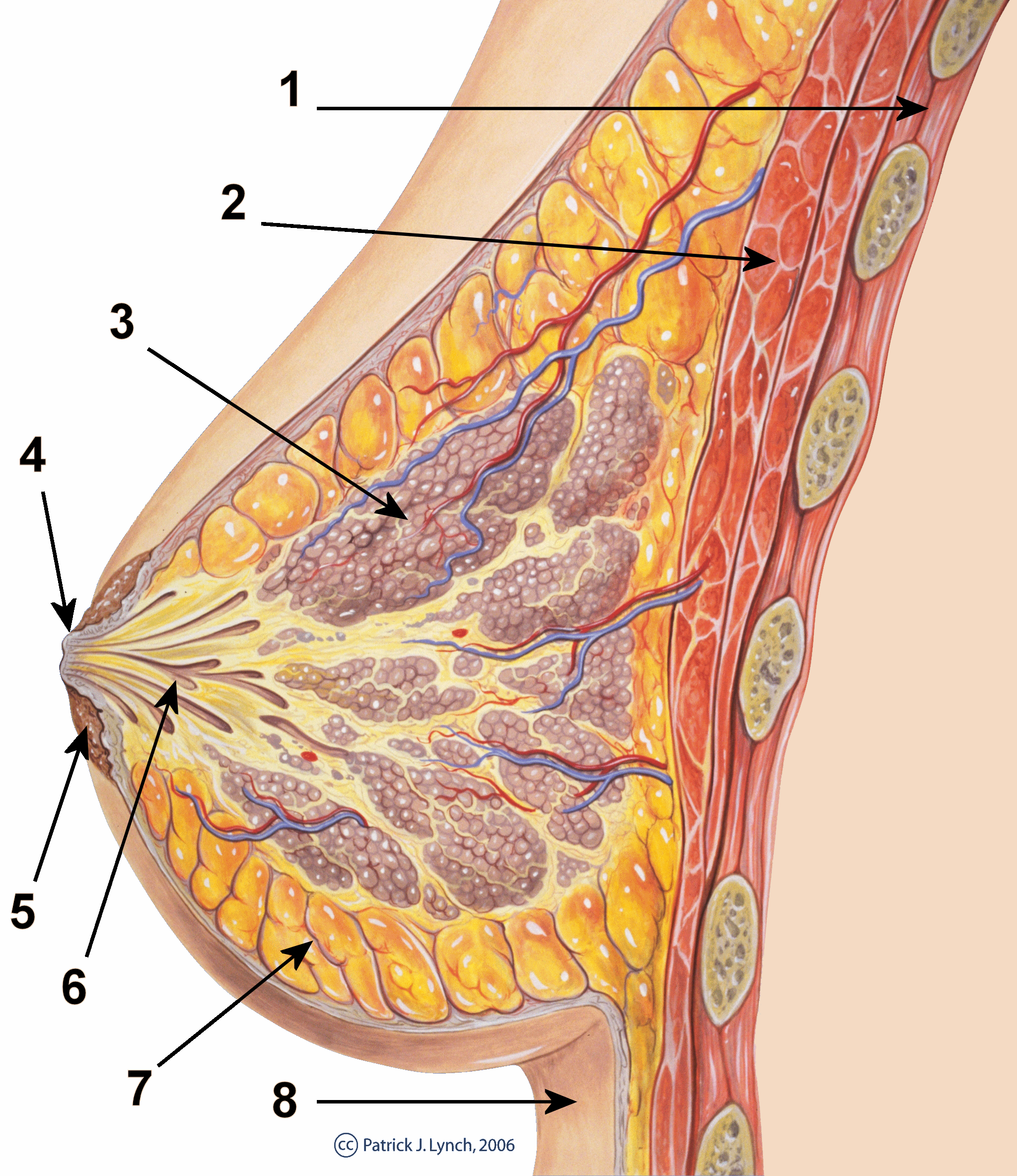
The nipple is a raised region of tissue on the surface of the breast from which, in females, milk leaves the breast through the lactiferous ducts to feed an infant. The milk can flow through the nipple passively or it can be ejected by smooth muscle contractions that occur along with the ductal system. The nipple is surrounded by the areola, which is often a darker colour than the surrounding skin. A nipple is often called a teat when referring to non-humans. Nipple or teat can also be used to describe the flexible mouthpiece of a baby bottle. In humans, the nipples of both males and females can be stimulated as part of sexual arousal. In many cultures, human female nipples are sexualized, or "regarded as sex objects and evaluated in terms of their physical characteristics and sexiness." Anatomy In mammals, a nipple (also called mammary papilla or teat) is a small projection of skin containing the outlets for 15–20 lactiferous ducts arranged cylindrically around the tip ... [...More Info...] [...Related Items...] OR: [Wikipedia] [Google] [Baidu] |
Perineal Sponge
The perineal sponge is a spongy cushion of tissue and blood vessels found in the lower genital area of women. It sits between the vaginal opening and rectum and is internal to the perineum and perineal body. Functions The perineal sponge is composed of erectile tissue; during arousal, it becomes swollen with blood compressing the outer third of the vagina along with the vestibular bulbs and urethral sponge thereby tightening the vagina and increasing stimulation for the vagina and a penis, if involved. Sexual stimulation The perineal sponge is erogenous tissue encompassing a large number of nerve endings, and can, therefore, be stimulated through the back wall of the vagina In mammals, the vagina is the elastic, muscular part of the female genital tract. In humans, it extends from the vestibule to the cervix. The outer vaginal opening is normally partly covered by a thin layer of mucosal tissue called the hy ... or the top wall of the rectum. It is most effectively ... [...More Info...] [...Related Items...] OR: [Wikipedia] [Google] [Baidu] |
Urethral Sponge
The urethral sponge is a spongy cushion of tissue, found in the lower genital area of females, that sits against both the pubic bone and vaginal wall, and surrounds the urethra. Functions The urethral sponge is composed of erectile tissue; during arousal, it becomes swollen with blood, compressing the urethra, helping, along with the pubococcygeus muscle, to prevent urination during sexual activity. Female ejaculation Additionally, the urethral sponge contains the Skene's glands, which may be involved in female ejaculation. Sexual stimulation The urethral sponge encompasses sensitive nerve endings, and can be stimulated through the front wall of the vagina. Some women experience intense pleasure from stimulation of the urethral sponge and others find the sensation irritating. The urethral sponge surrounds the clitoral nerve, and since the two are so closely interconnected, stimulation of the clitoris may stimulate the nerve endings of the urethral sponge and vice versa. ... [...More Info...] [...Related Items...] OR: [Wikipedia] [Google] [Baidu] |
Nasal Cycle
The nasal cycle is the unconscious alternating partial congestion and decongestion of the nasal cavities in humans and other animals. This results in greater airflow through one nostril with periodic alternation between the nostrils. It is a physiological congestion of the nasal conchae, also called the nasal turbinates (curled bony projections within the nasal cavities), due to selective activation of one half of the autonomic nervous system by the hypothalamus. It should not be confused with pathological nasal congestion. Description The nasal cycle was studied and discussed in the ancient Indian yoga of literature of pranayama. In the modern western literature, it was first described by the German physician Richard Kayser in 1895. In 1927, Heetderks described the alternating turgescence of the inferior turbinates in 80% of a normal population. According to Heetderks, the cycle is the result of alternating congestion and decongestion of the nasal conchae or turbinates, ... [...More Info...] [...Related Items...] OR: [Wikipedia] [Google] [Baidu] |
Nasal Concha
In anatomy, a nasal concha (), plural conchae (), also called a nasal turbinate or turbinal, is a long, narrow, curled shelf of bone that protrudes into the breathing passage of the nose in humans and various animals. The conchae are shaped like an elongated seashell, which gave them their name (Latin ''concha'' from Greek ''κόγχη''). A concha is any of the scrolled spongy bones of the nasal passages in vertebrates.''Anatomy of the Human Body'' Gray, Henry (1918) The Nasal Cavity. In humans, the conchae divide the nasal airway into four groove-like air passages, and are responsible for forcing inhaled air to flow in a steady, regular pattern around the largest possible of [...More Info...] [...Related Items...] OR: [Wikipedia] [Google] [Baidu] |
Nasal Septum
The nasal septum () separates the left and right airways of the nasal cavity, dividing the two nostrils. It is depressed by the depressor septi nasi muscle. Structure The fleshy external end of the nasal septum is called the columella or columella nasi, and is made up of cartilage and soft tissue. The nasal septum contains bone and hyaline cartilage. It is normally about 2 mm thick. The nasal septum is composed of four structures: * Perpendicular plate of ethmoid bone * Vomer bone * Septal nasal cartilage * Maxillary bone (the crest) The lowest part of the septum is a narrow strip of bone that projects from the maxilla and the palatine bones, and is the length of the septum. This strip of bone is called the maxillary crest; it articulates in front with the septal nasal cartilage, and at the back with the vomer. The maxillary crest is described in the anatomy of the nasal septum as having a maxillary component and a palatine component. Development At an early peri ... [...More Info...] [...Related Items...] OR: [Wikipedia] [Google] [Baidu] |
Corpus Spongiosum Penis
The corpus spongiosum is the mass of spongy tissue surrounding the male urethra within the penis. It is also called the corpus cavernosum urethrae in older texts. Anatomy The proximal part of the corpus spongiosum is expanded to form the urethral bulb, and lies in apposition with the inferior fascia of the urogenital diaphragm, from which it receives a fibrous investment. The urethra enters the bulb nearer to the superior than to the inferior surface. On the latter there is a median sulcus (groove), from which a thin fibrous septum (wall) projects into the substance of the bulb and divides it imperfectly into two lateral lobes or hemispheres. The portion of the corpus spongiosum in front of the bulb lies in a groove on the under surface of the conjoined corpora cavernosa penis. It is cylindrical in form and tapers slightly from behind forward. Its anterior end is expanded in the form of an obtuse cone, flattened from above downward. This expansion, termed the glans penis, ... [...More Info...] [...Related Items...] OR: [Wikipedia] [Google] [Baidu] |
Bulb Of Vestibule
In female anatomy, the vestibular bulbs, bulbs of the vestibule or clitoral bulbs are two elongated masses of erectile tissue typically described as being situated on either side of the vaginal opening. They are united to each other in front by a narrow median band. Some research indicates that they do not surround the vaginal opening, and are more closely related to the clitoris than to the vestibule. Structure Research indicates that the vestibular bulbs are more closely related to the clitoris than to the vestibule because of the similarity of the trabecular and erectile tissue within the clitoris and bulbs, and the absence of trabecular tissue in other genital organs, with the erectile tissue's trabecular nature allowing engorgement and expansion during sexual arousal. Ginger et al. state that although a number of texts report that they surround the vaginal opening, this does not appear to be the case and tunica albuginea does not envelop the erectile tissue of the bulb. T ... [...More Info...] [...Related Items...] OR: [Wikipedia] [Google] [Baidu] |


