|
Epidermal
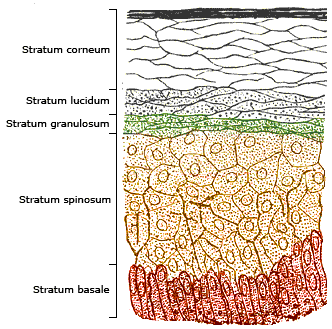
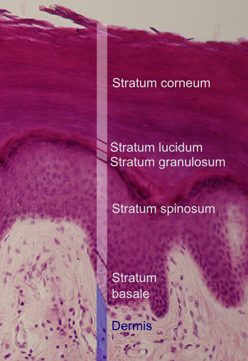
The epidermis is the outermost of the three layers that comprise the skin, the inner layers being the dermis and hypodermis. The epidermis layer provides a barrier to infection from environmental pathogens and regulates the amount of water released from the body into the atmosphere through transepidermal water loss. The epidermis is composed of multiple layers of flattened cells that overlie a base layer (stratum basale) composed of columnar cells arranged perpendicularly. The layers of cells develop from stem cells in the basal layer. The human epidermis is a familiar example of epithelium, particularly a stratified squamous epithelium. The word epidermis is derived through Latin , itself and . Something related to or part of the epidermis is termed epidermal. Structure Cellular components The epidermis primarily consists of keratinocytes ( proliferating basal and differentiated suprabasal), which comprise 90% of its cells, but also contains melanocytes, Langerhans cel ... [...More Info...] [...Related Items...] OR: [Wikipedia] [Google] [Baidu] |
Keratinocytes
Keratinocytes are the primary type of cell found in the epidermis, the outermost layer of the skin. In humans, they constitute 90% of epidermal skin cells. Basal cells in the basal layer (''stratum basale'') of the skin are sometimes referred to as basal keratinocytes. Keratinocytes form a barrier against environmental damage by heat, UV radiation, water loss, pathogenic bacteria, fungi, parasites, and viruses. A number of structural proteins, enzymes, lipids, and antimicrobial peptides contribute to maintain the important barrier function of the skin. Keratinocytes differentiate from epidermal stem cells in the lower part of the epidermis and migrate towards the surface, finally becoming corneocytes and eventually be shed off, which happens every 40 to 56 days in humans. Function The primary function of keratinocytes is the formation of a barrier against environmental damage by heat, UV radiation, water loss, pathogenic bacteria, fungi, parasites, and viruses. Pathogen ... [...More Info...] [...Related Items...] OR: [Wikipedia] [Google] [Baidu] |
Stratum Basale
The ''stratum basale'' (basal layer, sometimes referred to as ''stratum germinativum'') is the deepest layer of the five layers of the epidermis, the external covering of skin in mammals. The ''stratum basale'' is a single layer of columnar or cuboidal basal cells. The cells are attached to each other and to the overlying stratum spinosum cells by desmosomes and hemidesmosomes. The nucleus is large, ovoid and occupies most of the cell. Some basal cells can act like stem cells with the ability to divide and produce new cells, and these are sometimes called basal keratinocyte stem cells. Others serve to anchor the epidermis glabrous skin (hairless), and hyper-proliferative epidermis (from a skin disease).McGrath, J.A.; Eady, R.A.; Pope, F.M. (2004). ''Rook's Textbook of Dermatology'' (Seventh Edition). Blackwell Publishing. Pages 3.7. . They divide to form the keratinocytes of the stratum spinosum, which migrate superficially. Other types of cells found within the ''stratum bas ... [...More Info...] [...Related Items...] OR: [Wikipedia] [Google] [Baidu] |
Merkel Cell
Merkel cells, also known as Merkel-Ranvier cells or tactile epithelial cells, are oval-shaped mechanoreceptors essential for light touch sensation and found in the skin of vertebrates. They are abundant in highly sensitive skin like that of the fingertips in humans, and make synaptic contacts with somatosensory afferent nerve fibers. Though it has been reported that Merkel cells are derived from neural crest cells, more recent experiments in mammals have indicated that they are in fact epithelial in origin. Structure Merkel cells are found in the skin and some parts of the mucosa of all vertebrates. In mammalian skin, they are clear cells found in the ''stratum basale'' (at the bottom of sweat duct ridges) of the epidermis approximately 10 μm in diameter. They also occur in epidermal invaginations of the plantar foot surface called rete ridges. Most often, they are associated with sensory nerve endings, when they are known as Merkel nerve endings (also called a Merkel ... [...More Info...] [...Related Items...] OR: [Wikipedia] [Google] [Baidu] |
Histology
Histology, also known as microscopic anatomy or microanatomy, is the branch of biology which studies the microscopic anatomy of biological tissues. Histology is the microscopic counterpart to gross anatomy, which looks at larger structures visible without a microscope. Although one may divide microscopic anatomy into ''organology'', the study of organs, ''histology'', the study of tissues, and ''cytology'', the study of cells, modern usage places all of these topics under the field of histology. In medicine, histopathology is the branch of histology that includes the microscopic identification and study of diseased tissue. In the field of paleontology, the term paleohistology refers to the histology of fossil organisms. Biological tissues Animal tissue classification There are four basic types of animal tissues: muscle tissue, nervous tissue, connective tissue, and epithelial tissue. All animal tissues are considered to be subtypes of these four principal tissue types ... [...More Info...] [...Related Items...] OR: [Wikipedia] [Google] [Baidu] |
Melanocyte
Melanocytes are melanin-producing neural crest-derived cells located in the bottom layer (the stratum basale) of the skin's epidermis, the middle layer of the eye (the uvea), the inner ear, vaginal epithelium, meninges, bones, and heart. Melanin is a dark pigment primarily responsible for skin color. Once synthesized, melanin is contained in special organelles called melanosomes which can be transported to nearby keratinocytes to induce pigmentation. Thus darker skin tones have more melanosomes present than lighter skin tones. Functionally, melanin serves as protection against UV radiation. Melanocytes also have a role in the immune system. Function Through a process called melanogenesis, melanocytes produce melanin, which is a pigment found in the skin, eyes, hair, nasal cavity, and inner ear. This melanogenesis leads to a long-lasting pigmentation, which is in contrast to the pigmentation that originates from oxidation of already-existing melanin. There are both ... [...More Info...] [...Related Items...] OR: [Wikipedia] [Google] [Baidu] |
Cadherins
Cadherins (named for "calcium-dependent adhesion") are a type of cell adhesion molecule (CAM) that is important in the formation of adherens junctions to allow cells to adhere to each other . Cadherins are a class of type-1 transmembrane proteins, and they are dependent on calcium (Ca2+) ions to function, hence their name. Cell-cell adhesion is mediated by extracellular cadherin domains, whereas the Cadherin cytoplasmic region, intracellular cytoplasmic tail associates with numerous adaptors and signaling proteins, collectively referred to as the cadherin adhesome. The cadherin family is essential in maintaining the cell-cell contact and regulating cytoskeletal complexes. The cadherin superfamily includes cadherins, protocadherins, desmogleins, desmocollins, and more. In structure, they share ''cadherin repeats'', which are the extracellular Ca2+-binding domains. There are multiple classes of cadherin molecules, each designated with a prefix (in general, noting the types of tissue w ... [...More Info...] [...Related Items...] OR: [Wikipedia] [Google] [Baidu] |
Adherens Junction
Adherens junctions (or zonula adherens, intermediate junction, or "belt desmosome") are protein complexes that occur at cell–cell junctions, cell–matrix junctions in epithelial and endothelial tissues, usually more basal than tight junctions. An adherens junction is defined as a cell junction whose cytoplasmic face is linked to the actin cytoskeleton. They can appear as bands encircling the cell (zonula adherens) or as spots of attachment to the extracellular matrix (focal adhesion). Adherens junctions uniquely disassemble in uterine epithelial cells to allow the blastocyst to penetrate between epithelial cells. A similar cell junction in non-epithelial, non-endothelial cells is the fascia adherens. It is structurally the same, but appears in ribbonlike patterns that do not completely encircle the cells. One example is in cardiomyocytes. Proteins Adherens junctions are composed of the following proteins: * cadherins. The cadherins are a family of transmembrane proteins tha ... [...More Info...] [...Related Items...] OR: [Wikipedia] [Google] [Baidu] |
Epithelial Sodium Channel
The epithelial sodium channel (ENaC), (also known as amiloride-sensitive sodium channel) is a membrane-bound ion channel that is selectively permeable to sodium ions (). It is assembled as a heterotrimer composed of three homologous subunits α or δ, β, and γ, These subunits are encoded by four genes: SCNN1A, SCNN1B, SCNN1G, and SCNN1D. The ENaC is involved primarily in the reabsorption of sodium ions at the collecting ducts of the kidney's nephrons. In addition to being implicated in diseases where fluid balance across epithelial membranes is perturbed, including pulmonary edema, cystic fibrosis, COPD and COVID-19, proteolyzed forms of ENaC function as the human salt taste receptor. The apical membranes of many tight epithelia contain sodium channels that are characterized primarily by their high affinity for the diuretic blocker amiloride. These channels mediate the first step of active sodium reabsorption essential for the maintenance of body salt and water homeostasis. In ... [...More Info...] [...Related Items...] OR: [Wikipedia] [Google] [Baidu] |
Circulatory System
The blood circulatory system is a system of organs that includes the heart, blood vessels, and blood which is circulated throughout the entire body of a human or other vertebrate. It includes the cardiovascular system, or vascular system, that consists of the heart and blood vessels (from Greek ''kardia'' meaning ''heart'', and from Latin ''vascula'' meaning ''vessels''). The circulatory system has two divisions, a systemic circulation or circuit, and a pulmonary circulation or circuit. Some sources use the terms ''cardiovascular system'' and ''vascular system'' interchangeably with the ''circulatory system''. The network of blood vessels are the great vessels of the heart including large elastic arteries, and large veins; other arteries, smaller arterioles, capillaries that join with venules (small veins), and other veins. The Closed circulatory system, circulatory system is closed in vertebrates, which means that the blood never leaves the network of blood vessels. Some in ... [...More Info...] [...Related Items...] OR: [Wikipedia] [Google] [Baidu] |
Venule
A venule is a very small blood vessel in the microcirculation that allows blood to return from the capillary beds to drain into the larger blood vessels, the veins. Venules range from 7μm to 1mm in diameter. Veins contain approximately 70% of total blood volume, while about 25% is contained in the venules. Many venules unite to form a vein. Structure Venule walls have three layers: An inner endothelium composed of squamous endothelial cells that act as a membrane, a middle layer of muscle and elastic tissue and an outer layer of fibrous connective tissue. The middle layer is poorly developed so that venules have thinner walls than arterioles. They are porous so that fluid and blood cells can move easily from the bloodstream through their walls. Short portal venules between the neural and anterior pituitary lobes provide an avenue for rapid hormonal exchange via the blood. Specifically within and between the pituitary lobes is anatomical evidence for confluent interlobe ve ... [...More Info...] [...Related Items...] OR: [Wikipedia] [Google] [Baidu] |
Arteriole
An arteriole is a small-diameter blood vessel in the microcirculation that extends and branches out from an artery and leads to capillaries. Arterioles have muscular walls (usually only one to two layers of smooth muscle cells) and are the primary site of vascular resistance. The greatest change in blood pressure and velocity of blood flow occurs at the transition of arterioles to capillaries.This function is extremely important because it prevents the thin, one-layer capillaries from exploding upon pressure. The arterioles achieve this decrease in pressure, as they are the site with the highest resistance (a large contributor to total peripheral resistance) which translates to a large decrease in the pressure. Structure Microanatomy In a healthy vascular system the endothelium lines all blood-contacting surfaces, including arteries, arterioles, veins, venules, capillaries, and heart chambers. This healthy condition is promoted by the ample production of nitric oxide by the end ... [...More Info...] [...Related Items...] OR: [Wikipedia] [Google] [Baidu] |
Capillaries
A capillary is a small blood vessel from 5 to 10 micrometres (μm) in diameter. Capillaries are composed of only the tunica intima, consisting of a thin wall of simple squamous endothelial cells. They are the smallest blood vessels in the body: they convey blood between the arterioles and venules. These microvessels are the site of exchange of many substances with the interstitial fluid surrounding them. Substances which cross capillaries include water, oxygen, carbon dioxide, urea, glucose, uric acid, lactic acid and creatinine. Lymph capillaries connect with larger lymph vessels to drain lymphatic fluid collected in the microcirculation. During early embryonic development, new capillaries are formed through vasculogenesis, the process of blood vessel formation that occurs through a '' de novo'' production of endothelial cells that then form vascular tubes. The term ''angiogenesis'' denotes the formation of new capillaries from pre-existing blood vessels and already present endo ... [...More Info...] [...Related Items...] OR: [Wikipedia] [Google] [Baidu] |


