|
Enterostomy
An enterostomy ('' entero-'' + '' -stomy''; ) is either (1) a surgical procedure to create a durable opening (called a stoma) through the abdominal wall into an intestine ( small intestine or large intestine) or (2) the stoma thus created. The various types of enterostomy are named according to which intestinal segment is involved. Indications for surgery and complications are dependent on the site of the enterostomy. Gastrostomies and enterostomies can be used to provide nutrition in digestive disorders. Hernia development at both permanent and temporary enterostomy sites in a common complication. See also * Gastrostomy Gastrostomy is the creation of an artificial external opening into the stomach for nutritional support or gastric decompression. Typically this would include an incision in the patient's epigastrium as part of a formal operation. It can be perfor ... References {{reflist Medical procedures ... [...More Info...] [...Related Items...] OR: [Wikipedia] [Google] [Baidu] |
Ileostomy
Ileostomy is a stoma (surgical opening) constructed by bringing the end or loop of small intestine (the ileum) out onto the surface of the skin, or the surgical procedure which creates this opening. Intestinal waste passes out of the ileostomy and is collected in an external ostomy system which is placed next to the opening. Ileostomies are usually sited above the groin on the right hand side of the abdomen. Uses Ileostomies are necessary where injury or a surgical response to disease has meant the large intestine cannot safely process waste, typically because the colon and rectum have been partially or wholly removed. Diseases of the large intestine which may require surgical removal include Crohn's disease, ulcerative colitis, familial adenomatous polyposis, and total colonic Hirschsprung's disease.''Ileostomy Guide''< ... [...More Info...] [...Related Items...] OR: [Wikipedia] [Google] [Baidu] |
Colostomy
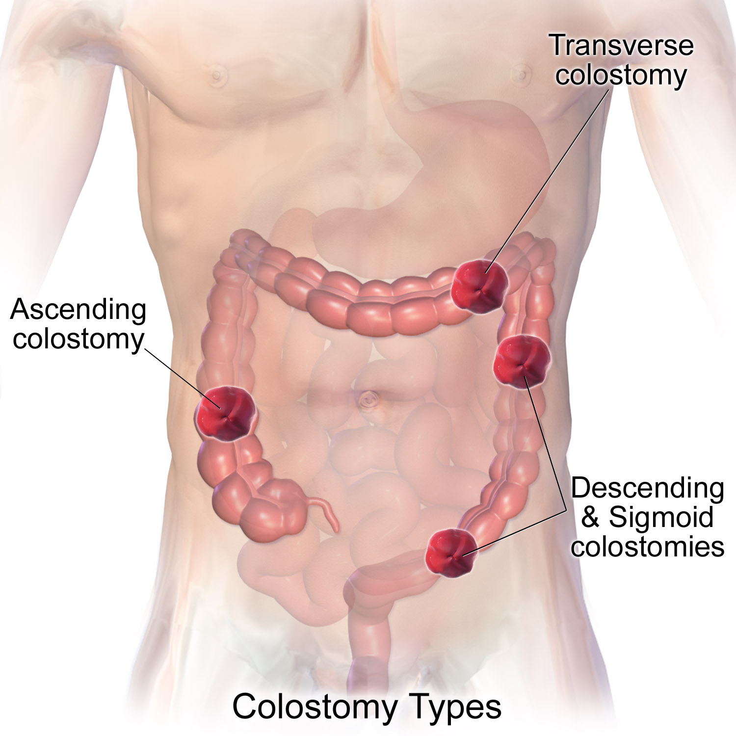
A colostomy is an opening ( stoma) in the large intestine (colon), or the surgical procedure that creates one. The opening is formed by drawing the healthy end of the colon through an incision in the anterior abdominal wall and suturing it into place. This opening, often in conjunction with an attached ostomy system, provides an alternative channel for feces to leave the body. Thus if the natural anus is unavailable for that function (for example, in cases where it has been removed in the fight against colorectal cancer or ulcerative colitis), an artificial anus takes over. It may be reversible or irreversible, depending on the circumstances. Uses There are many reasons for this procedure. Some common reasons are: * A part of the colon has been removed, e.g. due to colon cancer requiring a total mesorectal excision, diverticulitis, injury, etc., so that it is no longer possible for feces to exit via the anus. * A part of the colon has been operated upon and needs to be 'r ... [...More Info...] [...Related Items...] OR: [Wikipedia] [Google] [Baidu] |
Appendix (anatomy)
The appendix (or vermiform appendix; also cecal r caecalappendix; vermix; or vermiform process) is a finger-like, blind-ended tube connected to the cecum, from which it develops in the embryo. The cecum is a pouch-like structure of the large intestine, located at the junction of the small and the large intestines. The term "vermiform" comes from Latin and means "worm-shaped". The appendix was once considered a vestigial organ, but this view has changed since the early 2000s. Research suggests that the appendix may serve an important purpose. In particular, it may serve as a reservoir for beneficial gut bacteria. Structure The human appendix averages in length but can range from . The diameter of the appendix is , and more than is considered a thickened or inflamed appendix. The longest appendix ever removed was long. The appendix is usually located in the lower right quadrant of the abdomen, near the right hip bone. The base of the appendix is located beneath the ileoce ... [...More Info...] [...Related Items...] OR: [Wikipedia] [Google] [Baidu] |
Cecum
The cecum or caecum is a pouch within the peritoneum that is considered to be the beginning of the large intestine. It is typically located on the right side of the body (the same side of the body as the appendix (anatomy), appendix, to which it is joined). The word cecum (, plural ceca ) stems from the Latin ''wikt:caecus, caecus'' meaning blindness, blind. It receives chyme from the ileum, and connects to the ascending colon of the large intestine. It is separated from the ileum by the ileocecal valve (ICV) or Bauhin's valve. It is also separated from the Large intestine#Structure, colon by the cecocolic junction. While the cecum is usually intraperitoneal, the ascending colon is Retroperitoneal space, retroperitoneal. In herbivores, the cecum stores food material where bacteria are able to break down the cellulose. In humans, the cecum is involved in absorption of salts and electrolytes and lubricates the solid waste that passes into the large intestine. Structure Develo ... [...More Info...] [...Related Items...] OR: [Wikipedia] [Google] [Baidu] |
Cecostomy
A Malone antegrade continence enema is a surgical procedure used to create a continent pathway proximal to the anus that facilitates fecal evacuation using enemas. Description The operation involves connecting the appendix to the abdominal wall and fashioning a valve mechanism that allows catheterization of the appendix, but avoids leakage of stool through it. If the appendix was previously removed or is unusable, a neoappendix can be created with a cecal flap. Indications It is done to treat fecal incontinence unresponsive to treatment with medications. It is frequently done with a procedure (Mitrofanoff procedure) to treat urinary incontinence as the two often co-exist, such as in spina bifida. Cecostomy tube alternative A percutaneous cecostomy tube (C-tube)What is a Cecostomy Catheter? cecostomy.com. URLhttp://www.cecostomy.com/Introduction/cecostomy.htm. Accessed on: August 9, 2008. is an alternative to a MACE. It involves the surgical insertion of a catheter into the c ... [...More Info...] [...Related Items...] OR: [Wikipedia] [Google] [Baidu] |
Ileum
The ileum () is the final section of the small intestine in most higher vertebrates, including mammals, reptiles, and birds. In fish, the divisions of the small intestine are not as clear and the terms posterior intestine or distal intestine may be used instead of ileum. Its main function is to absorb vitamin B12, bile salts, and whatever products of digestion that were not absorbed by the jejunum. The ileum follows the duodenum and jejunum and is separated from the cecum by the ileocecal valve (ICV). In humans, the ileum is about 2–4 m long, and the pH is usually between 7 and 8 (neutral or slightly basic). ''Ileum ''is derived from the Greek word ''eilein'', meaning "to twist up tightly". Structure The ileum is the third and final part of the small intestine. It follows the jejunum and ends at the ileocecal junction, where the terminal ileum communicates with the cecum of the large intestine through the ileocecal valve. The ileum, along with the jejunum, is suspended ... [...More Info...] [...Related Items...] OR: [Wikipedia] [Google] [Baidu] |
Jejunum
The jejunum is the second part of the small intestine in humans and most higher vertebrates, including mammals, reptiles, and birds. Its lining is specialised for the absorption by enterocytes of small nutrient molecules which have been previously digested by enzymes in the duodenum. The jejunum lies between the duodenum and the ileum and is considered to start at the suspensory muscle of the duodenum, a location called the duodenojejunal flexure. The division between the jejunum and ileum is not anatomically distinct. In adult humans, the small intestine is usually long (post mortem), about two-fifths of which (about ) is the jejunum. Structure The interior surface of the jejunum—which is exposed to ingested food—is covered in finger–like projections of mucosa, called villi, which increase the surface area of tissue available to absorb nutrients from ingested foodstuffs. The epithelial cells which line these villi have microvilli. The transport of nutrients across epi ... [...More Info...] [...Related Items...] OR: [Wikipedia] [Google] [Baidu] |
Jejunostomy
Jejunostomy is the surgical creation of an opening (stoma) through the skin at the front of the abdomen and the wall of the jejunum (part of the small intestine). It can be performed either endoscopically, or with open surgery. A jejunostomy may be formed following bowel resection in cases where there is a need to bypass the distal small bowel and/or colon due to a bowel leak or perforation. Depending on the length of jejunum resected or bypassed the patient may have resultant short bowel syndrome and require parenteral nutrition. A jejunostomy is different from a jejunal feeding tube. A jejunal feeding tube is an alternative to a gastrostomy feeding tube and is commonly used when gastric enteral feeding is contraindicated or carries significant risks. The advantage over a gastrostomy is its low risk of aspiration due to its distal placement. Disadvantages include small bowel obstruction, ischemia, and requirement for continuous feeding. Techniques The Witzel jejunostomy is th ... [...More Info...] [...Related Items...] OR: [Wikipedia] [Google] [Baidu] |
Duodenum
The duodenum is the first section of the small intestine in most higher vertebrates, including mammals, reptiles, and birds. In fish, the divisions of the small intestine are not as clear, and the terms anterior intestine or proximal intestine may be used instead of duodenum. In mammals the duodenum may be the principal site for iron absorption. The duodenum precedes the jejunum and ileum and is the shortest part of the small intestine. In humans, the duodenum is a hollow jointed tube about 25–38 cm (10–15 inches) long connecting the stomach to the middle part of the small intestine. It begins with the duodenal bulb and ends at the suspensory muscle of duodenum. Duodenum can be divided into four parts: the first (superior), the second (descending), the third (horizontal) and the fourth (ascending) parts. Structure The duodenum is a C-shaped structure lying adjacent to the stomach. It is divided anatomically into four sections. The first part of the duodenum lies ... [...More Info...] [...Related Items...] OR: [Wikipedia] [Google] [Baidu] |


