|
Electroencephalographic
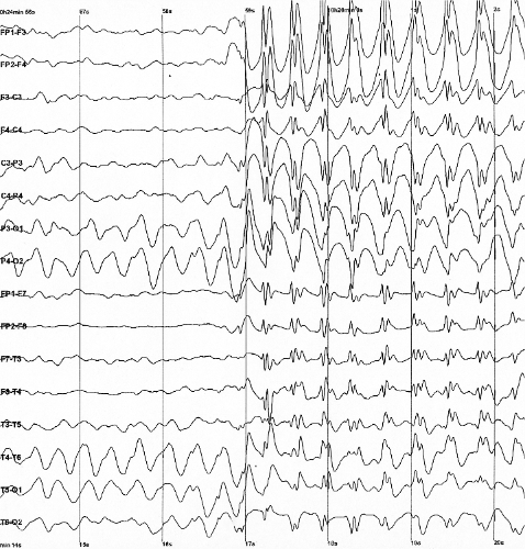
Electroencephalography (EEG) is a method to record an electrogram of the spontaneous electrical activity of the brain. The biosignals detected by EEG have been shown to represent the postsynaptic potentials of pyramidal neurons in the neocortex and allocortex. It is typically non-invasive, with the EEG electrodes placed along the scalp (commonly called "scalp EEG") using the International 10-20 system, or variations of it. Electrocorticography, involving surgical placement of electrodes, is sometimes called "intracranial EEG". Clinical interpretation of EEG recordings is most often performed by visual inspection of the tracing or quantitative EEG analysis. Voltage fluctuations measured by the EEG bioamplifier and electrodes allow the evaluation of normal brain activity. As the electrical activity monitored by EEG originates in neurons in the underlying brain tissue, the recordings made by the electrodes on the surface of the scalp vary in accordance with their orientation a ... [...More Info...] [...Related Items...] OR: [Wikipedia] [Google] [Baidu] |
10–20 System (EEG)
The 10–20 system or International 10–20 system is an internationally recognized method to describe and apply the location of scalp electrodes in the context of an EEG exam, polysomnograph sleep study, or voluntary lab research. This method was developed to maintain standardized testing methods ensuring that a subject's study outcomes (clinical or research) could be compiled, reproduced, and effectively analyzed and compared using the scientific method. The system is based on the relationship between the location of an electrode and the underlying area of the brain, specifically the cerebral cortex. During sleep and wake cycles, the brain produces different, objectively recognized and distinguishable electrical patterns, which can be detected by electrodes on the skin. (These patterns might vary, and can be affected by multiple extrinsic factors, i.e. age, prescription drugs, somatic diagnoses, hx of neurologic insults/injury/trauma, and substance abuse) The "10" and "20" re ... [...More Info...] [...Related Items...] OR: [Wikipedia] [Google] [Baidu] |
Spike-and-wave
Spike-and-wave is a pattern of the electroencephalogram (EEG) typically observed during epileptic seizures. A spike-and-wave discharge is a regular, symmetrical, generalized EEG pattern seen particularly during absence epilepsy, also known as ‘petit mal’ epilepsy. The basic mechanisms underlying these patterns are complex and involve part of the cerebral cortex, the thalamocortical network, and intrinsic neuronal mechanisms. The first spike-and-wave pattern was recorded in the early twentieth century by Hans Berger. Many aspects of the pattern are still being researched and discovered, and still many aspects are uncertain. The spike-and-wave pattern is most commonly researched in absence epilepsy, but is common in several epilepsies such as Lennox-Gastaut syndrome (LGS) and Ohtahara syndrome. Antiepileptic drugs (AEDs) are commonly prescribed to treat epileptic seizures, and new ones are being discovered with fewer adverse effects. Today, most of the research is focused o ... [...More Info...] [...Related Items...] OR: [Wikipedia] [Google] [Baidu] |
Electrography (other)
Electrography often refers to electrophotography, that is, Kirlian photography. Electrography may also refer to: * Measurement and recording of electrophysiologic activity for diagnostic purposes ** Electrocardiography (ECG or EKG), electrography of heart electrical activity and rhythm ** Electromyography (EMG), electrography of other muscle action potentials throughout the body ** Electroencephalography (EEG), electrography of brain waves (from outside the skull) *** Electrocorticography or intracranial EEG (iEEG or ECoG), EEG with direct contact to the cerebral cortex ** Electrooculography (EOG), electrography of intraocular potential differences ** Electroolfactography (EOG), electrography of olfaction (smell) ** Electroretinography (ERG), electrography of retinal cell action potentials ** Electronystagmography (ENG), electrography of eye muscle movements ** Electrocochleography (ECOG), electrography of cochlear auditory activity ** Electroantennography (EAG), electr ... [...More Info...] [...Related Items...] OR: [Wikipedia] [Google] [Baidu] |
Seizures
An epileptic seizure, informally known as a seizure, is a period of symptoms due to abnormally excessive or synchronous neuronal activity in the brain. Outward effects vary from uncontrolled shaking movements involving much of the body with loss of consciousness ( tonic-clonic seizure), to shaking movements involving only part of the body with variable levels of consciousness (focal seizure), to a subtle momentary loss of awareness (absence seizure). Most of the time these episodes last less than two minutes and it takes some time to return to normal. Loss of bladder control may occur. Seizures may be provoked and unprovoked. Provoked seizures are due to a temporary event such as low blood sugar, alcohol withdrawal, abusing alcohol together with prescription medication, low blood sodium, fever, brain infection, or concussion. Unprovoked seizures occur without a known or fixable cause such that ongoing seizures are likely. Unprovoked seizures may be exacerbated by stress or slee ... [...More Info...] [...Related Items...] OR: [Wikipedia] [Google] [Baidu] |
Computed Tomography
A computed tomography scan (CT scan; formerly called computed axial tomography scan or CAT scan) is a medical imaging technique used to obtain detailed internal images of the body. The personnel that perform CT scans are called radiographers or radiology technologists. CT scanners use a rotating X-ray tube and a row of detectors placed in a gantry to measure X-ray attenuations by different tissues inside the body. The multiple X-ray measurements taken from different angles are then processed on a computer using tomographic reconstruction algorithms to produce tomographic (cross-sectional) images (virtual "slices") of a body. CT scans can be used in patients with metallic implants or pacemakers, for whom magnetic resonance imaging (MRI) is contraindicated. Since its development in the 1970s, CT scanning has proven to be a versatile imaging technique. While CT is most prominently used in medical diagnosis, it can also be used to form images of non-living objects. The 1979 N ... [...More Info...] [...Related Items...] OR: [Wikipedia] [Google] [Baidu] |
Magnetic Resonance Imaging
Magnetic resonance imaging (MRI) is a medical imaging technique used in radiology to form pictures of the anatomy and the physiological processes inside the body. MRI scanners use strong magnetic fields, magnetic field gradients, and radio waves to generate images of the organs in the body. MRI does not involve X-rays or the use of ionizing radiation, which distinguishes it from computed tomography (CT) and positron emission tomography (PET) scans. MRI is a medical application of nuclear magnetic resonance (NMR) which can also be used for imaging in other NMR applications, such as NMR spectroscopy. MRI is widely used in hospitals and clinics for medical diagnosis, staging and follow-up of disease. Compared to CT, MRI provides better contrast in images of soft tissues, e.g. in the brain or abdomen. However, it may be perceived as less comfortable by patients, due to the usually longer and louder measurements with the subject in a long, confining tube, although "open ... [...More Info...] [...Related Items...] OR: [Wikipedia] [Google] [Baidu] |
Stroke
Stroke (also known as a cerebrovascular accident (CVA) or brain attack) is a medical condition in which poor blood flow to the brain causes cell death. There are two main types of stroke: ischemic, due to lack of blood flow, and hemorrhagic, due to bleeding. Both cause parts of the brain to stop functioning properly. Signs and symptoms of stroke may include an inability to move or feel on one side of the body, problems understanding or speaking, dizziness, or loss of vision to one side. Signs and symptoms often appear soon after the stroke has occurred. If symptoms last less than one or two hours, the stroke is a transient ischemic attack (TIA), also called a mini-stroke. Hemorrhagic stroke may also be associated with a severe headache. The symptoms of stroke can be permanent. Long-term complications may include pneumonia and loss of bladder control. The biggest risk factor for stroke is high blood pressure. Other risk factors include high blood cholesterol, to ... [...More Info...] [...Related Items...] OR: [Wikipedia] [Google] [Baidu] |
Tumor
A neoplasm () is a type of abnormal and excessive growth of tissue. The process that occurs to form or produce a neoplasm is called neoplasia. The growth of a neoplasm is uncoordinated with that of the normal surrounding tissue, and persists in growing abnormally, even if the original trigger is removed. This abnormal growth usually forms a mass, when it may be called a tumor. ICD-10 classifies neoplasms into four main groups: benign neoplasms, in situ neoplasms, malignant neoplasms, and neoplasms of uncertain or unknown behavior. Malignant neoplasms are also simply known as cancers and are the focus of oncology. Prior to the abnormal growth of tissue, as neoplasia, cells often undergo an abnormal pattern of growth, such as metaplasia or dysplasia. However, metaplasia or dysplasia does not always progress to neoplasia and can occur in other conditions as well. The word is from Ancient Greek 'new' and 'formation, creation'. Types A neoplasm can be benign, potenti ... [...More Info...] [...Related Items...] OR: [Wikipedia] [Google] [Baidu] |
Brain Death
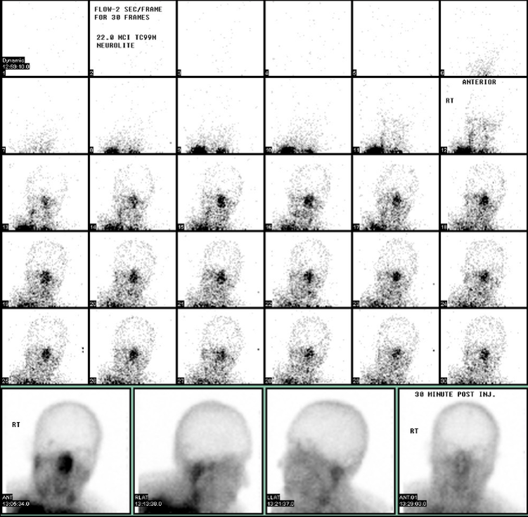
Brain death is the permanent, irreversible, and complete loss of brain function which may include cessation of involuntary activity necessary to sustain life. It differs from persistent vegetative state, in which the person is alive and some autonomic functions remain. It is also distinct from comas as long as some brain and bodily activity and function remain, and it is also not the same as the condition locked-in syndrome. A differential diagnosis can medically distinguish these differing conditions. Brain death is used as an indicator of legal death in many jurisdictions, but it is defined inconsistently and often confused by the public. Various parts of the brain may keep functioning when others do not anymore, and the term "brain death" has been used to refer to various combinations. For example, although one major medical dictionary considers "brain death" to be synonymous with "cerebral death" (death of the cerebrum), the US National Library of Medicine Medical Subje ... [...More Info...] [...Related Items...] OR: [Wikipedia] [Google] [Baidu] |
Cardiac Arrest
Cardiac arrest is when the heart suddenly and unexpectedly stops beating. It is a medical emergency that, without immediate medical intervention, will result in sudden cardiac death within minutes. Cardiopulmonary resuscitation (CPR) and possibly defibrillation are needed until further treatment can be provided. Cardiac arrest results in a rapid unconsciousness, loss of consciousness, and breathing may be respiratory arrest, abnormal or absent. While cardiac arrest may be caused by myocardial infarction, heart attack or heart failure, these are not the same, and in 15 to 25% of cases, there is a non-cardiac cause. Some individuals may experience chest pain, shortness of breath, nausea, an elevated heart rate, and a light-headed feeling immediately before entering cardiac arrest. The most common cause of cardiac arrest is an underlying heart problem like coronary artery disease that decreases the amount of oxygenated blood supplying the heart muscle. This, in turn, damages the ... [...More Info...] [...Related Items...] OR: [Wikipedia] [Google] [Baidu] |
Cerebral Hypoxia
Cerebral hypoxia is a form of hypoxia (reduced supply of oxygen), specifically involving the brain; when the brain is completely deprived of oxygen, it is called ''cerebral anoxia''. There are four categories of cerebral hypoxia; they are, in order of increasing severity: diffuse cerebral hypoxia (DCH), focal cerebral ischemia, cerebral infarction, and global cerebral ischemia. Prolonged hypoxia induces neuronal cell death via apoptosis, resulting in a hypoxic brain injury. Cases of total oxygen deprivation are termed "anoxia", which can be hypoxic in origin (reduced oxygen availability) or ischemic in origin (oxygen deprivation due to a disruption in blood flow). Brain injury as a result of oxygen deprivation either due to hypoxic or anoxic mechanisms are generally termed hypoxic/anoxic injuries (HAI). Hypoxic ischemic encephalopathy (HIE) is a condition that occurs when the entire brain is deprived of an adequate oxygen supply, but the deprivation is not total. While HIE is a ... [...More Info...] [...Related Items...] OR: [Wikipedia] [Google] [Baidu] |
Encephalopathies
Encephalopathy (; from grc, ἐνκέφαλος "brain" + πάθος "suffering") means any disorder or disease of the brain, especially chronic degenerative conditions. In modern usage, encephalopathy does not refer to a single disease, but rather to a syndrome of overall brain dysfunction; this syndrome has many possible organic and inorganic causes. Signs and symptoms The hallmark of encephalopathy is an altered mental state or delirium. Characteristic of the altered mental state is impairment of the cognition, attention, orientation, sleep–wake cycle and consciousness. An altered state of consciousness may range from failure of selective attention to drowsiness. Hypervigilance may be present; with or without: cognitive deficits, headache, epileptic seizures, myoclonus (involuntary twitching of a muscle or group of muscles) or asterixis ("flapping tremor" of the hand when wrist is extended). Depending on the type and severity of encephalopathy, common neurological sympto ... [...More Info...] [...Related Items...] OR: [Wikipedia] [Google] [Baidu] |


