|
Direct Pathway Of Movement
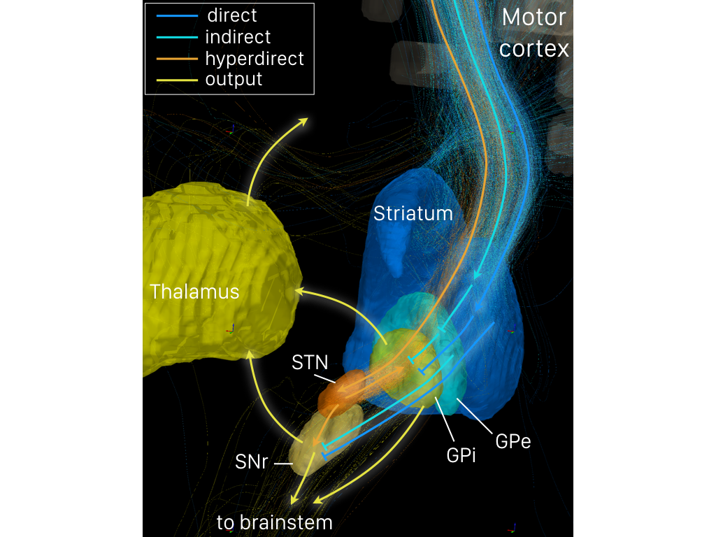
The direct pathway, sometimes known as the direct pathway of movement, is a neural pathway within the central nervous system (CNS) through the basal ganglia which facilitates the initiation and execution of voluntary movement. It works in conjunction with the indirect pathway. Both of these pathways are part of the cortico-basal ganglia-thalamo-cortical loop. Overview of neuronal connections and normal function The direct pathway passes through the caudate nucleus, putamen, and globus pallidus, which are parts of the basal ganglia. It also involves another basal ganglia component the substantia nigra, a part of the midbrain. In a resting individual, a specific region of the globus pallidus, the internal globus pallidus (GPi), and a part of the substantia nigra, the pars reticulata (SNpr), send spontaneous inhibitory signals to the ventral lateral nucleus (VL) of the thalamus, through the release of GABA, an inhibitory neurotransmitter. Inhibition of the inhibitory neurons tha ... [...More Info...] [...Related Items...] OR: [Wikipedia] [Google] [Baidu] |
Motor Loop
An engine or motor is a machine designed to convert one or more forms of energy into motion (physics), mechanical energy. Available energy sources include potential energy (e.g. energy of the Earth's gravitational field as exploited in hydroelectric power generation), heat energy (e.g. geothermal), chemical energy, electric potential and nuclear energy (from nuclear fission or nuclear fusion). Many of these processes generate heat as an intermediate energy form, so heat engines have special importance. Some natural processes, such as atmospheric convection cells convert environmental heat into motion (e.g. in the form of rising air currents). Mechanical energy is of particular importance in transportation, but also plays a role in many industrial processes such as cutting, grinding, crushing, and mixing. Mechanical heat engines convert heat into work via various thermodynamic processes. The internal combustion engine is perhaps the most common example of a mechanical heat eng ... [...More Info...] [...Related Items...] OR: [Wikipedia] [Google] [Baidu] |
Ventral Lateral Nucleus
The ventral lateral nucleus (VL) is a nucleus in the ventral nuclear group of the thalamus. Inputs and outputs It receives neuronal inputs from the basal ganglia which includes the substantia nigra and the globus pallidus (via the thalamic fasciculus). It also has inputs from the cerebellum (via the dentatothalamic tract). It sends neuronal output to the primary motor cortex and premotor cortex. The ventral lateral nucleus in the thalamus forms the motor functional division in the thalamic nuclei along with the ventral anterior nucleus. The ventral lateral nucleus receives motor information from the cerebellum and the globus pallidus. Output from the ventral lateral nucleus then goes to the primary motor cortex. Functions The function of the ventral lateral nucleus is to target efferents including the motor cortex, premotor cortex, and supplementary motor cortex. Therefore, its function helps the coordination and planning of movement. It also plays a role in the learning of mov ... [...More Info...] [...Related Items...] OR: [Wikipedia] [Google] [Baidu] |
Hypokinesia
Hypokinesia is one of the classifications of movement disorders, and refers to decreased bodily movement. Hypokinesia is characterized by a partial or complete loss of muscle movement due to a disruption in the basal ganglia. Hypokinesia is a symptom of Parkinson's disease shown as muscle rigidity and an inability to produce movement. It is also associated with mental health disorders and prolonged inactivity due to illness, amongst other diseases. The other category of movement disorder is hyperkinesia that features an exaggeration of unwanted movement, such as twitching or writhing in Huntington's disease or Tourette syndrome. Spectrum of disorders Hypokinesia describes a variety of more specific disorders: Causes The most common cause of Hypokinesia is Parkinson's disease, and conditions related to Parkinson's disease. Other conditions may also cause slowness of movements. These includhypothyroidism and severe depression.These conditions need to be carefully ruled out ... [...More Info...] [...Related Items...] OR: [Wikipedia] [Google] [Baidu] |
Striatum
The striatum, or corpus striatum (also called the striate nucleus), is a nucleus (a cluster of neurons) in the subcortical basal ganglia of the forebrain. The striatum is a critical component of the motor and reward systems; receives glutamatergic and dopaminergic inputs from different sources; and serves as the primary input to the rest of the basal ganglia. Functionally, the striatum coordinates multiple aspects of cognition, including both motor and action planning, decision-making, motivation, reinforcement, and reward perception. The striatum is made up of the caudate nucleus and the lentiform nucleus. The lentiform nucleus is made up of the larger putamen, and the smaller globus pallidus. Strictly speaking the globus pallidus is part of the striatum. It is common practice, however, to implicitly exclude the globus pallidus when referring to striatal structures. In primates, the striatum is divided into a ventral striatum, and a dorsal striatum, subdivisions that are ... [...More Info...] [...Related Items...] OR: [Wikipedia] [Google] [Baidu] |
Cerebral Cortex
The cerebral cortex, also known as the cerebral mantle, is the outer layer of neural tissue of the cerebrum of the brain in humans and other mammals. The cerebral cortex mostly consists of the six-layered neocortex, with just 10% consisting of allocortex. It is separated into two cortices, by the longitudinal fissure that divides the cerebrum into the left and right cerebral hemispheres. The two hemispheres are joined beneath the cortex by the corpus callosum. The cerebral cortex is the largest site of neural integration in the central nervous system. It plays a key role in attention, perception, awareness, thought, memory, language, and consciousness. The cerebral cortex is part of the brain responsible for cognition. In most mammals, apart from small mammals that have small brains, the cerebral cortex is folded, providing a greater surface area in the confined volume of the cranium. Apart from minimising brain and cranial volume, cortical folding is crucial for the brain ... [...More Info...] [...Related Items...] OR: [Wikipedia] [Google] [Baidu] |
Pre-frontal Cortex
In mammalian brain anatomy, the prefrontal cortex (PFC) covers the front part of the frontal lobe of the cerebral cortex. The PFC contains the Brodmann areas BA8, BA9, BA10, BA11, BA12, BA13, BA14, BA24, BA25, BA32, BA44, BA45, BA46, and BA47. The basic activity of this brain region is considered to be orchestration of thoughts and actions in accordance with internal goals. Many authors have indicated an integral link between a person's will to live, personality, and the functions of the prefrontal cortex. This brain region has been implicated in executive functions, such as planning, decision making, short-term memory, personality expression, moderating social behavior and controlling certain aspects of speech and language. Executive function relates to abilities to differentiate among conflicting thoughts, determine good and bad, better and best, same and different, future consequences of current activities, working toward a defined goal, prediction of outcome ... [...More Info...] [...Related Items...] OR: [Wikipedia] [Google] [Baidu] |
Telencephalon
The cerebrum, telencephalon or endbrain is the largest part of the brain containing the cerebral cortex (of the two cerebral hemispheres), as well as several subcortical structures, including the hippocampus, basal ganglia, and olfactory bulb. In the human brain, the cerebrum is the uppermost region of the central nervous system. The cerebrum develops prenatally from the forebrain (prosencephalon). In mammals, the dorsal telencephalon, or pallium, develops into the cerebral cortex, and the ventral telencephalon, or subpallium, becomes the basal ganglia. The cerebrum is also divided into approximately symmetric left and right cerebral hemispheres. With the assistance of the cerebellum, the cerebrum controls all voluntary actions in the human body. Structure The cerebrum is the largest part of the brain. Depending upon the position of the animal it lies either in front or on top of the brainstem. In humans, the cerebrum is the largest and best-developed of the five major divi ... [...More Info...] [...Related Items...] OR: [Wikipedia] [Google] [Baidu] |
Ventral Anterior Nucleus
The ventral anterior nucleus (VA) is a nucleus of the thalamus. It acts with the anterior part of the ventral lateral nucleus to modify signals from the basal ganglia. Inputs and outputs The ventral anterior nucleus receives neuronal inputs from the basal ganglia. Its main afferent fibres are from the globus pallidus. The efferent fibres from this nucleus pass into the premotor cortex The premotor cortex is an area of the motor cortex lying within the frontal lobe of the brain just anterior to the primary motor cortex. It occupies part of Brodmann's area 6. It has been studied mainly in primates, including monkeys and humans. ... for initiation and planning of movement. Functions It helps to function in movement by providing feedback for the outputs of the basal ganglia. Additional images File:Constudthal.gif, Thalamus File:Territoriostalamo.svg, Thalamus References {{DEFAULTSORT:Ventral Anterior Nucleus Thalamus ... [...More Info...] [...Related Items...] OR: [Wikipedia] [Google] [Baidu] |
Neurotransmitter
A neurotransmitter is a signaling molecule secreted by a neuron to affect another cell across a synapse. The cell receiving the signal, any main body part or target cell, may be another neuron, but could also be a gland or muscle cell. Neurotransmitters are released from synaptic vesicles into the synaptic cleft where they are able to interact with neurotransmitter receptors on the target cell. The neurotransmitter's effect on the target cell is determined by the receptor it binds. Many neurotransmitters are synthesized from simple and plentiful precursors such as amino acids, which are readily available and often require a small number of biosynthetic steps for conversion. Neurotransmitters are essential to the function of complex neural systems. The exact number of unique neurotransmitters in humans is unknown, but more than 100 have been identified. Common neurotransmitters include glutamate, GABA, acetylcholine, glycine and norepinephrine. Mechanism and cycle Synthes ... [...More Info...] [...Related Items...] OR: [Wikipedia] [Google] [Baidu] |
Thalamus
The thalamus (from Greek θάλαμος, "chamber") is a large mass of gray matter located in the dorsal part of the diencephalon (a division of the forebrain). Nerve fibers project out of the thalamus to the cerebral cortex in all directions, allowing hub-like exchanges of information. It has several functions, such as the relaying of sensory signals, including motor signals to the cerebral cortex and the regulation of consciousness, sleep, and alertness. Anatomically, it is a paramedian symmetrical structure of two halves (left and right), within the vertebrate brain, situated between the cerebral cortex and the midbrain. It forms during embryonic development as the main product of the diencephalon, as first recognized by the Swiss embryologist and anatomist Wilhelm His Sr. in 1893. Anatomy The thalamus is a paired structure of gray matter located in the forebrain which is superior to the midbrain, near the center of the brain, with nerve fibers projecting out to the ... [...More Info...] [...Related Items...] OR: [Wikipedia] [Google] [Baidu] |
Pars Reticulata
The pars reticulata (SNpr) is a portion of the substantia nigra and is located lateral to the pars compacta. Most of the neurons that project out of the pars reticulata are inhibitory GABAergic neurons (i.e., these neurons release GABA, which is an inhibitory neurotransmitter). Anatomy Neurons in the pars reticulata are much less densely packed than those in the pars compacta (they were sometimes named pars diffusa). They are smaller and thinner than the dopaminergic neurons and conversely identical and morphologically similar to the pallidal neurons (see primate basal ganglia). Their dendrites as well as the pallidal are preferentially perpendicular to the striatal afferents. The massive striatal afferents correspond to the medial end of the nigrostriatal bundle. Nigral neurons have the same peculiar synaptology with the striatal axonal endings. They make connections with the dopamine neurons of the pars compacta whose long dendrites plunge deeply in the pars reticulata. The ... [...More Info...] [...Related Items...] OR: [Wikipedia] [Google] [Baidu] |


