|
Descemet's Membrane
Descemet's membrane ( or the Descemet membrane) is the basement membrane that lies between the corneal proper substance, also called stroma, and the endothelial layer of the cornea. It is composed of different kinds of collagen (Type IV and VIII) than the stroma. The endothelial layer is located at the posterior of the cornea. Descemet's membrane, as the basement membrane for the endothelial layer, is secreted by the single layer of squamous epithelial cells that compose the endothelial layer of the cornea. Structure Its thickness ranges from 3 μm at birth to 8–10 μm in adults.Johnson DH, Bourne WM, Campbell RJ: The ultrastructure of Descemet's membrane. I. Changes with age in normal cornea. Arch Ophthalmol 100:1942, 1982 The corneal endothelium is a single layer of squamous cells covering the surface of the cornea that faces the anterior chamber. Clinical significance Significant damage to the membrane may require a corneal transplant. Damage caused by the hereditary co ... [...More Info...] [...Related Items...] OR: [Wikipedia] [Google] [Baidu] |
Epithelium
Epithelium or epithelial tissue is one of the four basic types of animal tissue, along with connective tissue, muscle tissue and nervous tissue. It is a thin, continuous, protective layer of compactly packed cells with a little intercellular matrix. Epithelial tissues line the outer surfaces of organs and blood vessels throughout the body, as well as the inner surfaces of cavities in many internal organs. An example is the epidermis, the outermost layer of the skin. There are three principal shapes of epithelial cell: squamous (scaly), columnar, and cuboidal. These can be arranged in a singular layer of cells as simple epithelium, either squamous, columnar, or cuboidal, or in layers of two or more cells deep as stratified (layered), or ''compound'', either squamous, columnar or cuboidal. In some tissues, a layer of columnar cells may appear to be stratified due to the placement of the nuclei. This sort of tissue is called pseudostratified. All glands are made up of epithe ... [...More Info...] [...Related Items...] OR: [Wikipedia] [Google] [Baidu] |
Basement Membrane
The basement membrane is a thin, pliable sheet-like type of extracellular matrix that provides cell and tissue support and acts as a platform for complex signalling. The basement membrane sits between Epithelium, epithelial tissues including mesothelium and endothelium, and the underlying connective tissue. Structure As seen with the electron microscope, the basement membrane is composed of two layers, the basal lamina and the reticular lamina. The underlying connective tissue attaches to the basal lamina with collagen VII anchoring fibrils and fibrillin microfibrils. The basal lamina layer can further be subdivided into two layers based on their visual appearance in electron microscopy. The lighter-colored layer closer to the epithelium is called the lamina lucida, while the denser-colored layer closer to the connective tissue is called the lamina densa. The Electron microscope, electron-dense lamina densa layer is about 30–70 nanometers thick and consists of an underlying ... [...More Info...] [...Related Items...] OR: [Wikipedia] [Google] [Baidu] |
Haab's Striae
Haab's striae, or Descemet's tears, are horizontal breaks in the Descemet membrane associated with congenital glaucoma Primary juvenile glaucoma is glaucoma that develops due to ocular hypertension and is evident either at birth or within the first few years of life. It is caused due to abnormalities in the anterior chamber angle development that obstruct aqueous o .... It is named after Otto Haab. These occur because descemet's membrane is less elastic than the corneal stroma. Tears are usually peripheral, concentric with limbus and appear as line with double contour. References External links * Ophthalmology Disorders of sclera and cornea {{Eye-disease-stub ... [...More Info...] [...Related Items...] OR: [Wikipedia] [Google] [Baidu] |
Jean Descemet
Jean Descemet was a French physician and botanist (1732–1810) who first described what is now known as Descemet's membrane of the eye. He studied medicine in Paris (doctorate in 1757) and in 1759 he published a ''Catalog of the plants in the Garden of the Apothecaries in Paris according to their genera and the characteristics of their flowers, using Mr. Tournefort's method'' ( French: ''Catalogue des plantes du jardin de MM. les apothicaires de Paris, suivant leurs genres et les caractères des fleurs, conformément à la méthode de M. Tournefort'') (1759). He is also known for his book ''Observations sur la choroide'', which describes the choroid The choroid, also known as the choroidea or choroid coat, is a part of the uvea, the vascular layer of the eye, and contains connective tissues, and lies between the retina and the sclera. The human choroid is thickest at the far extreme rear ... layer of the eye and was published in 1768 by L'Imprimerie Royale. References ... [...More Info...] [...Related Items...] OR: [Wikipedia] [Google] [Baidu] |
Kayser–Fleischer Ring
Kayser–Fleischer rings (KF rings) are dark rings that appear to encircle the cornea of the eye. They are due to copper deposition in the Descemet's membrane as a result of particular liver diseases. They are named after German ophthalmologists Bernhard Kayser and Bruno Fleischer who first described them in 1902 and 1903. Initially thought to be due to the accumulation of silver, they were first demonstrated to contain copper in 1934. Presentation The rings, which consist of copper deposits where the cornea meets the sclera, in Descemet's membrane, first appear as a crescent at the top of the cornea. Eventually, a second crescent forms below, at the "six o'clock position", and ultimately completely encircles the cornea. Associations Kayser–Fleischer rings are a sign of Wilson's disease, which involves abnormal copper handling by the liver resulting in copper accumulation in the body and is characterised by abnormalities of the basal ganglia of the brain, liver cirrhosis, sp ... [...More Info...] [...Related Items...] OR: [Wikipedia] [Google] [Baidu] |
Wilson's Disease
Wilson's disease is a genetic disorder in which excess copper builds up in the body. Symptoms are typically related to the brain and liver. Liver-related symptoms include vomiting, weakness, fluid build up in the abdomen, swelling of the legs, yellowish skin and itchiness. Brain-related symptoms include tremors, muscle stiffness, trouble in speaking, personality changes, anxiety, and psychosis. Wilson's disease is caused by a mutation in the Wilson disease protein (''ATP7B'') gene. This protein transports excess copper into bile, where it is excreted in waste products. The condition is autosomal recessive; for a person to be affected, they must inherit a mutated copy of the gene from both parents. Diagnosis may be difficult and often involves a combination of blood tests, urine tests and a liver biopsy. Genetic testing may be used to screen family members of those affected. Wilson's disease is typically treated with dietary changes and medication. Dietary changes involve e ... [...More Info...] [...Related Items...] OR: [Wikipedia] [Google] [Baidu] |
Corneal Transplantation
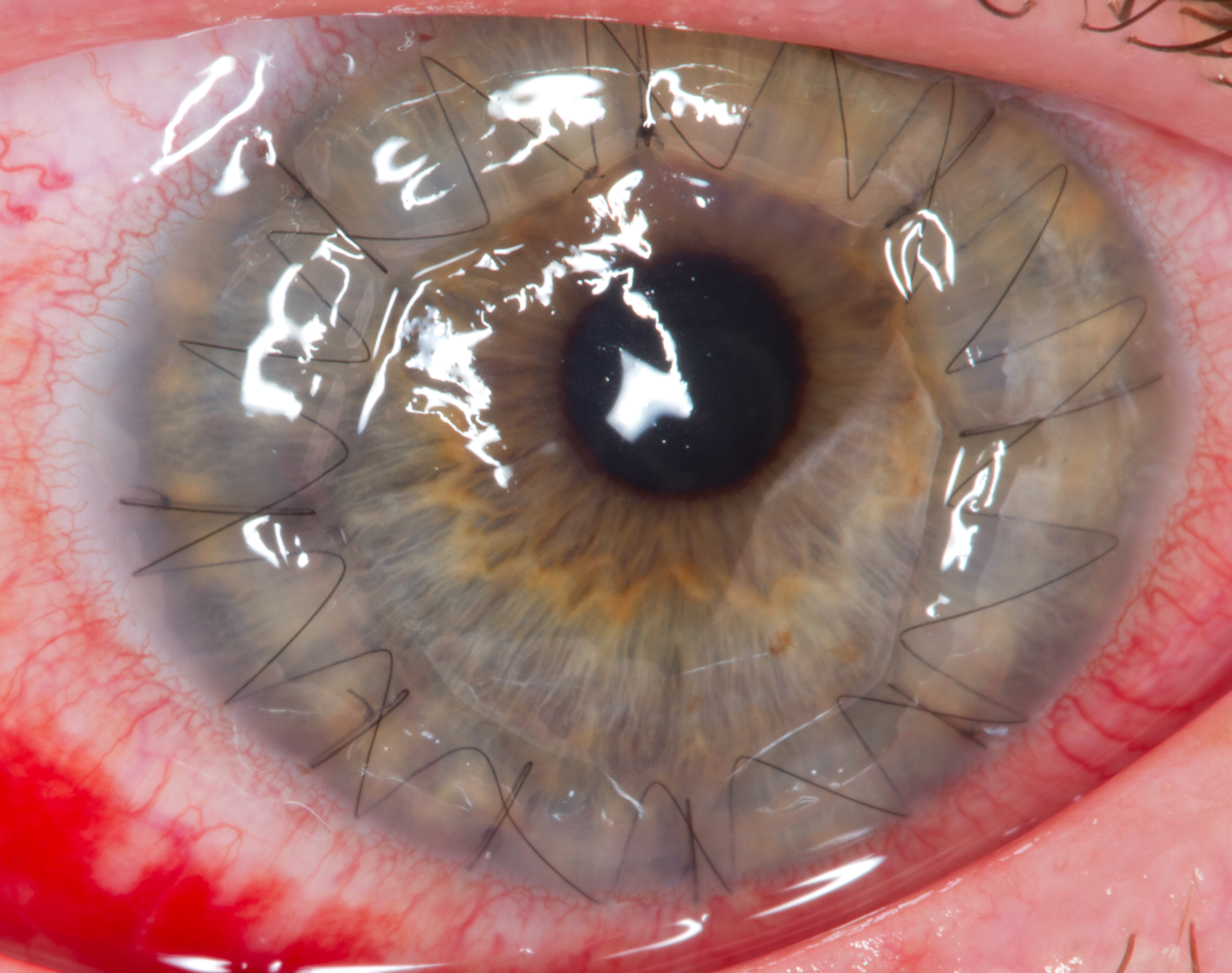
Corneal transplantation, also known as corneal grafting, is a surgical procedure where a damaged or diseased cornea is replaced by donated corneal tissue (the graft). When the entire cornea is replaced it is known as penetrating keratoplasty and when only part of the cornea is replaced it is known as lamellar keratoplasty. Keratoplasty simply means surgery to the cornea. The graft is taken from a recently deceased individual with no known diseases or other factors that may affect the chance of survival of the donated tissue or the health of the recipient. The cornea is the transparent front part of the eye that covers the iris, pupil and anterior chamber. The surgical procedure is performed by ophthalmologists, physicians who specialize in eyes, and is often done on an outpatient basis. Donors can be of any age, as is shown in the case of Janis Babson, who donated her eyes after dying at the age of 10. Corneal transplantation is performed when medicines, keratoconus conservat ... [...More Info...] [...Related Items...] OR: [Wikipedia] [Google] [Baidu] |
Fuchs Dystrophy
Fuchs dystrophy, also referred to as Fuchs endothelial corneal dystrophy (FECD) and Fuchs endothelial dystrophy (FED), is a slowly progressing corneal dystrophy that usually affects both eyes and is slightly more common in women than in men. Although early signs of Fuchs dystrophy are sometimes seen in people in their 30s and 40s, the disease rarely affects vision until people reach their 50s and 60s. Signs and symptoms As a progressive, chronic condition, signs and symptoms of Fuchs dystrophy gradually progress over decades of life, starting in middle age. Early symptoms include blurry vision upon wakening which improves during the morning, as fluid retained in the cornea is unable to evaporate through the surface of the eye when the lids are closed overnight. As the disease worsens, the interval of blurry morning vision extends from minutes to hours. In moderate stages of the disease, an increase in guttae and swelling in the cornea can contribute to changes in vision and decreas ... [...More Info...] [...Related Items...] OR: [Wikipedia] [Google] [Baidu] |
Stroma Of Cornea
The stroma of the cornea (or substantia propria) is a fibrous, tough, unyielding, perfectly transparent and the thickest layer of the cornea of the eye. It is between Bowman's membrane anteriorly, and Descemet's membrane posteriorly. At its centre, human corneal stroma is composed of about 200 flattened ''lamellæ'' (layers of collagen fibrils), superimposed one on another. They are each about 1.5-2.5 μm in thickness. The anterior lamellæ interweave more than posterior lamellæ. The fibrils of each lamella are parallel with one another, but at different angles to those of adjacent lamellæ. The lamellæ are produced by keratocytes (corneal connective tissue cells), which occupy about 10% of the substantia propria. Apart from the cells, the major non-aqueous constituents of the stroma are collagen fibrils and proteoglycans. The collagen fibrils are made of a mixture of type I and type V collagens. These molecules are tilted by about 15 degrees to the fibril axis, and because of ... [...More Info...] [...Related Items...] OR: [Wikipedia] [Google] [Baidu] |
Eponym
An eponym is a person, a place, or a thing after whom or which someone or something is, or is believed to be, named. The adjectives which are derived from the word eponym include ''eponymous'' and ''eponymic''. Usage of the word The term ''eponym'' functions in multiple related ways, all based on an explicit relationship between two named things. A person, place, or thing named after a particular person share an eponymous relationship. In this way, Elizabeth I of England is the eponym of the Elizabethan era. When Henry Ford is referred to as "the ''eponymous'' founder of the Ford Motor Company", his surname "Ford" serves as the eponym. The term also refers to the title character of a fictional work (such as Rocky Balboa of the Rocky film series, ''Rocky'' film series), as well as to ''self-titled'' works named after their creators (such as the album The Doors (album), ''The Doors'' by the band the Doors). Walt Disney created the eponymous The Walt Disney Company, Walt Disney Com ... [...More Info...] [...Related Items...] OR: [Wikipedia] [Google] [Baidu] |
Anterior Elastic Lamina
The Bowman layer (Bowman's membrane, anterior limiting lamina, anterior elastic lamina) is a smooth, acellular, nonregenerating layer, located between the superficial epithelium and the stroma in the cornea of the eye. It is composed of strong, randomly oriented collagen fibrils in which the smooth anterior surface faces the epithelial basement membrane and the posterior surface merges with the collagen lamellae of the corneal stroma proper. In adult humans, Bowman layer is 8-12 μm thick. With ageing, this layer becomes thinner. The function of the Bowman layer remains unclear and appears to have no critical function in corneal physiology. Recently, it is postulated that the layer may act as a physical barrier to protect the subepithelial nerve plexus and thereby hastens epithelial innervation and sensory recovery. Moreover, it may also serve as a barrier that prevents direct traumatic contact with the corneal stroma and hence it is highly involved in stromal wound healing an ... [...More Info...] [...Related Items...] OR: [Wikipedia] [Google] [Baidu] |


