|
Dermoepidermal Junction
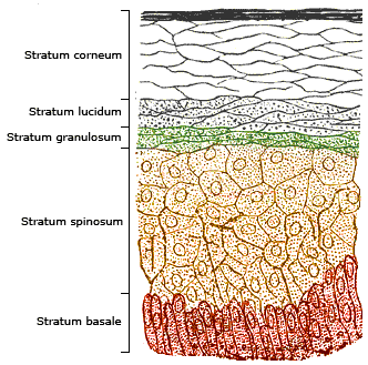
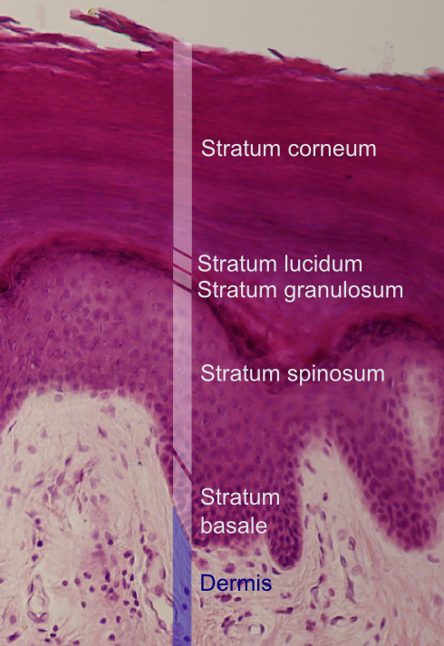
The dermoepidermal junction or dermal-epidermal junction (DEJ) is the area of tissue that joins the epidermal and the dermal layers of the skin. The basal cells in the stratum basale of the epidermis connect to the basement membrane by the anchoring filaments of hemidesmosomes; the cells of the papillary layer of the dermis are attached to the basement membrane by anchoring fibrils, which consist of type VII collagen. Stevens–Johnson syndrome and toxic epidermal necrolysis Toxic epidermal necrolysis (TEN) is a type of severe skin reaction. Together with Stevens–Johnson syndrome (SJS) it forms a spectrum of disease, with TEN being more severe. Early symptoms include fever and flu-like symptoms. A few days later ... are diseases where there is a breakdown of the dermoepidermal junction. References Skin anatomy {{dermatology-stub ... [...More Info...] [...Related Items...] OR: [Wikipedia] [Google] [Baidu] |
Epidermis (skin)
The epidermis is the outermost of the three layers that comprise the skin, the inner layers being the dermis and hypodermis. The epidermis layer provides a barrier to infection from environmental pathogens and regulates the amount of water released from the body into the atmosphere through transepidermal water loss. The epidermis is composed of multiple layers of flattened cells that overlie a base layer (stratum basale) composed of columnar cells arranged perpendicularly. The layers of cells develop from stem cells in the basal layer. The human epidermis is a familiar example of epithelium, particularly a stratified squamous epithelium. The word epidermis is derived through Latin , itself and . Something related to or part of the epidermis is termed epidermal. Structure Cellular components The epidermis primarily consists of keratinocytes ( proliferating basal and differentiated suprabasal), which comprise 90% of its cells, but also contains melanocytes, Langerhans c ... [...More Info...] [...Related Items...] OR: [Wikipedia] [Google] [Baidu] |
Dermis
The dermis or corium is a layer of skin between the epidermis (with which it makes up the cutis) and subcutaneous tissues, that primarily consists of dense irregular connective tissue and cushions the body from stress and strain. It is divided into two layers, the superficial area adjacent to the epidermis called the papillary region and a deep thicker area known as the reticular dermis.James, William; Berger, Timothy; Elston, Dirk (2005). ''Andrews' Diseases of the Skin: Clinical Dermatology'' (10th ed.). Saunders. Pages 1, 11–12. . The dermis is tightly connected to the epidermis through a basement membrane. Structural components of the dermis are collagen, elastic fibers, and extrafibrillar matrix.Marks, James G; Miller, Jeffery (2006). ''Lookingbill and Marks' Principles of Dermatology'' (4th ed.). Elsevier Inc. Page 8–9. . It also contains mechanoreceptors that provide the sense of touch and thermoreceptors that provide the sense of heat. In addition, hair follicles, ... [...More Info...] [...Related Items...] OR: [Wikipedia] [Google] [Baidu] |
Stratum Basale
The ''stratum basale'' (basal layer, sometimes referred to as ''stratum germinativum'') is the deepest layer of the five layers of the epidermis, the external covering of skin in mammals. The ''stratum basale'' is a single layer of columnar or cuboidal basal cells. The cells are attached to each other and to the overlying stratum spinosum cells by desmosomes and hemidesmosomes. The nucleus is large, ovoid and occupies most of the cell. Some basal cells can act like stem cells with the ability to divide and produce new cells, and these are sometimes called basal keratinocyte stem cells. Others serve to anchor the epidermis glabrous skin (hairless), and hyper-proliferative epidermis (from a skin disease).McGrath, J.A.; Eady, R.A.; Pope, F.M. (2004). ''Rook's Textbook of Dermatology'' (Seventh Edition). Blackwell Publishing. Pages 3.7. . They divide to form the keratinocytes of the stratum spinosum, which migrate superficially. Other types of cells found within the ''stratum bas ... [...More Info...] [...Related Items...] OR: [Wikipedia] [Google] [Baidu] |
Basement Membrane
The basement membrane is a thin, pliable sheet-like type of extracellular matrix that provides cell and tissue support and acts as a platform for complex signalling. The basement membrane sits between Epithelium, epithelial tissues including mesothelium and endothelium, and the underlying connective tissue. Structure As seen with the electron microscope, the basement membrane is composed of two layers, the basal lamina and the reticular lamina. The underlying connective tissue attaches to the basal lamina with collagen VII anchoring fibrils and fibrillin microfibrils. The basal lamina layer can further be subdivided into two layers based on their visual appearance in electron microscopy. The lighter-colored layer closer to the epithelium is called the lamina lucida, while the denser-colored layer closer to the connective tissue is called the lamina densa. The Electron microscope, electron-dense lamina densa layer is about 30–70 nanometers thick and consists of an underlying ... [...More Info...] [...Related Items...] OR: [Wikipedia] [Google] [Baidu] |
Hemidesmosomes
Hemidesmosomes are very small stud-like structures found in keratinocytes of the epidermis of skin that attach to the extracellular matrix. They are similar in form to desmosomes when visualized by electron microscopy, however, desmosomes attach to adjacent cells. Hemidesmosomes are also comparable to focal adhesions, as they both attach cells to the extracellular matrix. Instead of desmogleins and desmocollins in the extracellular space, hemidesmosomes utilize integrins. Hemidesmosomes are found in epithelial cells connecting the basal epithelial cells to the lamina lucida, which is part of the basal lamina. Hemidesmosomes are also involved in signaling pathways, such as keratinocyte migration or carcinoma cell intrusion. Structure Hemidesmosomes can be categorized into two types based on their protein constituents. Type 1 hemidesmosomes are found in stratified and pseudo-stratified epithelium. Type 1 hemidesmosomes have five main elements: integrin α6 β4, plectin in its i ... [...More Info...] [...Related Items...] OR: [Wikipedia] [Google] [Baidu] |
Papillary Dermis
The dermis or corium is a layer of skin between the epidermis (with which it makes up the cutis) and subcutaneous tissues, that primarily consists of dense irregular connective tissue and cushions the body from stress and strain. It is divided into two layers, the superficial area adjacent to the epidermis called the papillary region and a deep thicker area known as the reticular dermis.James, William; Berger, Timothy; Elston, Dirk (2005). ''Andrews' Diseases of the Skin: Clinical Dermatology'' (10th ed.). Saunders. Pages 1, 11–12. . The dermis is tightly connected to the epidermis through a basement membrane. Structural components of the dermis are collagen, elastic fibers, and extrafibrillar matrix.Marks, James G; Miller, Jeffery (2006). ''Lookingbill and Marks' Principles of Dermatology'' (4th ed.). Elsevier Inc. Page 8–9. . It also contains mechanoreceptors that provide the sense of touch and thermoreceptors that provide the sense of heat. In addition, hair follicles, swe ... [...More Info...] [...Related Items...] OR: [Wikipedia] [Google] [Baidu] |
Anchoring Fibrils
Anchoring fibrils (composed largely of type VII collagen) extend from the basal lamina of epithelial cells and attach to the lamina reticularis (also known as the reticular lamina) by wrapping around the reticular fiber ( collagen III) bundles. The basal lamina and lamina reticularis together make up the basement membrane. Anchoring fibrils are essential to the functional integrity of the dermoepidermal junction. Epidermolysis bullosa dystrophica Epidermolysis bullosa dystrophica, also known as Dystrophic EB (DEB) is a chronic skin condition caused when anchoring fibrils are abnormal, diminished, or absent. This causes a weak dermoepidermal junction, where the epidermis easily separates from the dermis causing much pain. This condition is caused by a mutation of COL7A1, the gene that codes for a type of collagen 7. See also *Epidermis (skin) The epidermis is the outermost of the three layers that comprise the skin, the inner layers being the dermis and hypodermis. Th ... [...More Info...] [...Related Items...] OR: [Wikipedia] [Google] [Baidu] |
Type VII Collagen
Collagen alpha-1(VII) chain is a protein that in humans is encoded by the ''COL7A1'' gene. It is composed of a triple helical, collagenous domain flanked by two non-collagenous domains, and functions as an anchoring fibril between the dermal-epidermal junction in the basement membrane. Mutations in COL7A1 cause all types of dystrophic epidermolysis bullosa, and the exact mutations vary based on the specific type or subtype. It has been shown that interactions between the NC-1 domain of collagen VII and several other proteins, including laminin-5 and collagen IV, contribute greatly to the overall stability of the basement membrane. Structure Type VII collagen is composed of three main domains in the following order: a non-collagenous domain, abbreviated NC-1; a collagenous domain; and a second non-collagenous domain, NC-2. The NC-1 domain has a cartilage matrix protein (CMP), nine fibronectin III (FNIII)-like subdomains, and a von Willebrand Factor A-like subdomain (VWFA1); a no ... [...More Info...] [...Related Items...] OR: [Wikipedia] [Google] [Baidu] |
Stevens–Johnson Syndrome
Stevens–Johnson syndrome (SJS) is a type of severe skin reaction. Together with toxic epidermal necrolysis (TEN) and Stevens–Johnson/toxic epidermal necrolysis (SJS/TEN), it forms a spectrum of disease, with SJS being less severe. Erythema multiforme (EM) is generally considered a separate condition. Early symptoms of SJS include fever and flu-like symptoms. A few days later, the skin begins to blister and peel, forming painful raw areas. Mucous membranes, such as the mouth, are also typically involved. Complications include dehydration, sepsis, pneumonia and multiple organ failure. The most common cause is certain medications such as lamotrigine, carbamazepine, allopurinol, sulfonamide antibiotics and nevirapine. Other causes can include infections such as ''Mycoplasma pneumoniae'' and cytomegalovirus, or the cause may remain unknown. Risk factors include HIV/AIDS and systemic lupus erythematosus. The diagnosis of Stevens–Johnson syndrome is based on involvement of les ... [...More Info...] [...Related Items...] OR: [Wikipedia] [Google] [Baidu] |
Toxic Epidermal Necrolysis
Toxic epidermal necrolysis (TEN) is a type of severe skin reaction. Together with Stevens–Johnson syndrome (SJS) it forms a spectrum of disease, with TEN being more severe. Early symptoms include fever and flu-like symptoms. A few days later the skin begins to blister and peel forming painful raw areas. Mucous membranes, such as the mouth, are also typically involved. Complications include dehydration, sepsis, pneumonia, and multiple organ failure. The most common cause is certain medications such as lamotrigine, carbamazepine, allopurinol, sulfonamide antibiotics, and nevirapine. Other causes can include infections such as ''Mycoplasma pneumoniae'' and cytomegalovirus or the cause may remain unknown. Risk factors include HIV/AIDS and systemic lupus erythematosus. Diagnosis is based on a skin biopsy and involvement of more than 30% of the skin. TEN is a type of severe cutaneous adverse reactions (SCARs), together with SJS, a SJS/TEN, and drug reaction with eosinophilia and sys ... [...More Info...] [...Related Items...] OR: [Wikipedia] [Google] [Baidu] |


