|
Desmoplasia
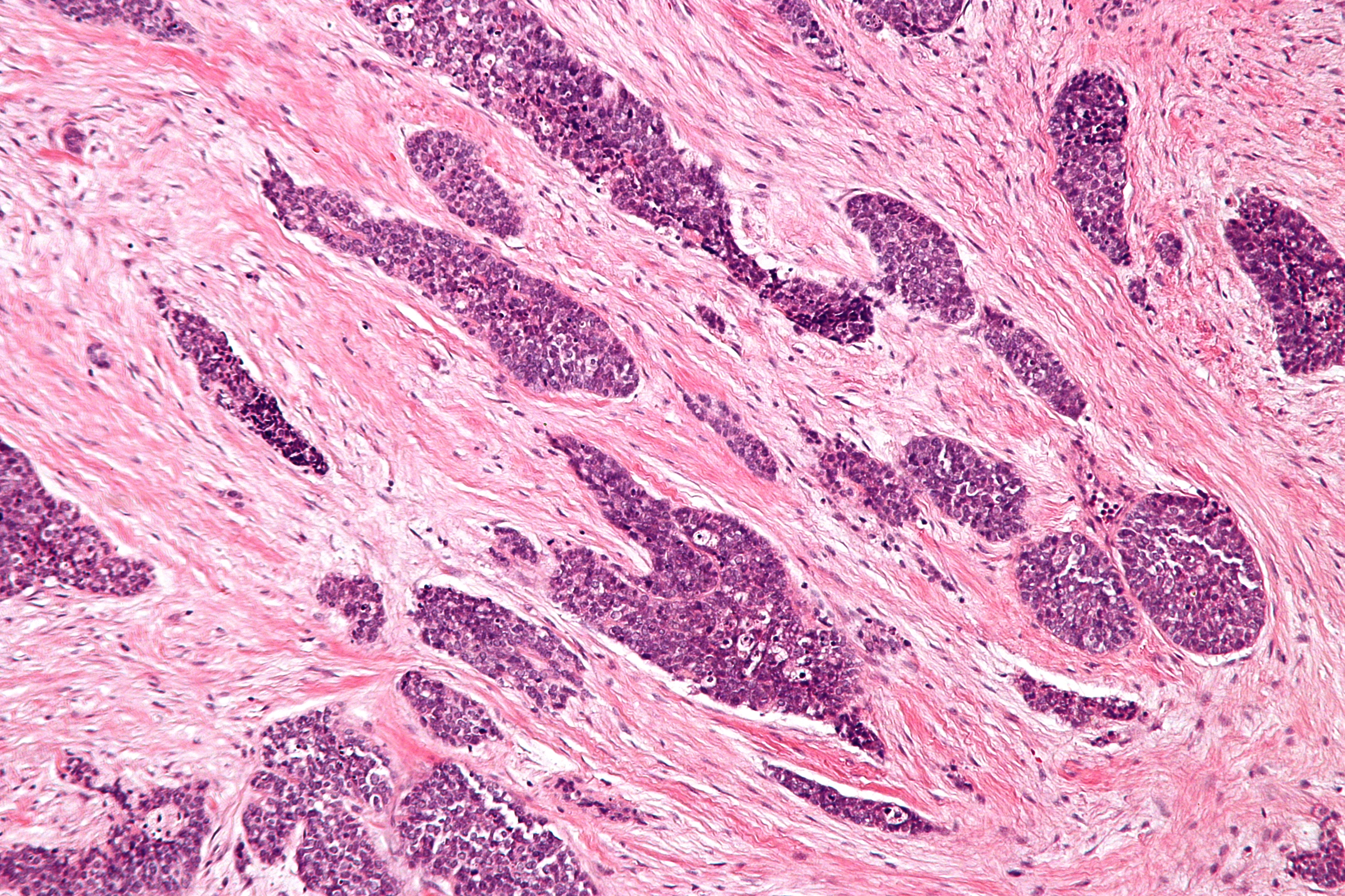
In medicine, desmoplasia is the growth of fibrous or connective tissue. It is also called desmoplastic reaction to emphasize that it is secondary to an insult. Desmoplasia may occur around a neoplasm, causing dense fibrosis around the tumor, or scar tissue (adhesions) within the abdomen after abdominal surgery. Desmoplasia is usually only associated with malignant neoplasms, which can evoke a fibrotic response invading healthy tissue. Invasive ductal carcinomas of the breast often have a stellate appearance caused by desmoplastic formations. Terminology Desmoplasia originates from the Ancient Greek δεσμός ''desmos'', "knot", "bond" and πλάσις ''plasis'', "formation". It is usually used in the description of desmoplastic small round cell tumors. Neoplasia is the medical term used for both benign and malignant tumors, or any abnormal, excessive, uncoordinated, and autonomous cellular or tissue growth. Desmoplasia refers to growth of dense connective tissue or strom ... [...More Info...] [...Related Items...] OR: [Wikipedia] [Google] [Baidu] |
Desmoplastic Small Round Cell Tumour - Intermed Mag
In medicine, a desmoplastic fibroma is a benign, but locally aggressive, fibrous and rare tumor of the bone, affecting children and young adults, potentially resulting in cortical bone destruction. It usually affects craniofacial bones, mandible most frequently, long bones (metaphyseal femur, tibia, humerus). The World Health Organization, 2020, reclassified these tumors as specific benign tumors in the category of fibroblastic and myofibroblastic tumors. Although it does not tend to metastatize, it has a high local recurrence and infiltrative growth. Treatment consists in wide local excision to prevent otherwise frequent recurrences. The role of radiotherapy and chemotherapy in this tumor still is unclear. Some cases have been described, in which an osteosarcoma has arisen from a desmoplastic fibroma. A famous occurrence of this particular form of the disease involved Italo-Australian Riccardo Torresan in 2011, with 18 cm of femur needing to be removed with the now widely ... [...More Info...] [...Related Items...] OR: [Wikipedia] [Google] [Baidu] |
Parenchymal Cell
Parenchyma () is the bulk of functional substance in an animal organ or structure such as a tumour. In zoology it is the name for the tissue that fills the interior of flatworms. Etymology The term ''parenchyma'' is New Latin from the word παρέγχυμα ''parenchyma'' meaning 'visceral flesh', and from παρεγχεῖν ''parenchyma'' meaning 'to pour in' from παρα- ''para-'' 'beside' + ἐν ''en-'' 'in' + χεῖν ''chyma'' 'to pour'. Originally, Erasistratus and other anatomists used it to refer to certain human tissues. Later, it was also applied to plant tissues by Nehemiah Grew. Structure The parenchyma is the ''functional'' parts of an organ, or of a structure such as a tumour in the body. This is in contrast to the stroma, which refers to the ''structural'' tissue of organs or of structures, namely, the connective tissues. Brain The brain parenchyma refers to the functional tissue in the brain that is made up of the two types of brain cell, neurons a ... [...More Info...] [...Related Items...] OR: [Wikipedia] [Google] [Baidu] |
Matrix Metalloproteinase
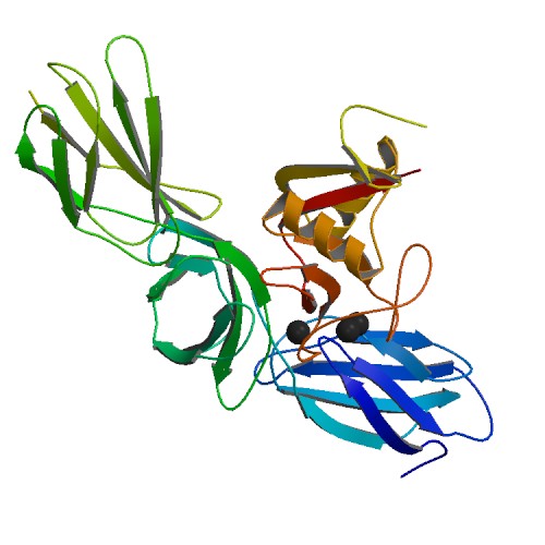
Matrix metalloproteinases (MMPs), also known as matrix metallopeptidases or matrixins, are metalloproteinases that are calcium-dependent zinc-containing endopeptidases; other family members are adamalysins, serralysins, and astacins. The MMPs belong to a larger family of proteases known as the metzincin superfamily. Collectively, these enzymes are capable of degrading all kinds of extracellular matrix proteins, but also can process a number of bioactive molecules. They are known to be involved in the cleavage of cell surface receptors, the release of apoptotic ligands (such as the FAS ligand), and chemokine/cytokine inactivation. MMPs are also thought to play a major role in cell behaviors such as cell proliferation, migration (adhesion/dispersion), differentiation, angiogenesis, apoptosis, and host defense. They were first described in vertebrates (1962), including humans, but have since been found in invertebrates and plants. They are distinguished from other endopeptida ... [...More Info...] [...Related Items...] OR: [Wikipedia] [Google] [Baidu] |
Cytokine
Cytokines are a broad and loose category of small proteins (~5–25 kDa) important in cell signaling. Cytokines are peptides and cannot cross the lipid bilayer of cells to enter the cytoplasm. Cytokines have been shown to be involved in autocrine, paracrine and endocrine signaling as immunomodulating agents. Cytokines include chemokines, interferons, interleukins, lymphokines, and tumour necrosis factors, but generally not hormones or growth factors (despite some overlap in the terminology). Cytokines are produced by a broad range of cells, including immune cells like macrophages, B lymphocytes, T lymphocytes and mast cells, as well as endothelial cells, fibroblasts, and various stromal cells; a given cytokine may be produced by more than one type of cell. They act through cell surface receptors and are especially important in the immune system; cytokines modulate the balance between humoral and cell-based immune responses, and they regulate the maturation, growth, and res ... [...More Info...] [...Related Items...] OR: [Wikipedia] [Google] [Baidu] |
Glycosaminoglycans
Glycosaminoglycans (GAGs) or mucopolysaccharides are long, linear polysaccharides consisting of repeating disaccharide units (i.e. two-sugar units). The repeating two-sugar unit consists of a uronic sugar and an amino sugar, except in the case of the sulfated glycosaminoglycan keratan, where, in place of the uronic sugar there is a galactose unit. GAGs are found in vertebrates, invertebrates and bacteria. Because GAGs are highly polar molecules and attract water; the body uses them as lubricants or shock absorbers. Mucopolysaccharidoses are a group of metabolic disorders in which abnormal accumulations of glycosaminoglycans occur due to enzyme deficiencies. Production Glycosaminoglycans vary greatly in molecular mass, disaccharide structure, and sulfation. This is because GAG synthesis is not template driven, as are proteins or nucleic acids, but constantly altered by processing enzymes. GAGs are classified into four groups, based on their core disaccharide structures. Hepa ... [...More Info...] [...Related Items...] OR: [Wikipedia] [Google] [Baidu] |
Proteoglycans
Proteoglycans are proteins that are heavily glycosylated. The basic proteoglycan unit consists of a "core protein" with one or more covalently attached glycosaminoglycan (GAG) chain(s). The point of attachment is a serine (Ser) residue to which the glycosaminoglycan is joined through a tetrasaccharide bridge (e.g. chondroitin sulfate- GlcA- Gal-Gal- Xyl-PROTEIN). The Ser residue is generally in the sequence -Ser-Gly-X-Gly- (where X can be any amino acid residue but proline), although not every protein with this sequence has an attached glycosaminoglycan. The chains are long, linear carbohydrate polymers that are negatively charged under physiological conditions due to the occurrence of sulfate and uronic acid groups. Proteoglycans occur in connective tissue. Types Proteoglycans are categorized by their relative size (large and small) and the nature of their glycosaminoglycan chains. Types include: Certain members are considered members of the "small leucine-rich proteoglyc ... [...More Info...] [...Related Items...] OR: [Wikipedia] [Google] [Baidu] |
Growth Factor
A growth factor is a naturally occurring substance capable of stimulating cell proliferation, wound healing, and occasionally cellular differentiation. Usually it is a secreted protein or a steroid hormone. Growth factors are important for regulating a variety of cellular processes. Growth factors typically act as signaling molecules between cells. Examples are cytokines and hormones that bind to specific receptors on the surface of their target cells. They often promote cell differentiation and maturation, which varies between growth factors. For example, epidermal growth factor (EGF) enhances osteogenic differentiation, while fibroblast growth factors and vascular endothelial growth factors stimulate blood vessel differentiation (angiogenesis). Comparison to cytokines ''Growth factor'' is sometimes used interchangeably among scientists with the term ''cytokine.'' Historically, cytokines were associated with hematopoietic (blood and lymph forming) cells and immune syst ... [...More Info...] [...Related Items...] OR: [Wikipedia] [Google] [Baidu] |
Extracellular Matrix
In biology, the extracellular matrix (ECM), also called intercellular matrix, is a three-dimensional network consisting of extracellular macromolecules and minerals, such as collagen, enzymes, glycoproteins and hydroxyapatite that provide structural and biochemical support to surrounding cells. Because multicellularity evolved independently in different multicellular lineages, the composition of ECM varies between multicellular structures; however, cell adhesion, cell-to-cell communication and differentiation are common functions of the ECM. The animal extracellular matrix includes the interstitial matrix and the basement membrane. Interstitial matrix is present between various animal cells (i.e., in the intercellular spaces). Gels of polysaccharides and fibrous proteins fill the Interstitial fluid, interstitial space and act as a compression buffer against the stress placed on the ECM. Basement membranes are sheet-like depositions of ECM on which various epithelial cells rest ... [...More Info...] [...Related Items...] OR: [Wikipedia] [Google] [Baidu] |
Microenvironment (biology)
The tumor microenvironment (TME) is the environment around a tumor, including the surrounding blood vessels, immune cells, fibroblasts, signaling molecules and the extracellular matrix (ECM). The tumor and the surrounding microenvironment are closely related and interact constantly. Tumors can influence the microenvironment by releasing extracellular signals, promoting tumor angiogenesis and inducing peripheral immune tolerance, while the immune cells in the microenvironment can affect the growth and evolution of cancerous cells. History The importance of a stromal microenvironment, especially "wound" or regenerating tissue, has been recognized since the late 1800s. The interplay between the tumor and its microenvironment was part of Stephen Paget's 1889 "seed and soil" theory, in which he postulated that metastases of a particular type of cancer ("the seed") often metastasizes to certain sites ("the soil") based on the similarity of the original and secondary tumor sites. ... [...More Info...] [...Related Items...] OR: [Wikipedia] [Google] [Baidu] |
Micrograph Of Loose, Moderate And Dense Desmoplastic Stroma In Pancreatic Ductal Adenocarcinoma
A micrograph or photomicrograph is a photograph or digital image taken through a microscope or similar device to show a magnified image of an object. This is opposed to a macrograph or photomacrograph, an image which is also taken on a microscope but is only slightly magnified, usually less than 10 times. Micrography is the practice or art of using microscopes to make photographs. A micrograph contains extensive details of microstructure. A wealth of information can be obtained from a simple micrograph like behavior of the material under different conditions, the phases found in the system, failure analysis, grain size estimation, elemental analysis and so on. Micrographs are widely used in all fields of microscopy. Types Photomicrograph A light micrograph or photomicrograph is a micrograph prepared using an optical microscope, a process referred to as ''photomicroscopy''. At a basic level, photomicroscopy may be performed simply by connecting a camera to a microscope, t ... [...More Info...] [...Related Items...] OR: [Wikipedia] [Google] [Baidu] |
Metastasis
Metastasis is a pathogenic agent's spread from an initial or primary site to a different or secondary site within the host's body; the term is typically used when referring to metastasis by a cancerous tumor. The newly pathological sites, then, are metastases (mets). It is generally distinguished from cancer invasion, which is the direct extension and penetration by cancer cells into neighboring tissues. Cancer occurs after cells are genetically altered to proliferate rapidly and indefinitely. This uncontrolled proliferation by mitosis produces a primary heterogeneic tumour. The cells which constitute the tumor eventually undergo metaplasia, followed by dysplasia then anaplasia, resulting in a malignant phenotype. This malignancy allows for invasion into the circulation, followed by invasion to a second site for tumorigenesis. Some cancer cells known as circulating tumor cells acquire the ability to penetrate the walls of lymphatic or blood vessels, after which they are abl ... [...More Info...] [...Related Items...] OR: [Wikipedia] [Google] [Baidu] |
Wound
A wound is a rapid onset of injury that involves laceration, lacerated or puncture wound, punctured skin (an ''open'' wound), or a bruise, contusion (a ''closed'' wound) from blunt force physical trauma, trauma or compression. In pathology, a ''wound'' is an acute injury that damages the epidermis of the skin. To healing, heal a wound, the body undertakes a series of actions collectively known as the wound healing process. Classification According to level of contamination, a wound can be classified as: * Clean wound – made under sterile conditions where there are no organisms present, and the skin is likely to heal without complications. * Contaminated wound – usually resulting from accidental injury; there are pathogenic organisms and foreign bodies in the wound. * Infected wound – the wound has pathogenic organisms present and multiplying, exhibiting clinical signs of infection (yellow appearance, soreness, redness, oozing pus). * Colonized wound – a chronic situation, ... [...More Info...] [...Related Items...] OR: [Wikipedia] [Google] [Baidu] |


