|
Descending Branch Of Lateral Femoral Circumflex Artery
The lateral circumflex femoral artery, also known as the lateral femoral circumflex artery, or the external circumflex artery, is an artery in the upper thigh. It is usually a branch of the profunda femoris artery, and produces three branches. It is mostly distributed to the muscles of the lateral thigh. Structure Origin The lateral femoral circumflex artery usually arises from the lateral side of the profunda femoris artery, but may occasionally arise directly from the femoral artery. It is the largest branch of the profunda femoris artery. Course and relations The lateral circumflex femoral artery usually courses anterior to the femoral neck. It passes horizontally between the divisions of the femoral nerve. It passes posterior to the sartorius muscle and rectus femoris muscle. It passes laterally across the hip joint capsule. It divides into ascending, transverse, and descending branches. Branches The lateral circumflex femoral artery has three branches: # The ascen ... [...More Info...] [...Related Items...] OR: [Wikipedia] [Google] [Baidu] |
Deep Femoral Artery
The deep artery of the thigh, (profunda femoris artery or deep femoral artery) is a large branch of the femoral artery. It travels more deeply (posteriorly) than the rest of the femoral artery. Structure The deep artery of the thigh branches off the posterolateral side of the femoral artery soon after its origin. It travels down the thigh closer to the femur than the femoral artery. It runs between the pectineus muscle and the adductor longus muscle. It runs on the posterior side of adductor longus muscle. It pierces the adductor magnus muscle, and may be known as the fourth perforating artery as it continues. The deep femoral artery does not leave the thigh. Branches The deep artery of the thigh gives off the following branches: * Lateral circumflex femoral artery. * Medial circumflex femoral artery. * 3 Perforating arteries - perforate the adductor magnus muscle to the posterior and medial compartments of the thigh to connect with the branches of the popliteal artery behind t ... [...More Info...] [...Related Items...] OR: [Wikipedia] [Google] [Baidu] |
Thigh
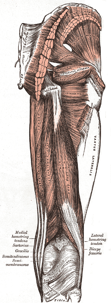
In human anatomy, the thigh is the area between the hip (pelvis) and the knee. Anatomically, it is part of the lower limb. The single bone in the thigh is called the femur. This bone is very thick and strong (due to the high proportion of bone tissue), and forms a ball and socket joint at the hip, and a modified hinge joint at the knee. Structure Bones The femur is the only bone in the thigh and serves as an attachment site for all muscles in the thigh. The head of the femur articulates with the acetabulum in the pelvic bone forming the hip joint, while the distal part of the femur articulates with the tibia and patella forming the knee. By most measures, the femur is the strongest bone in the body. The femur is also the longest bone in the body. The femur is categorised as a long bone and comprises a diaphysis, the shaft (or body) and two epiphysis or extremities that articulate with adjacent bones in the hip and knee. Muscular compartments In cross-section, the thigh is ... [...More Info...] [...Related Items...] OR: [Wikipedia] [Google] [Baidu] |
Orthopedic Surgery
Orthopedic surgery or orthopedics ( alternatively spelt orthopaedics), is the branch of surgery concerned with conditions involving the musculoskeletal system. Orthopedic surgeons use both surgical and nonsurgical means to treat musculoskeletal trauma, spine diseases, sports injuries, degenerative diseases, infections, tumors, and congenital disorders. Etymology Nicholas Andry coined the word in French as ', derived from the Ancient Greek words ὀρθός ''orthos'' ("correct", "straight") and παιδίον ''paidion'' ("child"), and published ''Orthopedie'' (translated as ''Orthopædia: Or the Art of Correcting and Preventing Deformities in Children'') in 1741. The word was assimilated into English as ''orthopædics''; the ligature ''æ'' was common in that era for ''ae'' in Greek- and Latin-based words. As the name implies, the discipline was initially developed with attention to children, but the correction of spinal and bone deformities in all stages of life eventually ... [...More Info...] [...Related Items...] OR: [Wikipedia] [Google] [Baidu] |
Centimetre
330px, Different lengths as in respect to the Electromagnetic spectrum, measured by the Metre and its deriveds scales. The Microwave are in-between 1 meter to 1 millimeter. A centimetre (international spelling) or centimeter (American spelling) (SI symbol cm) is a Units of measurement, unit of length in the International System of Units (SI), equal to one hundredth of a metre, ''centi'' being the SI prefix for a factor of . The centimetre was the base unit of length in the now deprecated centimetre–gram–second (CGS) system of units. Though for many physical quantities, SI prefixes for factors of 103—like ''milli-'' and ''kilo-''—are often preferred by technicians, the centimetre remains a practical unit of length for many everyday measurements. A centimetre is approximately the width of the fingernail of an average adult person. Equivalence to other units of length : One millilitre is defined as one cubic centimetre, under the SI system of units. Other uses In ... [...More Info...] [...Related Items...] OR: [Wikipedia] [Google] [Baidu] |
Medial Circumflex Femoral Artery
The medial circumflex femoral artery (internal circumflex artery, medial femoral circumflex artery) is an artery in the upper thigh that arises from the profunda femoris artery''.'' Damage to the artery following a femoral neck fracture may lead to avascular necrosis (ischemic) of the femoral neck/head. Structure Origin The medial femoral circumflex artery arises from the posterior medial aspect of the profunda femoris artery''.'' The medial femoral circumflex artery may occasionally arise directly from the femoral artery. Course and relations It winds around the medial side of the femur, passing first between the pectineus and iliopsoas muscles, and then between the obturator externus and the adductor brevis muscles. Branches At the upper border of the adductor brevis it gives off two branches: * The '' ascending branch'' * The ''descending branch'' descends beneath the adductor brevis, to supply it and the adductor magnus; the continuation of the vessel passes backward ... [...More Info...] [...Related Items...] OR: [Wikipedia] [Google] [Baidu] |
Neck Of The Femur
The femoral neck (femur neck or neck of the femur) is a flattened pyramidal process of bone, connecting the femoral head with the femoral shaft, and forming with the latter a wide angle opening medialward. Structure The neck is flattened from before backward, contracted in the middle, and broader laterally than medially. The vertical diameter of the lateral half is increased by the obliquity of the lower edge, which slopes downward to join the body at the level of the lesser trochanter, so that it measures one-third more than the antero-posterior diameter. The medial half is smaller and of a more circular shape. The anterior surface of the neck is perforated by numerous vascular foramina. Along the upper part of the line of junction of the anterior surface with the head is a shallow groove, best marked in elderly subjects; this groove lodges the orbicular fibers of the capsule of the hip joint. The posterior surface is smooth, and is broader and more concave than the anter ... [...More Info...] [...Related Items...] OR: [Wikipedia] [Google] [Baidu] |
Femoral Head
The femoral head (femur head or head of the femur) is the highest part of the thigh bone (femur). It is supported by the femoral neck. Structure The head is globular and forms rather more than a hemisphere, is directed upward, medialward, and a little forward, the greater part of its convexity being above and in front. The femoral head's surface is smooth. It is coated with cartilage in the fresh state, except over an ovoid depression, the fovea capitis, which is situated a little below and behind the center of the femoral head, and gives attachment to the ligament of head of femur. The thickest region of the articular cartilage is at the centre of the femoral head, measuring up to 2.8 mm. The diameter of the femoral head is usually larger in men than in women. Fovea capitis The fovea capitis is a small, concave depression within the head of the femur that serves as an attachment point for the ligamentum teres (Saladin). It is slightly ovoid in shape and is oriented "superior ... [...More Info...] [...Related Items...] OR: [Wikipedia] [Google] [Baidu] |
Perforating Arteries
The perforating arteries, usually three in number, are so named because they perforate the tendon of the Adductor magnus to reach the back of the thigh. They pass backward close to the linea aspera of the femur under cover of small tendinous arches in the muscle. The first is given off above the Adductor brevis, the second in front of that muscle, and the third immediately below it. First The ''first perforating artery'' (a. perforans prima) passes posteriorly between the Pectineus and Adductor brevis (sometimes it perforates the latter); it then pierces the Adductor magnus close to the linea aspera. It gives branches to the Adductores brevis and magnus, Biceps femoris, and Gluteus maximus, and anastomoses with the inferior gluteal, medial and lateral femoral circumflex and second perforating arteries. Second The ''second perforating artery'' (a. perforans secunda), larger than the first, pierces the tendons of the Adductores brevis and magnus, and divides into ascending and de ... [...More Info...] [...Related Items...] OR: [Wikipedia] [Google] [Baidu] |
Inferior Gluteal Artery
The inferior gluteal artery (sciatic artery), the smaller of the two terminal branches of the anterior trunk of the internal iliac artery, is distributed chiefly to the buttock and back of the thigh. It passes down on the sacral plexus of nerves and the piriformis muscle, behind the internal pudendal artery. It passes through the lower part of the greater sciatic foramen. It escapes from the pelvis between piriformis muscle and coccygeus muscle. It then descends in the interval between the greater trochanter of the femur and tuberosity of the ischium. It is accompanied by the sciatic nerve and the posterior femoral cutaneous nerves, and covered by the gluteus maximus. It continues down the back of the thigh, supplying the skin, and anastomosing with branches of the perforating arteries. Additional images File:Gray544.png, The arteries of the gluteal and posterior femoral regions. File:Gray829.png, Dissection of side wall of pelvis showing sacral and pudendal plexuses. See a ... [...More Info...] [...Related Items...] OR: [Wikipedia] [Google] [Baidu] |
Medial Femoral Circumflex Artery
The medial circumflex femoral artery (internal circumflex artery, medial femoral circumflex artery) is an artery in the upper thigh that arises from the profunda femoris artery''.'' Damage to the artery following a femoral neck fracture may lead to avascular necrosis (ischemic) of the femoral neck/head. Structure Origin The medial femoral circumflex artery arises from the posterior medial aspect of the profunda femoris artery''.'' The medial femoral circumflex artery may occasionally arise directly from the femoral artery. Course and relations It winds around the medial side of the femur, passing first between the pectineus and iliopsoas muscles, and then between the obturator externus and the adductor brevis muscles. Branches At the upper border of the adductor brevis it gives off two branches: * The '' ascending branch'' * The ''descending branch'' descends beneath the adductor brevis, to supply it and the adductor magnus; the continuation of the vessel passes backward a ... [...More Info...] [...Related Items...] OR: [Wikipedia] [Google] [Baidu] |
Greater Trochanter
The greater trochanter of the femur is a large, irregular, quadrilateral eminence and a part of the skeletal system. It is directed lateral and medially and slightly posterior. In the adult it is about 2–4 cm lower than the femoral head.Standring, Susan, editor. ''Gray’s Anatomy: The Anatomical Basis of Clinical Practice''. Forty-First edition, Elsevier Limited, 2016, p. 1327. Because the pelvic outlet in the female is larger than in the male, there is a greater distance between the greater trochanters in the female. It has two surfaces and four borders. It is a traction epiphysis. Surfaces The ''lateral surface'', quadrilateral in form, is broad, rough, convex, and marked by a diagonal impression, which extends from the postero-superior to the antero-inferior angle, and serves for the insertion of the tendon of the gluteus medius. Above the impression is a triangular surface, sometimes rough for part of the tendon of the same muscle, sometimes smooth for the interposi ... [...More Info...] [...Related Items...] OR: [Wikipedia] [Google] [Baidu] |
Femur
The femur (; ), or thigh bone, is the proximal bone of the hindlimb in tetrapod vertebrates. The head of the femur articulates with the acetabulum in the pelvic bone forming the hip joint, while the distal part of the femur articulates with the tibia (shinbone) and patella (kneecap), forming the knee joint. By most measures the two (left and right) femurs are the strongest bones of the body, and in humans, the largest and thickest. Structure The femur is the only bone in the upper leg. The two femurs converge medially toward the knees, where they articulate with the proximal ends of the tibiae. The angle of convergence of the femora is a major factor in determining the femoral-tibial angle. Human females have thicker pelvic bones, causing their femora to converge more than in males. In the condition ''genu valgum'' (knock knee) the femurs converge so much that the knees touch one another. The opposite extreme is ''genu varum'' (bow-leggedness). In the general populatio ... [...More Info...] [...Related Items...] OR: [Wikipedia] [Google] [Baidu] |


