|
Decapentaplegic Homolog 1
Decapentaplegic (Dpp) is a key morphogen involved in the development of the fruit fly ''Drosophila melanogaster'' and is the first validated secreted morphogen. It is known to be necessary for the correct patterning and development of the early ''Drosophila'' embryo and the fifteen imaginal discs, which are tissues that will become limbs and other organs and structures in the adult fly. It has also been suggested that Dpp plays a role in regulating the growth and size of tissues. Flies with mutations in decapentaplegic fail to form these structures correctly, hence the name (''decapenta''-, fifteen, -''plegic'', paralysis). Dpp is the Drosophila homolog of the vertebrate bone morphogenetic proteins (BMPs), which are members of the TGF-β superfamily, a class of proteins that are often associated with their own specific signaling pathway. Studies of Dpp in Drosophila have led to greater understanding of the function and importance of their homologs in vertebrates like humans. Fu ... [...More Info...] [...Related Items...] OR: [Wikipedia] [Google] [Baidu] |
Morphogen
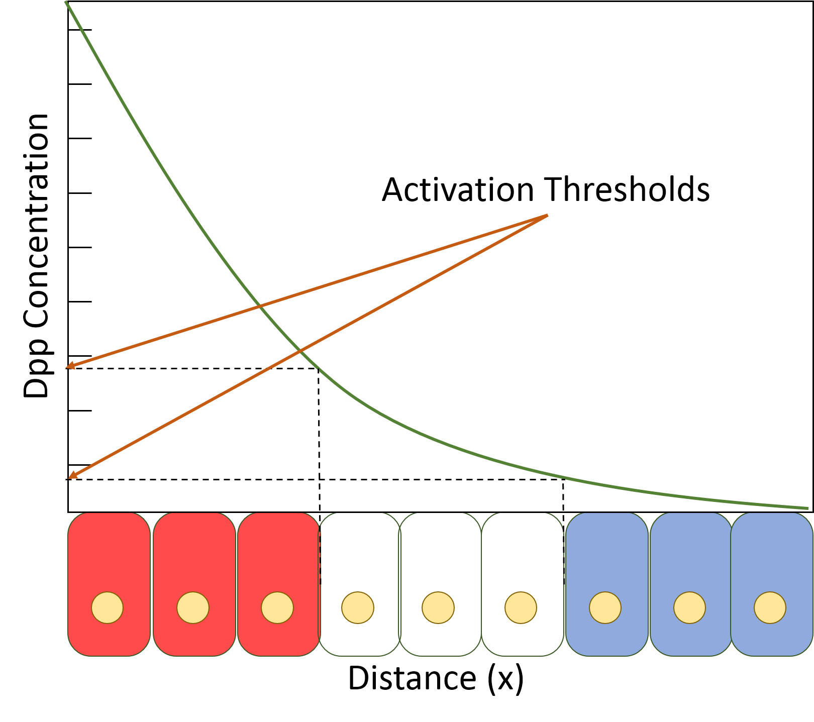
A morphogen is a substance whose non-uniform distribution governs the pattern of tissue development in the process of morphogenesis or pattern formation, one of the core processes of developmental biology, establishing positions of the various specialized cell types within a tissue. More specifically, a morphogen is a signaling molecule that acts directly on cells to produce specific cellular responses depending on its local concentration. Typically, morphogens are produced by source cells and diffuse through surrounding tissues in an embryo during early development, such that concentration gradients are set up. These gradients drive the process of differentiation of unspecialised stem cells into different cell types, ultimately forming all the tissues and organs of the body. The control of morphogenesis is a central element in evolutionary developmental biology (evo-devo). History The term was coined by Alan Turing in the paper "The Chemical Basis of Morphogenesis", where he ... [...More Info...] [...Related Items...] OR: [Wikipedia] [Google] [Baidu] |
Hedgehog (cell Signaling)
The Hedgehog signaling pathway is a signaling pathway that transmits information to embryonic cells required for proper cell differentiation. Different parts of the embryo have different concentrations of hedgehog signaling proteins. The pathway also has roles in the adult. Diseases associated with the malfunction of this pathway include cancer. The Hedgehog signaling pathway is one of the key regulators of animal development and is present in all bilaterians. The pathway takes its name from its polypeptide ligand (biochemistry), ligand, an intracellular signaling molecule called Hedgehog (''Hh'') found in fruit flies of the genus ''Drosophila''; fruit fly larva lacking the ''Hh'' gene are said to resemble hedgehogs. ''Hh'' is one of Drosophila's segment polarity gene products, involved in establishing the basis of the fly body plan. Larvae without ''Hh'' are short and spiny, resembling the hedgehog animal. The molecule remains important during later stages of embryogenesis and M ... [...More Info...] [...Related Items...] OR: [Wikipedia] [Google] [Baidu] |
Filopodia
Filopodia (singular filopodium) are slender cytoplasmic projections that extend beyond the leading edge of lamellipodia in migrating cells. Within the lamellipodium, actin ribs are known as ''microspikes'', and when they extend beyond the lamellipodia, they're known as filopodia. They contain microfilaments (also called actin filaments) cross-linked into bundles by actin-bundling proteins, such as fascin and fimbrin. Filopodia form focal adhesions with the substratum, linking them to the cell surface. Many types of migrating cells display filopodia, which are thought to be involved in both sensation of chemotropic cues, and resulting changes in directed locomotion. Activation of the Rho family of GTPases, particularly cdc42 and their downstream intermediates, results in the polymerization of actin fibers by Ena/Vasp homology proteins. Growth factors bind to receptor tyrosine kinases resulting in the polymerization of actin filaments, which, when cross-linked, make up the sup ... [...More Info...] [...Related Items...] OR: [Wikipedia] [Google] [Baidu] |
Actin
Actin is a family of globular multi-functional proteins that form microfilaments in the cytoskeleton, and the thin filaments in muscle fibrils. It is found in essentially all eukaryotic cells, where it may be present at a concentration of over 100 μM; its mass is roughly 42 kDa, with a diameter of 4 to 7 nm. An actin protein is the monomeric subunit of two types of filaments in cells: microfilaments, one of the three major components of the cytoskeleton, and thin filaments, part of the contractile apparatus in muscle cells. It can be present as either a free monomer called G-actin (globular) or as part of a linear polymer microfilament called F-actin (filamentous), both of which are essential for such important cellular functions as the mobility and contraction of cells during cell division. Actin participates in many important cellular processes, including muscle contraction, cell motility, cell division and cytokinesis, vesicle and organelle movement, cell sign ... [...More Info...] [...Related Items...] OR: [Wikipedia] [Google] [Baidu] |
Dynamin
Dynamin is a GTPase responsible for endocytosis in the eukaryotic cell. Dynamin is part of the "dynamin superfamily", which includes classical dynamins, dynamin-like proteins, Mx proteins, OPA1, mitofusins, and GBPs. Members of the dynamin family are principally involved in the scission of newly formed vesicles from the membrane of one cellular compartment and their targeting to, and fusion with, another compartment, both at the cell surface (particularly caveolae internalization) as well as at the Golgi apparatus.Hinshaw, J"Research statement, Jenny E. Hinshaw, Ph.D."National Institute of Diabetes & Digestive & Kidney Diseases, Laboratory of Cell Biochemistry and Biology. Accessed 19 March 2013. Dynamin family members also play a role in many processes including division of organelles, cytokinesis and microbial pathogen resistance. Structure Dynamin itself is a 96 kDa enzyme, and was first isolated when researchers were attempting to isolate new microtubule-based motors ... [...More Info...] [...Related Items...] OR: [Wikipedia] [Google] [Baidu] |
Dally (gene)
Dally (division abnormally delayed) is the name of a gene that encodes a HS-modified-protein found in the fruit fly (''Drosophila melanogaster''). The protein has to be processed after being codified, and in its mature form it is composed by 626 amino acids,Uniprot KB forming a proteoglycan rich in heparin sulfate which is anchored to the cell surface via covalent linkage to (GPI), so we can define it as a . For its normal biosynthesis it requires sugarless (''sgl''), a gene that encodes an enzyme which plays a critical role in the process of modification of da ... [...More Info...] [...Related Items...] OR: [Wikipedia] [Google] [Baidu] |
Heparan Sulfate
Heparan sulfate (HS) is a linear polysaccharide found in all animal tissues. It occurs as a proteoglycan (HSPG, i.e. Heparan Sulfate ProteoGlycan) in which two or three HS chains are attached in close proximity to cell surface or extracellular matrix proteins. It is in this form that HS binds to a variety of protein ligands, including Wnt, and regulates a wide range of biological activities, including developmental processes, angiogenesis, blood coagulation, abolishing detachment activity by GrB (Granzyme B), and tumour metastasis. HS has also been shown to serve as cellular receptor for a number of viruses, including the respiratory syncytial virus. One study suggests that cellular heparan sulfate has a role in SARS-CoV-2 Infection, particularly when the virus attaches with ACE2. Proteoglycans The major cell membrane HSPGs are the transmembrane syndecans and the glycosylphosphatidylinositol (GPI) anchored glypicans. Other minor forms of membrane HSPG include betaglycan and the ... [...More Info...] [...Related Items...] OR: [Wikipedia] [Google] [Baidu] |
Glypican
Glypicans constitute one of the two major families of heparan sulfate proteoglycans, with the other major family being syndecans. Six glypicans have been identified in mammals, and are referred to as GPC1 through GPC6. In ''Drosophila'' two glypicans have been identified, and these are referred to as dally (division abnormally delayed) and dally-like. One glypican has been identified in ''C. elegans''. Glypicans seem to play a vital role in developmental morphogenesis, and have been suggested as regulators for the Wnt and Hedgehog cell signaling pathways. They have additionally been suggested as regulators for fibroblast growth factor and bone morphogenic protein signaling. Structure While six glypicans have been identified in mammals, several characteristics remain consistent between these different proteins. First, the core protein of all glypicans is similar in size, approximately ranging between 60 and 70 kDa. Additionally, in terms of amino acid sequence, the location of fo ... [...More Info...] [...Related Items...] OR: [Wikipedia] [Google] [Baidu] |
Fluorescence Correlation Spectroscopy
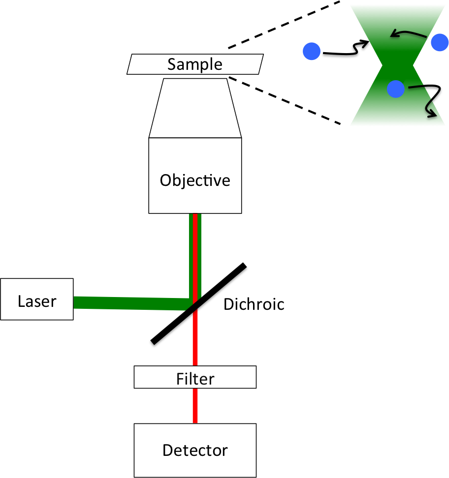
Fluorescence correlation spectroscopy (FCS) is a statistical analysis, via time correlation, of stationary fluctuations of the fluorescence intensity. Its theoretical underpinning originated from L. Onsager's regression hypothesis. The analysis provides kinetic parameters of the physical processes underlying the fluctuations. One of the interesting applications of this is an analysis of the concentration fluctuations of fluorescent particles (molecules) in solution. In this application, the fluorescence emitted from a very tiny space in solution containing a small number of fluorescent particles (molecules) is observed. The fluorescence intensity is fluctuating due to Brownian motion of the particles. In other words, the number of the particles in the sub-space defined by the optical system is randomly changing around the average number. The analysis gives the average number of fluorescent particles and average diffusion time, when the particle is passing through the space. Eventua ... [...More Info...] [...Related Items...] OR: [Wikipedia] [Google] [Baidu] |
Cytoneme
Cytonemes are thin, cellular projections that are specialized for exchange of signaling proteins between cells. Cytonemes emanate from cells that make signaling proteins, extending directly to cells that receive signaling proteins. Cytonemes also extend directly from cells that receive signaling proteins to cells that make them. A cytoneme is a type of filopodium - a thin, tubular extension of a cell’s plasma membrane that has a core composed of tightly bundled, parallel actin filaments. Filopodia can extend more than 100 μm and have been measured as thin as 0.1 μm and as thick as 0.5 μm. Cytonemes with a diameter of approximately 0.2 μm and as long as 80 μm have been observed in the Drosophila wing imaginal disc. Many cell types have filopodia. The functions of filopodia have been attributed to pathfinding of neurons, early stages of synapse formation, antigen presentation by dendritic cells of the immune system, force generation by macrophages and virus transmission. The ... [...More Info...] [...Related Items...] OR: [Wikipedia] [Google] [Baidu] |
Transcytosis
Transcytosis (also known as cytopempsis) is a type of transcellular transport in which various macromolecules are transported across the interior of a cell. Macromolecules are captured in vesicles on one side of the cell, drawn across the cell, and ejected on the other side. Examples of macromolecules transported include IgA, transferrin, and insulin. While transcytosis is most commonly observed in epithelial cells, the process is also present elsewhere. Blood capillaries are a well-known site for transcytosis, though it occurs in other cells, including neurons, osteoclasts and M cells of the intestine. Regulation The regulation of transcytosis varies greatly due to the many different tissues in which this process is observed. Various tissue-specific mechanisms of transcytosis have been identified. Brefeldin A, a commonly used inhibitor of ER-to-Golgi apparatus transport, has been shown to inhibit transcytosis in dog kidney cells, which provided the first clues as to the nature ... [...More Info...] [...Related Items...] OR: [Wikipedia] [Google] [Baidu] |


