|
Cytoneme
Cytonemes are thin, cellular projections that are specialized for exchange of signaling proteins between cells. Cytonemes emanate from cells that make signaling proteins, extending directly to cells that receive signaling proteins. Cytonemes also extend directly from cells that receive signaling proteins to cells that make them. A cytoneme is a type of filopodium - a thin, tubular extension of a cell’s plasma membrane that has a core composed of tightly bundled, parallel actin filaments. Filopodia can extend more than 100 μm and have been measured as thin as 0.1 μm and as thick as 0.5 μm. Cytonemes with a diameter of approximately 0.2 μm and as long as 80 μm have been observed in the Drosophila wing imaginal disc. Many cell types have filopodia. The functions of filopodia have been attributed to pathfinding of neurons, early stages of synapse formation, antigen presentation by dendritic cells of the immune system, force generation by macrophages and virus transmission. The ... [...More Info...] [...Related Items...] OR: [Wikipedia] [Google] [Baidu] |
Decapentaplegic
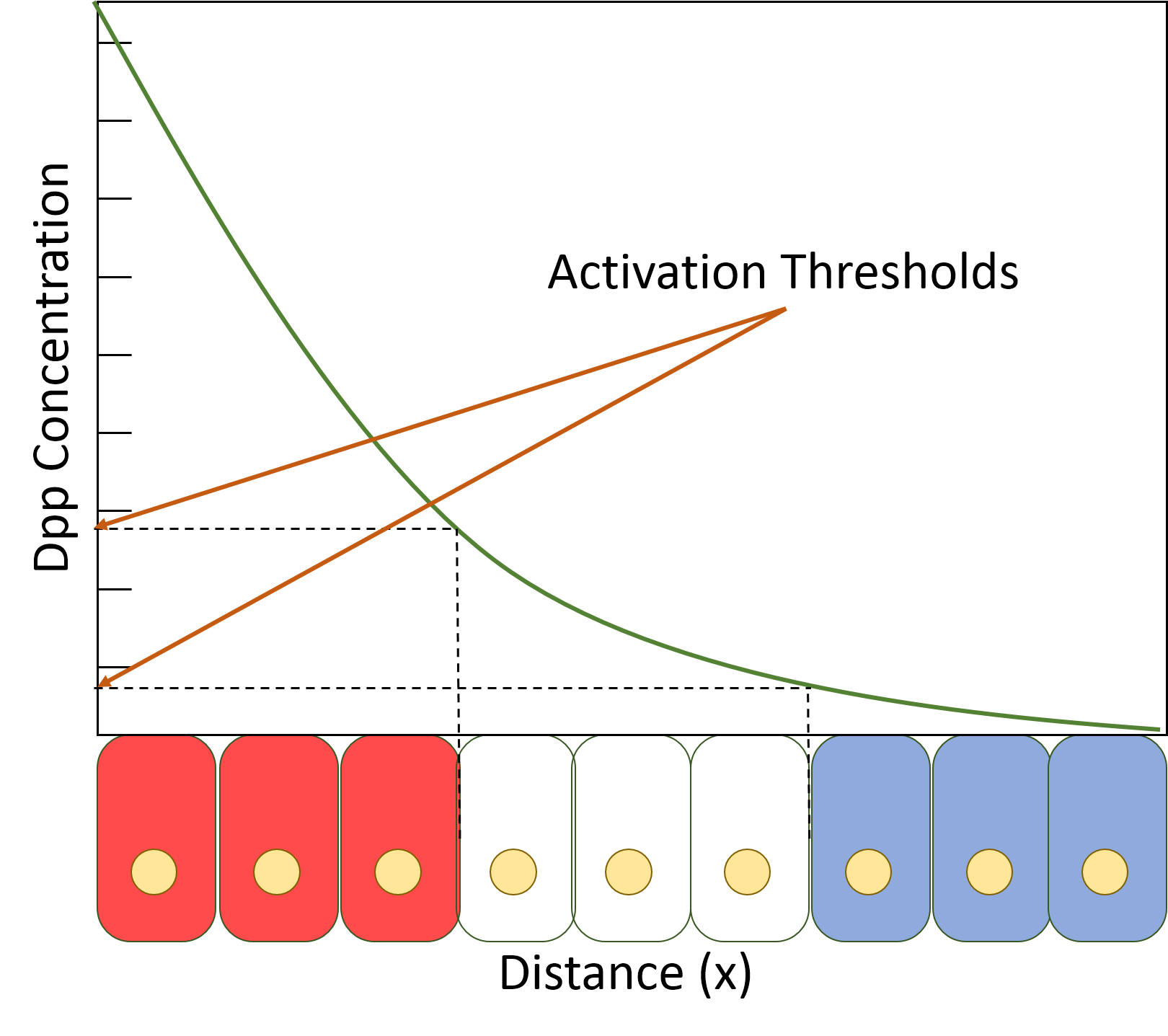
Decapentaplegic (Dpp) is a key morphogen involved in the development of the fruit fly ''Drosophila melanogaster'' and is the first validated secreted morphogen. It is known to be necessary for the correct patterning and development of the early ''Drosophila'' embryo and the fifteen imaginal discs, which are tissues that will become limbs and other organs and structures in the adult fly. It has also been suggested that Dpp plays a role in regulating the growth and size of tissues. Flies with mutations in decapentaplegic fail to form these structures correctly, hence the name (''decapenta''-, fifteen, -''plegic'', paralysis). Dpp is the Drosophila homolog of the vertebrate bone morphogenetic proteins (BMPs), which are members of the TGF-β superfamily, a class of proteins that are often associated with their own specific signaling pathway. Studies of Dpp in Drosophila have led to greater understanding of the function and importance of their homologs in vertebrates like humans. Func ... [...More Info...] [...Related Items...] OR: [Wikipedia] [Google] [Baidu] |
Tunneling Nanotubes
A tunneling nanotube (TNT) or membrane nanotube is a term that has been applied to protrusions that extend from the plasma membrane which enable different animal cells to touch over long distances, sometimes over 100 μm between T cells. Two types of structures have been called nanotubes. The first type are less than 0.7 micrometers in diameter, contain actin and carry portions of plasma membrane between cells in both directions. The second type are larger (>0.7 μm), contain both actin and microtubules, and can carry components of the cytoplasm such as vesicles and organelles between cells, including whole mitochondria. The diameter of TNTs ranges from 50 to 200 nm and they can reach lengths of several cell diameters. These structures may be involved in cell-to-cell communication, transfer of nucleic acids such as mRNA and miRNA between cells in culture or in a tissue, and the spread of pathogens or toxins such as HIV and prions. TNTs have observed lifetimes rangin ... [...More Info...] [...Related Items...] OR: [Wikipedia] [Google] [Baidu] |
Tracheal Cytonemes
The trachea, also known as the windpipe, is a cartilaginous tube that connects the larynx to the bronchi of the lungs, allowing the passage of air, and so is present in almost all air- breathing animals with lungs. The trachea extends from the larynx and branches into the two primary bronchi. At the top of the trachea the cricoid cartilage attaches it to the larynx. The trachea is formed by a number of horseshoe-shaped rings, joined together vertically by overlying ligaments, and by the trachealis muscle at their ends. The epiglottis closes the opening to the larynx during swallowing. The trachea begins to form in the second month of embryo development, becoming longer and more fixed in its position over time. It is epithelium lined with column-shaped cells that have hair-like extensions called cilia, with scattered goblet cells that produce protective mucins. The trachea can be affected by inflammation or infection, usually as a result of a viral illness affecting other pa ... [...More Info...] [...Related Items...] OR: [Wikipedia] [Google] [Baidu] |
Vasculogenesis
Vasculogenesis is the process of blood vessel formation, occurring by a '' de novo'' production of endothelial cells. It is sometimes paired with angiogenesis, as the first stage of the formation of the vascular network, closely followed by angiogenesis. Process In the sense distinguished from angiogenesis, vasculogenesis is different in one aspect: whereas angiogenesis is the formation of new blood vessels from pre-existing ones, vasculogenesis is the formation of new blood vessels, in blood islands, when there are no pre-existing ones. For example, if a monolayer of endothelial cells begins sprouting to form capillaries, angiogenesis is occurring. Vasculogenesis, in contrast, is when endothelial precursor cells (angioblasts) migrate and differentiate in response to local cues (such as growth factors and extracellular matrices) to form new blood vessels. These vascular trees are then pruned and extended through angiogenesis. Occurrences Vasculogenesis occurs during embryologic ... [...More Info...] [...Related Items...] OR: [Wikipedia] [Google] [Baidu] |
B-lymphocytes
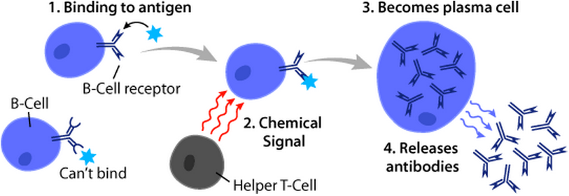
B cells, also known as B lymphocytes, are a type of white blood cell of the lymphocyte subtype. They function in the humoral immunity component of the adaptive immune system. B cells produce antibody molecules which may be either secreted or inserted into the plasma membrane where they serve as a part of B-cell receptors. When a naïve or memory B cell is activated by an antigen, it proliferates and differentiates into an antibody-secreting effector cell, known as a plasmablast or plasma cell. Additionally, B cells Antigen presentation, present antigens (they are also classified as professional Antigen-presenting cell, antigen-presenting cells (APCs)) and secrete cytokines. In mammals, B cells Cellular differentiation, mature in the bone marrow, which is at the core of most bones. In birds, B cells mature in the bursa of Fabricius, a lymphoid organ where they were first discovered by Chang and Glick, which is why the 'B' stands for bursa and not bone marrow as commonly believed ... [...More Info...] [...Related Items...] OR: [Wikipedia] [Google] [Baidu] |
Mast Cells
A mast cell (also known as a mastocyte or a labrocyte) is a resident cell of connective tissue that contains many granules rich in histamine and heparin. Specifically, it is a type of granulocyte derived from the myeloid stem cell that is a part of the immune and neuroimmune systems. Mast cells were discovered by Paul Ehrlich in 1877. Although best known for their role in allergy and anaphylaxis, mast cells play an important protective role as well, being intimately involved in wound healing, angiogenesis, immune tolerance, defense against pathogens, and vascular permeability in brain tumours. The mast cell is very similar in both appearance and function to the basophil, another type of white blood cell. Although mast cells were once thought to be tissue-resident basophils, it has been shown that the two cells develop from different hematopoietic lineages and thus cannot be the same cells. Structure Mast cells are very similar to basophil granulocytes (a class of white blo ... [...More Info...] [...Related Items...] OR: [Wikipedia] [Google] [Baidu] |
Trachea
The trachea, also known as the windpipe, is a Cartilage, cartilaginous tube that connects the larynx to the bronchi of the lungs, allowing the passage of air, and so is present in almost all air-breathing animals with lungs. The trachea extends from the larynx and branches into the two primary bronchi. At the top of the trachea the cricoid cartilage attaches it to the larynx. The trachea is formed by a number of horseshoe-shaped rings, joined together vertically by overlying annular ligaments of trachea, ligaments, and by the trachealis muscle at their ends. The epiglottis closes the opening to the larynx during swallowing. The trachea begins to form in the second month of embryo development, becoming longer and more fixed in its position over time. It is epithelium lined with columnar epithelium, column-shaped cells that have hair-like extensions called cilia, with scattered goblet cells that produce protective mucins. The trachea can be affected by inflammation or infection, us ... [...More Info...] [...Related Items...] OR: [Wikipedia] [Google] [Baidu] |
Morphogen
A morphogen is a substance whose non-uniform distribution governs the pattern of tissue development in the process of morphogenesis or pattern formation, one of the core processes of developmental biology, establishing positions of the various specialized cell types within a tissue. More specifically, a morphogen is a signaling molecule that acts directly on cells to produce specific cellular responses depending on its local concentration. Typically, morphogens are produced by source cells and diffuse through surrounding tissues in an embryo during early development, such that concentration gradients are set up. These gradients drive the process of differentiation of unspecialised stem cells into different cell types, ultimately forming all the tissues and organs of the body. The control of morphogenesis is a central element in evolutionary developmental biology (evo-devo). History The term was coined by Alan Turing in the paper "The Chemical Basis of Morphogenesis", where he ... [...More Info...] [...Related Items...] OR: [Wikipedia] [Google] [Baidu] |
Podosomes
Podosomes are conical, actin-rich structures found on the outer surface of the plasma membrane of animal cells. Their size ranges from approximately 0.5 µm to 2.0 µm in diameter. While usually situated on the periphery of the cellular membrane, these unique structures display a polarized pattern of distribution in migrating cells, situating at the front border between the lamellipodium and lamellum. Their primary purpose is connected to cellular motility and invasion; therefore, they serve as both sites of attachment and degradation along the extracellular matrix. Many different specialized cells exhibit these dynamic structures such as invasive cancer cells, osteoclasts, vascular smooth muscle cells, endothelial cells, and certain immune cells like macrophages and dendritic cells. Characteristics A podosome consists of a core rich in actin surrounded by adhesion and scaffolding proteins. The actin filaments within these structures are highly regulated by many ac ... [...More Info...] [...Related Items...] OR: [Wikipedia] [Google] [Baidu] |
Invadopodia
Invadopodia are actin-rich protrusions of the plasma membrane that are associated with degradation of the extracellular matrix in cancer invasiveness and metastasis. Very similar to podosomes, invadopodia are found in invasive cancer cells and are important for their ability to invade through the extracellular matrix, especially in cancer cell extravasation. Invadopodia are generally visualized by the holes they create in Extracellular matrix, ECM (fibronectin, collagen etc.)-coated plates, in combination with immunohistochemistry for the invadopodia localizing proteins such as cortactin, actin, Tks5 etc. Invadopodia can also be used as a marker to quantify the invasiveness of cancer cell lines ''in vitro'' using a hyaluronic acid hydrogel assay. History and controversy In the early 1980s, researchers noticed protrusions coming from the ventral membrane of cells that had been transformed by the Rous Sarcoma Virus and that they were at the sites of cell-to-extracellular matrix In ... [...More Info...] [...Related Items...] OR: [Wikipedia] [Google] [Baidu] |
Pseudopods
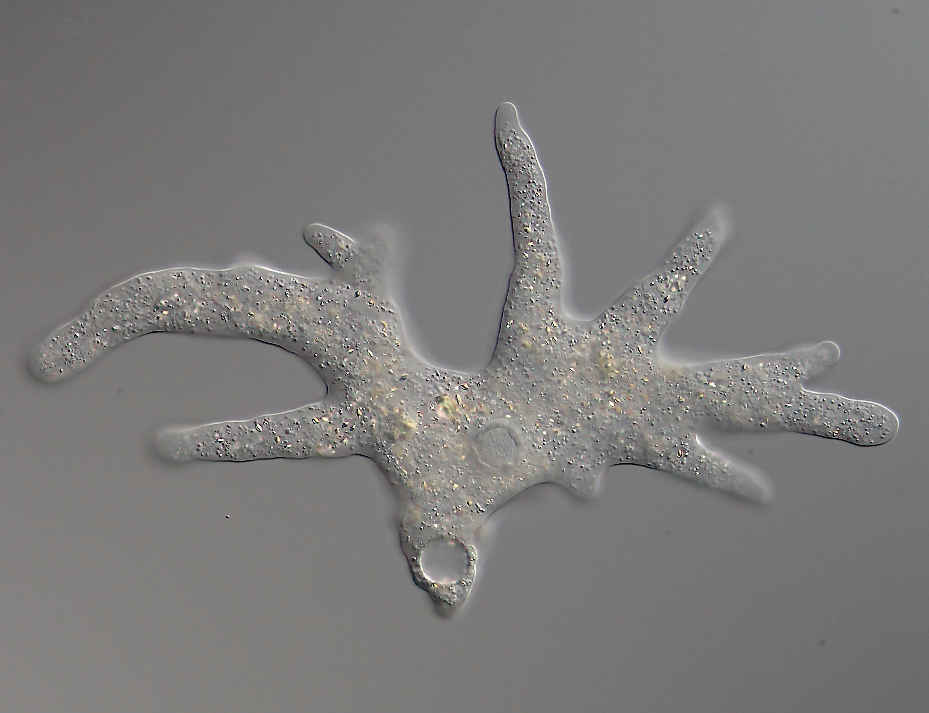
A pseudopod or pseudopodium (plural: pseudopods or pseudopodia) is a temporary arm-like projection of a eukaryotic cell membrane that is emerged in the direction of movement. Filled with cytoplasm, pseudopodia primarily consist of actin filaments and may also contain microtubules and intermediate filaments. Pseudopods are used for motility and ingestion. They are often found in amoebas. Different types of pseudopodia can be classified by their distinct appearances. Lamellipodia are broad and thin. Filopodia are slender, thread-like, and are supported largely by microfilaments. Lobopodia are bulbous and amoebic. Reticulopodia are complex structures bearing individual pseudopodia which form irregular nets. Axopodia are the phagocytosis type with long, thin pseudopods supported by complex microtubule arrays enveloped with cytoplasm; they respond rapidly to physical contact. Some pseudopodial cells are able to use multiple types of pseudopodia depending on the situation: Most of t ... [...More Info...] [...Related Items...] OR: [Wikipedia] [Google] [Baidu] |
Cancer Metastasis
Metastasis is a pathogenic agent's spread from an initial or primary site to a different or secondary site within the host's body; the term is typically used when referring to metastasis by a cancerous tumor. The newly pathological sites, then, are metastases (mets). It is generally distinguished from cancer invasion, which is the direct extension and penetration by cancer cells into neighboring tissues. Cancer occurs after cells are genetically altered to proliferate rapidly and indefinitely. This uncontrolled proliferation by mitosis produces a primary heterogeneic tumour. The cells which constitute the tumor eventually undergo metaplasia, followed by dysplasia then anaplasia, resulting in a malignant phenotype. This malignancy allows for invasion into the circulation, followed by invasion to a second site for tumorigenesis. Some cancer cells known as circulating tumor cells acquire the ability to penetrate the walls of lymphatic or blood vessels, after which they are abl ... [...More Info...] [...Related Items...] OR: [Wikipedia] [Google] [Baidu] |


