|
Cytoarchitectonics
Cytoarchitecture (Greek '' κύτος''= "cell" + '' ἀρχιτεκτονική''= "architecture"), also known as cytoarchitectonics, is the study of the cellular composition of the central nervous system's tissues under the microscope. Cytoarchitectonics is one of the ways to parse the brain, by obtaining sections of the brain using a microtome and staining them with chemical agents which reveal where different neurons are located. The study of the parcellation of ''nerve fibers'' (primarily axons) into layers forms the subject of myeloarchitectonics ( History of the cerebral cytoarchitecture Defining cerebral cytoarchitecture began with the advent of —the science of slicing a ...[...More Info...] [...Related Items...] OR: [Wikipedia] [Google] [Baidu] |
Theodor Meynert
Theodor Hermann Meynert (15 June 1833 – 31 May 1892) was a German-Austrian psychiatrist, neuropathologist and anatomist born in Dresden. Meynert believed that disturbances in brain development could be a predisposition for psychiatric illness and that certain psychoses are reversible. In 1861 he earned his medical doctorate, and in 1875 became director of the psychiatric clinic associated with the University of Vienna. Some of his better known students in Vienna were Josef Breuer, Sigmund Freud, who in 1883 worked at Meynert's psychiatric clinic, and Julius Wagner-Jauregg, who introduced fever treatment for syphilis. Meynert later distanced himself from Freud because of the latter's involvement with practices such as hypnosis. Meynert also ridiculed Freud's idea of male hysteria; though some authors believe this to be due to his own hidden suffering of the illness, prompting a reconciliation with Freud near to his death. Other famous students of Meynert's were Russian neuropsychi ... [...More Info...] [...Related Items...] OR: [Wikipedia] [Google] [Baidu] |
Constantin Von Economo
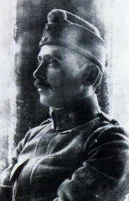
Constantin Freiherr von Economo ( gr, Κωνσταντίνος Οικονόμου; 21 August 1876 – 21 October 1931) was an Austrian psychiatrist and neurologist of Greek descent, born in modern-day Romania (then Ottoman Empire). He is mostly known for his discovery of encephalitis lethargica and his atlas of cytoarchitectonics of the cerebral cortex. Biography Youth and schooling Constantin Economo von San Serff was born in Brăila, Romania, to Johannes and Helene Economo, a wealthy family with large holdings in Thessaly and Macedonia. The Economo (Οικονόμου, '' Oikonomou'') family originated from Edessa, in the Ottoman Sanjak of Salonica (modern Edessa, Central Macedonia, Greece) where some of Constantin's ancestors were notables, and his family included many bishops. In 1877, the family moved to Trieste, Austria-Hungary,Economo, K. (1932). ''Constantin Freiherr von Economo''. Wien: Mayer & Co. and Constantin spent his childhood and youth in Trieste. He wa ... [...More Info...] [...Related Items...] OR: [Wikipedia] [Google] [Baidu] |
Korbinian Brodmann
Korbinian Brodmann (17 November 1868 – 22 August 1918) was a German neurologist who became famous for mapping the cerebral cortex and defining 52 distinct regions, known as Brodmann areas, based on their cytoarchitectonic (histological) characteristics. Life and career Brodmann was born in Liggersdorf, Province of Hohenzollern. He studied medicine in Munich, Würzburg, Berlin, and Freiburg, where he received his medical diploma in 1895. Subsequently he studied at the Medical School in the University of Lausanne in Switzerland, and then worked in the University Clinic in Munich. He received a doctor of medicine degree from the University of Leipzig in 1898, with a thesis on chronic ependymal sclerosis. From 1900 to 1901, Brodmann also worked in the Psychiatric Clinic at the University of Jena, with Ludwig Binswanger, and in the Municipal Mental Asylum in Frankfurt. There, he met Alois Alzheimer, who was influential in his decision to pursue basic neuroscience research. Fol ... [...More Info...] [...Related Items...] OR: [Wikipedia] [Google] [Baidu] |
Neutral Red
Neutral red (toluylene red, Basic Red 5, or C.I. 50040) is a eurhodin dye used for staining in histology. It stains lysosomes red. It is used as a general stain in histology, as a counterstain in combination with other dyes, and for many staining methods. Together with Janus Green B, it is used to stain embryonal tissues and supravital staining of blood. Can be used for staining Golgi apparatus in cells and Nissl granules in neurons. In microbiology, it is used in the MacConkey agar to differentiate bacteria for lactose fermentation.Neutral redcan be used as a vital stain. The Neutral Red Cytotoxicity Assay was first developed by Ellen Borenfreund in 1984. In the Neutral Red Assay live cells incorporate neutral red into their lysosomes. As cells begin to die, their ability to incorporate neutral red diminishes. Thus, loss of neutral red uptake corresponds to loss of cell viability. The neutral red is also used to stain cell cultures for plate titration of viruses. Neutral red ... [...More Info...] [...Related Items...] OR: [Wikipedia] [Google] [Baidu] |
Nissl Bodies
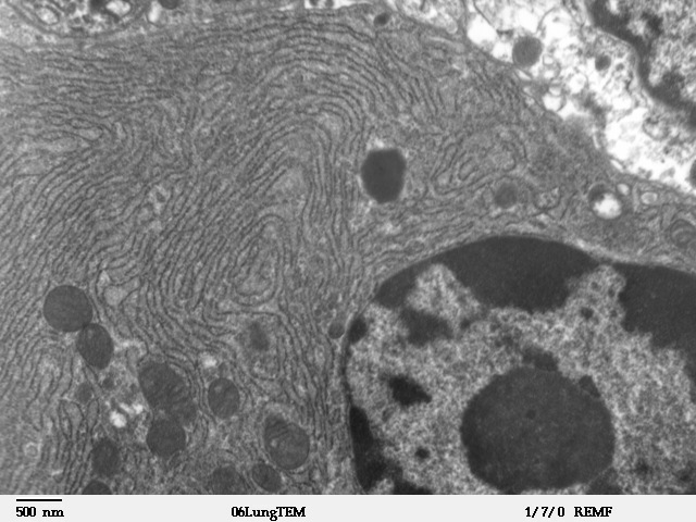
Nissl bodies (also called Nissl granules, Nissl substance or tigroid substance) are discrete granular structures in neurons that consist of rough endoplasmic reticulum, a collection of parallel, membrane-bound cisternae studded with ribosomes on the cystosolic surface of the membranes. Nissl bodies were named after Franz Nissl, a German neuropathologist who invented the staining method bearing his name ( Nissl staining). The term "Nissl bodies" generally refers to discrete clumps of rough endoplasmic reticulum in nerve cells. Masses of rough endoplasmic reticulum also occur in some non-neuronal cells, where they are referred to as ergastoplasm, basophilic bodies, or chromophilic substance. While these organelles differ in some ways from Nissl bodies in neurons, large amounts of rough endoplasmic reticulum are generally linked to the copious production of proteins. Staining "Nissl stains" refers to various basic dyes that selectively label negatively charged molecules such as DNA ... [...More Info...] [...Related Items...] OR: [Wikipedia] [Google] [Baidu] |
Rough Endoplasmic Reticulum
The endoplasmic reticulum (ER) is, in essence, the transportation system of the eukaryotic cell, and has many other important functions such as protein folding. It is a type of organelle made up of two subunits – rough endoplasmic reticulum (RER), and smooth endoplasmic reticulum (SER). The endoplasmic reticulum is found in most eukaryotic cells and forms an interconnected network of flattened, membrane-enclosed sacs known as cisternae (in the RER), and tubular structures in the SER. The membranes of the ER are continuous with the outer nuclear membrane. The endoplasmic reticulum is not found in red blood cells, or spermatozoa. The two types of ER share many of the same proteins and engage in certain common activities such as the synthesis of certain lipids and cholesterol. Different types of cells contain different ratios of the two types of ER depending on the activities of the cell. RER is found mainly toward the nucleus of cell and SER towards the cell membrane or plasma ... [...More Info...] [...Related Items...] OR: [Wikipedia] [Google] [Baidu] |
H&E Stain
Hematoxylin and eosin stain ( or haematoxylin and eosin stain or hematoxylin-eosin stain; often abbreviated as H&E stain or HE stain) is one of the principal tissue stains used in histology. It is the most widely used stain in medical diagnosis and is often the gold standard. For example, when a pathologist looks at a biopsy of a suspected cancer, the histological section is likely to be stained with H&E. H&E is the combination of two histological stains: hematoxylin and eosin. The hematoxylin stains cell nuclei a purplish blue, and eosin stains the extracellular matrix and cytoplasm pink, with other structures taking on different shades, hues, and combinations of these colors. Hence a pathologist can easily differentiate between the nuclear and cytoplasmic parts of a cell, and additionally, the overall patterns of coloration from the stain show the general layout and distribution of cells and provides a general overview of a tissue sample's structure. Thus, pattern recogniti ... [...More Info...] [...Related Items...] OR: [Wikipedia] [Google] [Baidu] |
Glia
Glia, also called glial cells (gliocytes) or neuroglia, are non-neuronal cells in the central nervous system (brain and spinal cord) and the peripheral nervous system that do not produce electrical impulses. They maintain homeostasis, form myelin in the peripheral nervous system, and provide support and protection for neurons. In the central nervous system, glial cells include oligodendrocytes, astrocytes, ependymal cells, and microglia, and in the peripheral nervous system they include Schwann cells and satellite cells. Function They have four main functions: *to surround neurons and hold them in place *to supply nutrients and oxygen to neurons *to insulate one neuron from another *to destroy pathogens and remove dead neurons. They also play a role in neurotransmission and synaptic connections, and in physiological processes such as breathing. While glia were thought to outnumber neurons by a ratio of 10:1, recent studies using newer methods and reappraisal of historical quan ... [...More Info...] [...Related Items...] OR: [Wikipedia] [Google] [Baidu] |
Thionine
Thionine, also known as Lauth's violet, is the salt of a heterocyclic compound A heterocyclic compound or ring structure is a cyclic compound that has atoms of at least two different elements as members of its ring(s). Heterocyclic chemistry is the branch of organic chemistry dealing with the synthesis, properties, and .... It was firstly synthesised by Charles Lauth. A variety of salts are known including the chloride and acetate, called respectively thionine chloride and thionine acetate. The dye is structurally related to methylene blue, which also features a phenothiazine core. The dye's name is frequently misspelled with omission of the e, and is not to be confused with the plant protein thionin. The -ine ending indicates that the compound is an amine. Dye properties and use Thionine is a strongly staining metachromatic dye, which is widely used for histologic stain, biological staining. Thionine can also be used in place of Schiff reagent in quantitative Feulgen st ... [...More Info...] [...Related Items...] OR: [Wikipedia] [Google] [Baidu] |
Immunohistochemistry
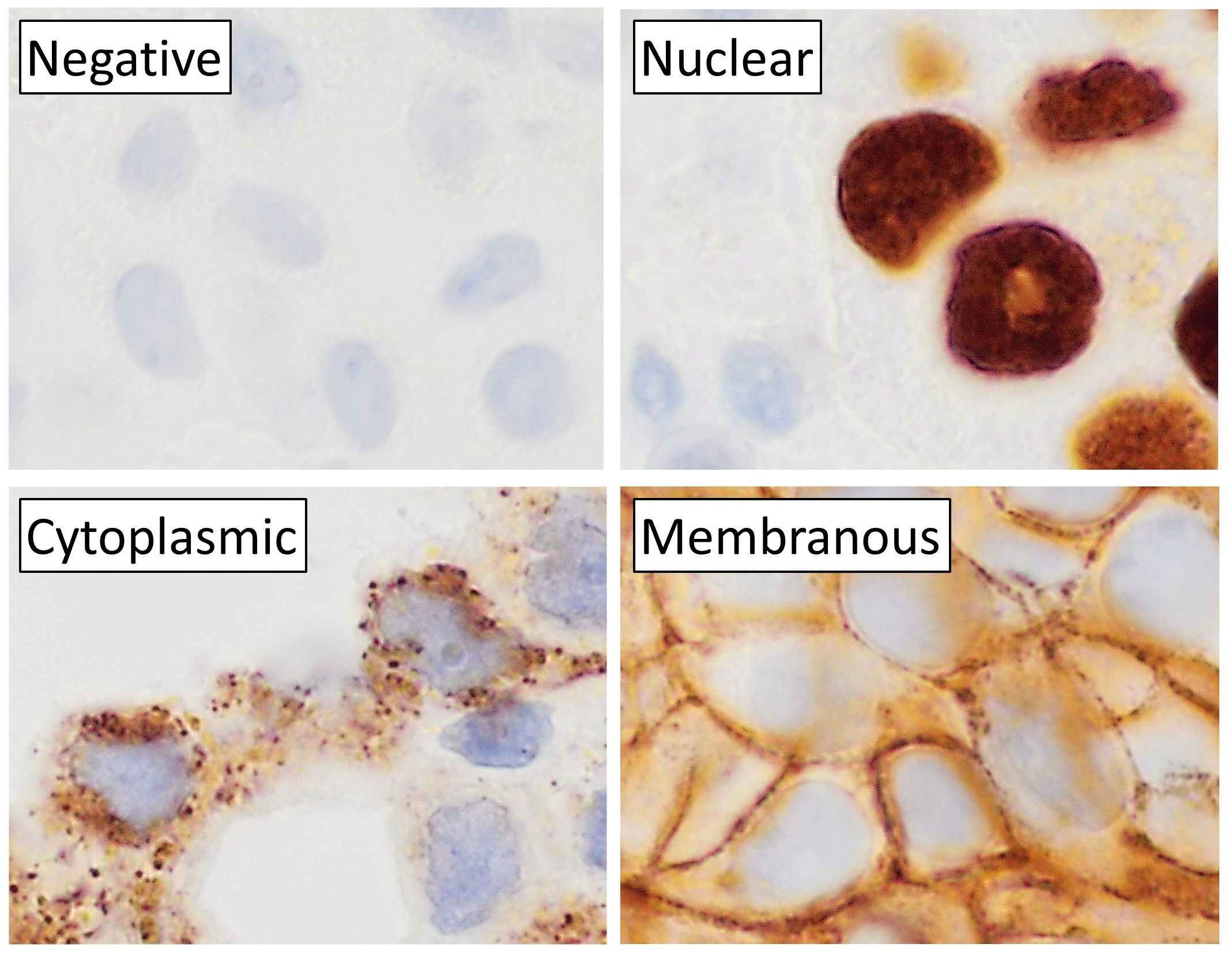
Immunohistochemistry (IHC) is the most common application of immunostaining. It involves the process of selectively identifying antigens (proteins) in cells of a tissue section by exploiting the principle of antibodies binding specifically to antigens in biological tissues. IHC takes its name from the roots "immuno", in reference to antibodies used in the procedure, and "histo", meaning tissue (compare to immunocytochemistry). Albert Coons conceptualized and first implemented the procedure in 1941. Visualising an antibody-antigen interaction can be accomplished in a number of ways, mainly either of the following: * ''Chromogenic immunohistochemistry'' (CIH), wherein an antibody is conjugated to an enzyme, such as peroxidase (the combination being termed immunoperoxidase), that can catalyse a colour-producing reaction. * '' Immunofluorescence'', where the antibody is tagged to a fluorophore, such as fluorescein or rhodamine. Immunohistochemical staining is widely used in the dia ... [...More Info...] [...Related Items...] OR: [Wikipedia] [Google] [Baidu] |
In Situ Hybridization
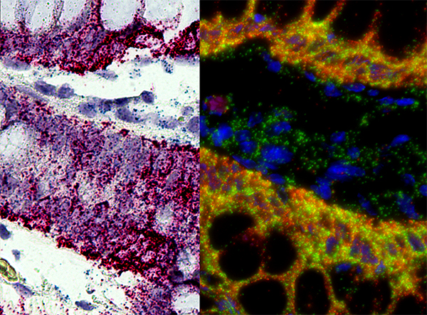
''In situ'' hybridization (ISH) is a type of hybridization that uses a labeled complementary DNA, RNA or modified nucleic acids strand (i.e., probe) to localize a specific DNA or RNA sequence in a portion or section of tissue (''in situ'') or if the tissue is small enough (e.g., plant seeds, ''Drosophila'' embryos), in the entire tissue (whole mount ISH), in cells, and in circulating tumor cells (CTCs). This is distinct from immunohistochemistry, which usually localizes proteins in tissue sections. In situ hybridization is used to reveal the location of specific nucleic acid sequences on chromosomes or in tissues, a crucial step for understanding the organization, regulation, and function of genes. The key techniques currently in use include ''in situ'' hybridization to mRNA with oligonucleotide and RNA probes (both radio-labeled and hapten-labeled), analysis with light and electron microscopes, whole mount ''in situ'' hybridization, double detection of RNAs and RNA plus prote ... [...More Info...] [...Related Items...] OR: [Wikipedia] [Google] [Baidu] |


