|
Craniopagus
Craniopagus twins are conjoined twins that are fused at the cranium. The union may occur on any portion of the cranium, but does not primarily involve either the face or the foramen magnum; their brains are usually separate, but they may share some brain tissue. Conjoined twins are genetically identical and always share the same sex. The thorax and abdomen are separate and each twin has its own umbilicus and umbilical cord. The condition is extremely rare, with an incidence of approximately 1 in 2.5 million live births. An estimated 50 craniopagus twins born around the world every year as of 2021, with only 15 twins surviving beyond the first 30 days of life. Relatively few craniopagus twins survive the perinatal period; approximately 40% of conjoined twins are stillborn and an additional 33% die within the immediate perinatal period, usually from organ abnormalities and failure. However, 25% of craniopagus twins survive and may be considered for a surgical separation; several su ... [...More Info...] [...Related Items...] OR: [Wikipedia] [Google] [Baidu] |
Conjoined Twins
Conjoined twins – sometimes popularly referred to as Siamese twins – are twins joined ''in utero''. A very rare phenomenon, the occurrence is estimated to range from 1 in 49,000 births to 1 in 189,000 births, with a somewhat higher incidence in Southwest Asia and Africa. Approximately half are stillborn, and an additional one-third die within 24 hours. Most live births are female, with a ratio of 3:1. Two theories exist to explain the origins of conjoined twins. The more generally accepted theory is ''fission'', in which the fertilized egg splits partially. The other theory, no longer believed to be the basis of conjoined twinning, is ''fusion'', in which a fertilized egg completely separates, but stem cells (which search for similar cells) find similar stem cells on the other twin and fuse the twins together. Conjoined twins share a single common chorion, placenta, and amniotic sac, although these characteristics are not exclusive to conjoined twins, as there are some monozyg ... [...More Info...] [...Related Items...] OR: [Wikipedia] [Google] [Baidu] |
Neurosurgery
Neurosurgery or neurological surgery, known in common parlance as brain surgery, is the medical specialty concerned with the surgical treatment of disorders which affect any portion of the nervous system including the brain, spinal cord and peripheral nervous system. Education and context In different countries, there are different requirements for an individual to legally practice neurosurgery, and there are varying methods through which they must be educated. In most countries, neurosurgeon training requires a minimum period of seven years after graduating from medical school. United States In the United States, a neurosurgeon must generally complete four years of undergraduate education, four years of medical school, and seven years of residency (PGY-1-7). Most, but not all, residency programs have some component of basic science or clinical research. Neurosurgeons may pursue additional training in the form of a fellowship after residency, or, in some cases, as a senior resid ... [...More Info...] [...Related Items...] OR: [Wikipedia] [Google] [Baidu] |
Occipital Lobe
The occipital lobe is one of the four major lobes of the cerebral cortex in the brain of mammals. The name derives from its position at the back of the head, from the Latin ''ob'', "behind", and ''caput'', "head". The occipital lobe is the visual processing center of the mammalian brain containing most of the anatomical region of the visual cortex. The primary visual cortex is Brodmann area 17, commonly called V1 (visual one). Human V1 is located on the medial side of the occipital lobe within the calcarine sulcus; the full extent of V1 often continues onto the occipital pole. V1 is often also called striate cortex because it can be identified by a large stripe of myelin, the Stria of Gennari. Visually driven regions outside V1 are called extrastriate cortex. There are many extrastriate regions, and these are specialized for different visual tasks, such as visuospatial processing, color differentiation, and motion perception. Bilateral lesions of the occipital lobe can lead ... [...More Info...] [...Related Items...] OR: [Wikipedia] [Google] [Baidu] |
Fission (biology)
Fission, in biology, is the division of a single entity into two or more parts and the regeneration of those parts to separate entities resembling the original. The object experiencing fission is usually a cell, but the term may also refer to how organisms, bodies, populations, or species split into discrete parts. The fission may be ''binary fission'', in which a single organism produces two parts, or ''multiple fission'', in which a single entity produces multiple parts. Binary fission Organisms in the domains of Archaea and Bacteria reproduce with binary fission. This form of asexual reproduction and cell division is also used by some organelles within eukaryotic organisms (e.g., mitochondria). Binary fission results in the reproduction of a living prokaryotic cell (or organelle) by dividing the cell into two parts, each with the potential to grow to the size of the original. Fission of prokaryotes The single DNA molecule first replicates, then attaches each copy to a differ ... [...More Info...] [...Related Items...] OR: [Wikipedia] [Google] [Baidu] |
Gyri
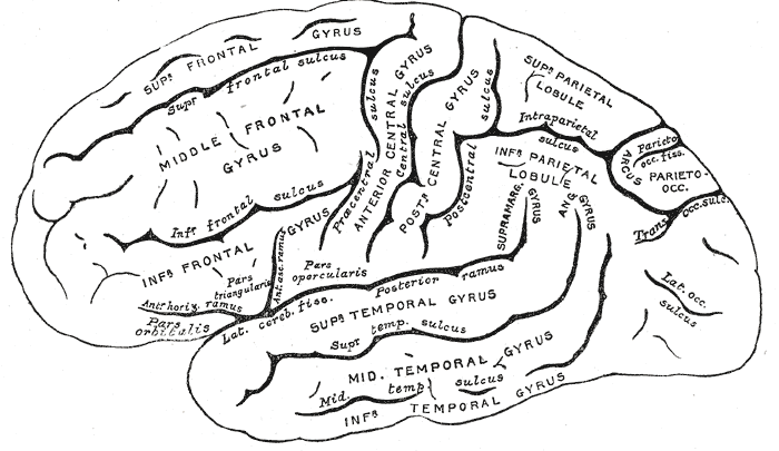
In neuroanatomy, a gyrus (pl. gyri) is a ridge on the cerebral cortex. It is generally surrounded by one or more sulci (depressions or furrows; sg. ''sulcus''). Gyri and sulci create the folded appearance of the brain in humans and other mammals. Structure The gyri are part of a system of folds and ridges that create a larger surface area for the human brain and other mammalian brains. Because the brain is confined to the skull, brain size is limited. Ridges and depressions create folds allowing a larger cortical surface area, and greater cognitive function, to exist in the confines of a smaller cranium. Development The human brain undergoes gyrification during fetal and neonatal development. In embryonic development, all mammalian brains begin as smooth structures derived from the neural tube. A cerebral cortex without surface convolutions is lissencephalic, meaning 'smooth-brained'. As development continues, gyri and sulci begin to take shape on the fetal brain, with ... [...More Info...] [...Related Items...] OR: [Wikipedia] [Google] [Baidu] |
Dura Mater
In neuroanatomy, dura mater is a thick membrane made of dense irregular connective tissue that surrounds the brain and spinal cord. It is the outermost of the three layers of membrane called the meninges that protect the central nervous system. The other two meningeal layers are the arachnoid mater and the pia mater. It envelops the arachnoid mater, which is responsible for keeping in the cerebrospinal fluid. It is derived primarily from the neural crest cell population, with postnatal contributions of the paraxial mesoderm. Structure The dura mater has several functions and layers. The dura mater is a membrane that envelops the arachnoid mater. It surrounds and supports the dural sinuses (also called dural venous sinuses, cerebral sinuses, or cranial sinuses) and carries blood from the brain toward the heart. Cranial dura mater has two layers called ''lamellae'', a superficial layer (also called the periosteal layer), which serves as the skull's inner periosteum, called the ... [...More Info...] [...Related Items...] OR: [Wikipedia] [Google] [Baidu] |
Calvaria (skull)
The calvaria is the top part of the skull. It is the upper part of the neurocranium and covers the cranial cavity containing the brain. It forms the main component of the skull roof. The calvaria is made up of the superior portions of the frontal bone, occipital bone, and parietal bones. In the human skull, the sutures between the bones normally remain flexible during the first few years of postnatal development, and fontanelles are palpable. Premature complete ossification of these sutures is called craniosynostosis. In Latin, the word ''calvaria'' is used as a feminine noun with plural ''calvariae''; however, many medical texts list the word as ''calvarium'', a neuter Latin noun with plural ''calvaria''. Structure The outer surface of the skull possesses a number of landmarks. The point at which the frontal bone and the two parietal bones meet is known as "Bregma". The point at which the two parietal bones and the occipital bone meet is known as "Lambda". Not only do t ... [...More Info...] [...Related Items...] OR: [Wikipedia] [Google] [Baidu] |
Occiput
The occipital bone () is a cranial dermal bone and the main bone of the occiput (back and lower part of the skull). It is trapezoidal in shape and curved on itself like a shallow dish. The occipital bone overlies the occipital lobes of the cerebrum. At the base of skull in the occipital bone, there is a large oval opening called the foramen magnum, which allows the passage of the spinal cord. Like the other cranial bones, it is classed as a flat bone. Due to its many attachments and features, the occipital bone is described in terms of separate parts. From its front to the back is the basilar part, also called the basioccipital, at the sides of the foramen magnum are the lateral parts, also called the exoccipitals, and the back is named as the squamous part. The basilar part is a thick, somewhat quadrilateral piece in front of the foramen magnum and directed towards the pharynx. The squamous part is the curved, expanded plate behind the foramen magnum and is the largest part o ... [...More Info...] [...Related Items...] OR: [Wikipedia] [Google] [Baidu] |
Artery
An artery (plural arteries) () is a blood vessel in humans and most animals that takes blood away from the heart to one or more parts of the body (tissues, lungs, brain etc.). Most arteries carry oxygenated blood; the two exceptions are the pulmonary and the umbilical arteries, which carry deoxygenated blood to the organs that oxygenate it (lungs and placenta, respectively). The effective arterial blood volume is that extracellular fluid which fills the arterial system. The arteries are part of the circulatory system, that is responsible for the delivery of oxygen and nutrients to all cells, as well as the removal of carbon dioxide and waste products, the maintenance of optimum blood pH, and the circulation of proteins and cells of the immune system. Arteries contrast with veins, which carry blood back towards the heart. Structure The anatomy of arteries can be separated into gross anatomy, at the macroscopic level, and microanatomy, which must be studied with a microscop ... [...More Info...] [...Related Items...] OR: [Wikipedia] [Google] [Baidu] |
Cerebral Hemispheres
The vertebrate cerebrum (brain) is formed by two cerebral hemispheres that are separated by a groove, the longitudinal fissure. The brain can thus be described as being divided into left and right cerebral hemispheres. Each of these hemispheres has an outer layer of grey matter, the cerebral cortex, that is supported by an inner layer of white matter. In eutherian (placental) mammals, the hemispheres are linked by the corpus callosum, a very large bundle of nerve fibers. Smaller commissures, including the anterior commissure, the posterior commissure and the fornix, also join the hemispheres and these are also present in other vertebrates. These commissures transfer information between the two hemispheres to coordinate localized functions. There are three known poles of the cerebral hemispheres: the ''occipital pole'', the ''frontal pole'', and the ''temporal pole''. The central sulcus is a prominent fissure which separates the parietal lobe from the frontal lobe and the prim ... [...More Info...] [...Related Items...] OR: [Wikipedia] [Google] [Baidu] |
Septum
In biology, a septum (Latin for ''something that encloses''; plural septa) is a wall, dividing a cavity or structure into smaller ones. A cavity or structure divided in this way may be referred to as septate. Examples Human anatomy * Interatrial septum, the wall of tissue that is a sectional part of the left and right atria of the heart * Interventricular septum, the wall separating the left and right ventricles of the heart * Lingual septum, a vertical layer of fibrous tissue that separates the halves of the tongue. *Nasal septum: the cartilage wall separating the nostrils of the nose * Alveolar septum: the thin wall which separates the alveoli from each other in the lungs * Orbital septum, a palpebral ligament in the upper and lower eyelids * Septum pellucidum or septum lucidum, a thin structure separating two fluid pockets in the brain * Uterine septum, a malformation of the uterus * Vaginal septum, a lateral or transverse partition inside the vagina * Intermuscular sep ... [...More Info...] [...Related Items...] OR: [Wikipedia] [Google] [Baidu] |


_-_inferiror_view.png)