|
Cranial Nerve Examination
The cranial nerve exam is a type of neurological examination. It is used to identify problems with the cranial nerves by physical examination. It has nine components. Each test is designed to assess the status of one or more of the twelve cranial nerves (I-XII). These components correspond to testing the sense of smell (I), visual fields and acuity (II), eye movements (III, IV, VI) and pupils (III, sympathetic and parasympathetic), sensory function of face (V), strength of facial (VII) and shoulder girdle muscles (XI), hearing (VII, VIII), taste (VII, IX, X), pharyngeal movement and reflex (IX, X), tongue movements (XII). Components See also * Cranial nerves * Cranial nerve nucleus References External links NeurologyExam.com Free neurology exam videos by Cleveland Clinic trained neurologist. {{Medical records and physical exam Neurology procedures Physical examination ... [...More Info...] [...Related Items...] OR: [Wikipedia] [Google] [Baidu] |
Neurological Examination
A neurological examination is the assessment of sensory neuron and motor responses, especially reflexes, to determine whether the nervous system is impaired. This typically includes a physical examination and a review of the patient's medical history, but not deeper investigation such as neuroimaging. It can be used both as a screening tool and as an investigative tool, the former of which when examining the patient when there is no expected neurological deficit and the latter of which when examining a patient where you do expect to find abnormalities. If a problem is found either in an investigative or screening process, then further tests can be carried out to focus on a particular aspect of the nervous system (such as lumbar punctures and blood tests). In general, a neurological examination is focused on finding out whether there are lesions in the central and peripheral nervous systems or there is another diffuse process that is troubling the patient. Once the patient has been ... [...More Info...] [...Related Items...] OR: [Wikipedia] [Google] [Baidu] |
Human Eye
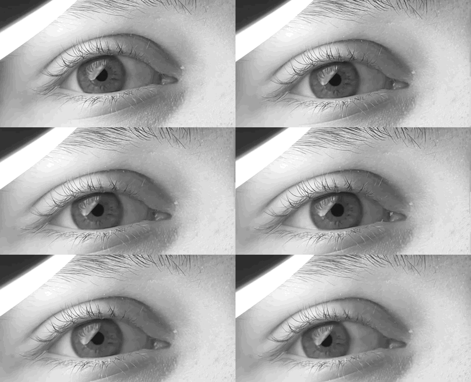
The human eye is a sensory organ, part of the sensory nervous system, that reacts to visible light and allows humans to use visual information for various purposes including seeing things, keeping balance, and maintaining circadian rhythm. The eye can be considered as a living optical device. It is approximately spherical in shape, with its outer layers, such as the outermost, white part of the eye (the sclera) and one of its inner layers (the pigmented choroid) keeping the eye essentially light tight except on the eye's optic axis. In order, along the optic axis, the optical components consist of a first lens (the cornea—the clear part of the eye) that accomplishes most of the focussing of light from the outside world; then an aperture (the pupil) in a diaphragm (the iris—the coloured part of the eye) that controls the amount of light entering the interior of the eye; then another lens (the crystalline lens) that accomplishes the remaining focussing of light into ... [...More Info...] [...Related Items...] OR: [Wikipedia] [Google] [Baidu] |
Physiologic Nystagmus
Nystagmus is a condition of involuntary (or voluntary, in some cases) eye movement. Infants can be born with it but more commonly acquire it in infancy or later in life. In many cases it may result in reduced or limited vision. Due to the involuntary movement of the eye, it has been called "dancing eyes". In normal eyesight, while the head rotates about an axis, distant visual images are sustained by rotating eyes in the opposite direction of the respective axis. The semicircular canals in the vestibule of the ear sense angular acceleration, and send signals to the nuclei for eye movement in the brain. From here, a signal is relayed to the extraocular muscles to allow one's gaze to fix on an object as the head moves. Nystagmus occurs when the semicircular canals are stimulated (e.g., by means of the caloric test, or by disease) while the head is stationary. The direction of ocular movement is related to the semicircular canal that is being stimulated. There are two key forms of ... [...More Info...] [...Related Items...] OR: [Wikipedia] [Google] [Baidu] |
Ptosis (eyelid)
Ptosis, also known as blepharoptosis, is a drooping or falling of the upper eyelid. The drooping may be worse after being awake longer when the individual's muscles are tired. This condition is sometimes called "lazy eye", but that term normally refers to the condition amblyopia. If severe enough and left untreated, the drooping eyelid can cause other conditions, such as amblyopia or astigmatism. This is why it is especially important for this disorder to be treated in children at a young age, before it can interfere with vision development. The term is from Greek 'fall, falling'. Signs and symptoms Signs and symptoms typically seen in this condition include: * The eyelid(s) may appear to droop. * Droopy eyelids can give the face a false appearance of being fatigued, disinterested, or even sinister. * The eyelid may not protect the eye as effectively, allowing it to dry out. * Sagging upper eyelids can partially block the person's field of view. * Obstructed vision may cause ... [...More Info...] [...Related Items...] OR: [Wikipedia] [Google] [Baidu] |
Extraocular Muscles
The extraocular muscles (extrinsic ocular muscles), are the seven extrinsic muscles of the human eye. Six of the extraocular muscles, the four recti muscles, and the superior and inferior oblique muscles, control movement of the eye and the other muscle, the levator palpebrae superioris, controls eyelid elevation. The actions of the six muscles responsible for eye movement depend on the position of the eye at the time of muscle contraction. Structure Since only a small part of the eye called the fovea provides sharp vision, the eye must move to follow a target. Eye movements must be precise and fast. This is seen in scenarios like reading, where the reader must shift gaze constantly. Although under voluntary control, most eye movement is accomplished without conscious effort. Precisely how the integration between voluntary and involuntary control of the eye occurs is a subject of continuing research."eye, human."Encyclopædia Britannica from Encyclopædia Britannica 2006 Ulti ... [...More Info...] [...Related Items...] OR: [Wikipedia] [Google] [Baidu] |
Accommodation Reflex
Accommodation may refer to: * A dwelling * A place for temporary lodging * The technique of adaptation to local cultures that the Jesuits used in their missions to spread Christianity among non-Christian peoples. * Reasonable accommodation, a legal doctrine protecting religious minorities or people with disabilities * Accommodation (religion), a theological principle linked to divine revelation within the Christian church * Accommodationism, a judicial interpretation with respect to Church and state issues * Accommodation bridge, a bridge provided to re-connect private land, separated by a new road or railway * Accommodation (law), a term used in US contract law * Accommodation (geology), the space available for sedimentation * Accommodation (eye), the process by which the eye increases optical power to maintain a clear image (focus) on an object as it draws near * Accommodation in psychology, the process by which existing mental structures and behaviors are modified to adapt to ... [...More Info...] [...Related Items...] OR: [Wikipedia] [Google] [Baidu] |
Diplopia
Diplopia is the simultaneous perception of two images of a single object that may be displaced horizontally or vertically in relation to each other. Also called double vision, it is a loss of visual focus under regular conditions, and is often voluntary. However, when occurring involuntarily, it results in impaired function of the extraocular muscles, where both eyes are still functional, but they cannot turn to target the desired object. Problems with these muscles may be due to mechanical problems, disorders of the neuromuscular junction, disorders of the cranial nerves ( III, IV, and VI) that innervate the muscles, and occasionally disorders involving the supranuclear oculomotor pathways or ingestion of toxins. Diplopia can be one of the first signs of a systemic disease, particularly to a muscular or neurological process, and it may disrupt a person's balance, movement, or reading abilities. Causes Diplopia has a diverse range of ophthalmologic, infectious, autoimmune, neu ... [...More Info...] [...Related Items...] OR: [Wikipedia] [Google] [Baidu] |
Eye Movements
Eye movement includes the voluntary or involuntary movement of the eyes. Eye movements are used by a number of organisms (e.g. primates, rodents, flies, birds, fish, cats, crabs, octopus) to fixate, inspect and track visual objects of interests. A special type of eye movement, rapid eye movement, occurs during REM sleep. The eyes are the visual organs of the human body, and move using a system of six muscles. The retina, a specialised type of tissue containing photoreceptors, senses light. These specialised cells convert light into electrochemical signals. These signals travel along the optic nerve fibers to the brain, where they are interpreted as vision in the visual cortex. Primates and many other vertebrates use three types of voluntary eye movement to track objects of interest: smooth pursuit, vergence shifts and saccades. These types of movements appear to be initiated by a small cortical region in the brain's frontal lobe. This is corroborated by removal of the ... [...More Info...] [...Related Items...] OR: [Wikipedia] [Google] [Baidu] |
Abducens Nerve
The abducens nerve or abducent nerve, also known as the sixth cranial nerve, cranial nerve VI, or simply CN VI, is a cranial nerve in humans and various other animals that controls the movement of the lateral rectus muscle, one of the extraocular muscles responsible for outward gaze. It is a somatic efferent nerve. Structure Nucleus The abducens nucleus is located in the pons, on the floor of the fourth ventricle, at the level of the facial colliculus. Axons from the facial nerve loop around the abducens nucleus, creating a slight bulge (the facial colliculus) that is visible on the dorsal surface of the floor of the fourth ventricle. The abducens nucleus is close to the midline, like the other motor nuclei that control eye movements (the oculomotor and trochlear nuclei). Motor axons leaving the abducens nucleus run ventrally and caudally through the pons. They pass lateral to the corticospinal tract (which runs longitudinally through the pons at this level) before exiting t ... [...More Info...] [...Related Items...] OR: [Wikipedia] [Google] [Baidu] |
Trochlear Nerve
The trochlear nerve (), ( lit. ''pulley-like'' nerve) also known as the fourth cranial nerve, cranial nerve IV, or CN IV, is a cranial nerve that innervates just one muscle: the superior oblique muscle of the eye, which operates through the pulley-like trochlea. CN IV is a motor nerve only (a somatic efferent nerve), unlike most other CNs. The trochlear nerve is unique among the cranial nerves in several respects: * It is the ''smallest'' nerve in terms of the number of axons it contains. * It has the greatest intracranial length. * It is the only cranial nerve that exits from the dorsal (rear) aspect of the brainstem. * It innervates a muscle, the superior oblique muscle, on the opposite side (contralateral) from its nucleus. The trochlear nerve decussates within the brainstem before emerging on the contralateral side of the brainstem (at the level of the inferior colliculus). An injury to the trochlear nucleus in the brainstem will result in an contralateral superior obliqu ... [...More Info...] [...Related Items...] OR: [Wikipedia] [Google] [Baidu] |
Oculomotor Nerve
The oculomotor nerve, also known as the third cranial nerve, cranial nerve III, or simply CN III, is a cranial nerve that enters the orbit through the superior orbital fissure and innervates extraocular muscles that enable most movements of the eye and that raise the eyelid. The nerve also contains fibers that innervate the intrinsic eye muscles that enable pupillary constriction and accommodation (ability to focus on near objects as in reading). The oculomotor nerve is derived from the basal plate of the embryonic midbrain. Cranial nerves IV and VI also participate in control of eye movement. Structure The oculomotor nerve originates from the third nerve nucleus at the level of the superior colliculus in the midbrain. The third nerve nucleus is located ventral to the cerebral aqueduct, on the pre-aqueductal grey matter. The fibers from the two third nerve nuclei located laterally on either side of the cerebral aqueduct then pass through the red nucleus. From the red nuc ... [...More Info...] [...Related Items...] OR: [Wikipedia] [Google] [Baidu] |


