|
Costovertebral Angle
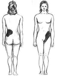
The costovertebral angle ( la, arcus costovertebralis) is the acute angle formed on either side of the human back between the twelfth rib and the vertebral column. The kidney lies directly below this area, so is the place where, with percussion A percussion instrument is a musical instrument that is sounded by being struck or scraped by a beater including attached or enclosed beaters or rattles struck, scraped or rubbed by hand or struck against another similar instrument. Exc ... ( la, sucussio renalis), pain is elicited when the person has kidney inflammation. The presence of pain is marked as a positive Murphy's punch sign or as costovertebral angle tenderness. References Back anatomy {{musculoskeletal-stub ... [...More Info...] [...Related Items...] OR: [Wikipedia] [Google] [Baidu] |
Human Back
The human back, also called the dorsum, is the large posterior area of the human body, rising from the top of the buttocks to the back of the neck. It is the surface of the body opposite from the chest and the abdomen. The vertebral column runs the length of the back and creates a central area of recession. The breadth of the back is created by the shoulders at the top and the pelvis at the bottom. Back pain is a common medical condition, generally benign in origin. Structure The central feature of the human back is the vertebral column, specifically the length from the top of the thoracic vertebrae to the bottom of the lumbar vertebrae, which houses the spinal cord in its spinal canal, and which generally has some curvature that gives shape to the back. The ribcage extends from the spine at the top of the back (with the top of the ribcage corresponding to the T1 vertebra), more than halfway down the length of the back, leaving an area with less protection between the bottom of ... [...More Info...] [...Related Items...] OR: [Wikipedia] [Google] [Baidu] |
Twelfth Rib
The rib cage, as an enclosure that comprises the ribs, vertebral column and sternum in the thorax of most vertebrates, protects vital organs such as the heart, lungs and great vessels. The sternum, together known as the thoracic cage, is a semi-rigid bony and cartilaginous structure which surrounds the thoracic cavity and supports the shoulder girdle to form the core part of the human skeleton. A typical human thoracic cage consists of 12 pairs of ribs and the adjoining costal cartilages, the sternum (along with the manubrium and xiphoid process), and the 12 thoracic vertebrae articulating with the ribs. Together with the skin and associated fascia and muscles, the thoracic cage makes up the thoracic wall and provides attachments for extrinsic skeletal muscles of the neck, upper limbs, upper abdomen and back. The rib cage intrinsically holds the muscles of respiration ( diaphragm, intercostal muscles, etc.) that are crucial for active inhalation and forced exhalation, and t ... [...More Info...] [...Related Items...] OR: [Wikipedia] [Google] [Baidu] |
Vertebral Column
The vertebral column, also known as the backbone or spine, is part of the axial skeleton. The vertebral column is the defining characteristic of a vertebrate in which the notochord (a flexible rod of uniform composition) found in all chordata, chordates has been replaced by a segmented series of bone: vertebrae separated by intervertebral discs. Individual vertebrae are named according to their region and position, and can be used as anatomical landmarks in order to guide procedures such as Lumbar puncture, lumbar punctures. The vertebral column houses the spinal canal, a cavity that encloses and protects the spinal cord. There are about 50,000 species of animals that have a vertebral column. The human vertebral column is one of the most-studied examples. Many different diseases in humans can affect the spine, with spina bifida and scoliosis being recognisable examples. The general structure of human vertebrae is fairly typical of that found in mammals, reptiles, and birds. Th ... [...More Info...] [...Related Items...] OR: [Wikipedia] [Google] [Baidu] |
Kidney
The kidneys are two reddish-brown bean-shaped organs found in vertebrates. They are located on the left and right in the retroperitoneal space, and in adult humans are about in length. They receive blood from the paired renal arteries; blood exits into the paired renal veins. Each kidney is attached to a ureter, a tube that carries excreted urine to the bladder. The kidney participates in the control of the volume of various body fluids, fluid osmolality, acid–base balance, various electrolyte concentrations, and removal of toxins. Filtration occurs in the glomerulus: one-fifth of the blood volume that enters the kidneys is filtered. Examples of substances reabsorbed are solute-free water, sodium, bicarbonate, glucose, and amino acids. Examples of substances secreted are hydrogen, ammonium, potassium and uric acid. The nephron is the structural and functional unit of the kidney. Each adult human kidney contains around 1 million nephrons, while a mouse kidney contains on ... [...More Info...] [...Related Items...] OR: [Wikipedia] [Google] [Baidu] |
Percussion (medicine)
Percussion is a technique of clinical examination. Overview Percussion is a method of tapping on a surface to determine the underlying structures, and is used in clinical examinations to assess the condition of the thorax or abdomen. It is one of the four methods of clinical examination, together with inspection, palpation, auscultation, and inquiry. It is done with the middle finger of one hand tapping on the middle finger of the other hand using a wrist action. The nonstriking finger (known as the pleximeter) is placed firmly on the body over tissue. When percussing boney areas such as the clavicle, the pleximeter can be omitted and the bone is tapped directly such as when percussing an apical cavitary lung lesion typical of tuberculosis. There are two types of percussion: direct, which uses only one or two fingers; and indirect, which uses only the middle/flexor finger. Broadly classifying, there are four types of percussion sounds: resonant, hyper-resonant, stony dull or dul ... [...More Info...] [...Related Items...] OR: [Wikipedia] [Google] [Baidu] |
Pyelonephritis
Pyelonephritis is inflammation of the kidney, typically due to a bacterial infection. Symptoms most often include fever and flank tenderness. Other symptoms may include nausea, burning with urination, and frequent urination. Complications may include pus around the kidney, sepsis, or kidney failure. It is typically due to a bacterial infection, most commonly ''Escherichia coli''. Risk factors include sexual intercourse, prior urinary tract infections, diabetes, structural problems of the urinary tract, and spermicide use. The mechanism of infection is usually spread up the urinary tract. Less often infection occurs through the bloodstream. Diagnosis is typically based on symptoms and supported by urinalysis. If there is no improvement with treatment, medical imaging may be recommended. Pyelonephritis may be preventable by urination after sex and drinking sufficient fluids. Once present it is generally treated with antibiotics, such as ciprofloxacin or ceftriaxone. Thos ... [...More Info...] [...Related Items...] OR: [Wikipedia] [Google] [Baidu] |
Costovertebral Angle Tenderness
Costovertebral angle (CVA) tenderness is pain that results from touching the region inside of the costovertebral angle.Bickley, Lynn S., Peter G. Szilagyi, and Richard M. Hoffman. Bates' guide to physical examination and history taking. Philadelphia: Wolters Kluwer, 2017. Print. The CVA is formed by the 12th rib and the spine. Assessing for CVA tenderness is part of the abdominal exam, and CVA tenderness often indicates kidney pathology. Anatomy The CVA is an anatomic concept of the relationship of the 12th rib to the transverse processes of the lumbar vertebrae. There is one CVA on each side of the spine.Moore, Keith L., A. M. R. Agur, and Arthur F. Dalley. Clinically oriented anatomy. Philadelphia: Wolters Kluwer, 2018. Print. The lateral part of the CVA is formed by the lower border of the 12th rib, and the medial part of the CVA is formed by the transverse processes of the lumbar vertebrae. The CVA is distinct from the costovertebral joints. The lower poles of the kidney ... [...More Info...] [...Related Items...] OR: [Wikipedia] [Google] [Baidu] |


