|
Costocervical Artery
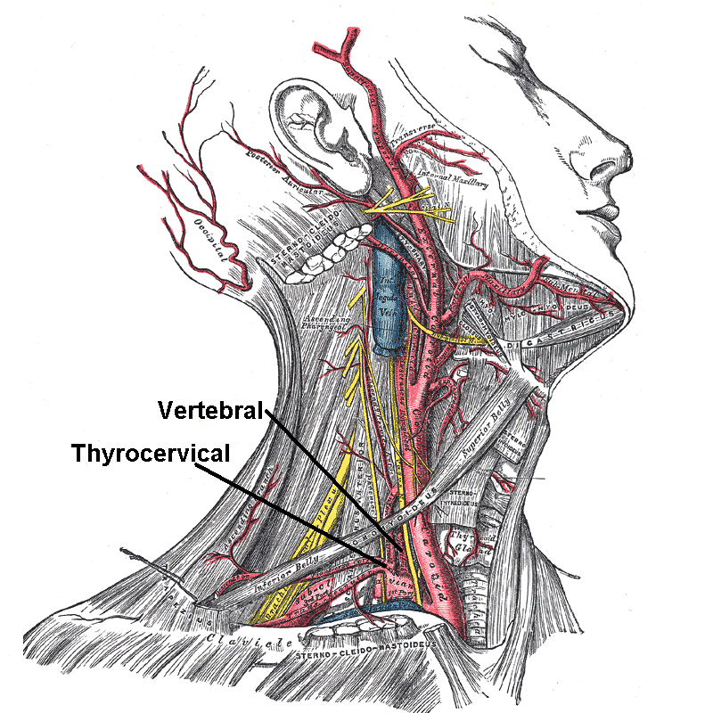
The costocervical trunk arises from the upper and back part of the second part of subclavian artery, behind the scalenus anterior on the right side, and medial to that muscle on the left side. Passing backward, it splits into the deep cervical artery and the supreme intercostal artery (highest intercostal artery), which descends behind the pleura in front of the necks of the first and second ribs, and anastomoses with the first aortic intercostal (3rd posterior intercostal artery). As it crosses the neck of the first rib it lies medial to the anterior division of the first thoracic nerve, and lateral to the first thoracic ganglion of the sympathetic trunk. In the first intercostal space, it gives off a branch which is distributed in a manner similar to the distribution of the aortic intercostals. The branch for the second intercostal space usually joins with one from the highest aortic intercostal artery. This branch is not constant, but is more commonly found on the right ... [...More Info...] [...Related Items...] OR: [Wikipedia] [Google] [Baidu] |
Subclavian Artery
In human anatomy, the subclavian arteries are paired major arteries of the upper thorax, below the clavicle. They receive blood from the aortic arch. The left subclavian artery supplies blood to the left arm and the right subclavian artery supplies blood to the right arm, with some branches supplying the head and thorax. On the left side of the body, the subclavian comes directly off the aortic arch, while on the right side it arises from the relatively short brachiocephalic artery when it bifurcates into the subclavian and the right common carotid artery. The usual branches of the subclavian on both sides of the body are the vertebral artery, the internal thoracic artery, the thyrocervical trunk, the costocervical trunk and the dorsal scapular artery, which may branch off the transverse cervical artery, which is a branch of the thyrocervical trunk. The subclavian becomes the axillary artery at the lateral border of the first rib. Structure From its origin, the subclavian artery t ... [...More Info...] [...Related Items...] OR: [Wikipedia] [Google] [Baidu] |
Deep Cervical Artery
The deep cervical artery (Profunda cervicalis) is an artery of the neck. Course It arises, in most cases, from the costocervical trunk, and is analogous to the posterior branch of an aortic intercostal artery: occasionally it is a separate branch from the subclavian artery. Passing backward, above the eighth cervical nerve and between the transverse process of the seventh cervical vertebra and the neck of the first rib, it runs up the back of the neck, between the semispinalis capitis and semispinalis cervicis, as high as the axis vertebra, supplying these and adjacent muscles, and anastomosing with the deep division of the descending branch of the occipital, and with branches of the vertebral. It gives off a spinal twig which enters the canal through the intervertebral foramen between the seventh cervical and first thoracic vertebrae In vertebrates, thoracic vertebrae compose the middle segment of the vertebral column, between the cervical vertebrae and the lumbar vert ... [...More Info...] [...Related Items...] OR: [Wikipedia] [Google] [Baidu] |
Superior Intercostal Artery
The intercostal arteries are a group of arteries that supply the area between the ribs ("costae"), called the intercostal space. The highest intercostal artery (supreme intercostal artery or superior intercostal artery) is an artery in the human body that usually gives rise to the first and second posterior intercostal arteries, which supply blood to their corresponding intercostal space. It usually arises from the costocervical trunk, which is a branch of the subclavian artery. Some anatomists may contend that there is no supreme intercostal artery, only a supreme intercostal vein. The anterior intercostal branches of internal thoracic artery supply the upper five or six intercostal spaces. The internal thoracic artery (previously called as internal mammary artery) then divides into the superior epigastric artery and musculophrenic artery. The latter gives out the remaining anterior intercostal branches. Two in number in each space, these small vessels pass lateralward, one ly ... [...More Info...] [...Related Items...] OR: [Wikipedia] [Google] [Baidu] |
Subclavian Artery
In human anatomy, the subclavian arteries are paired major arteries of the upper thorax, below the clavicle. They receive blood from the aortic arch. The left subclavian artery supplies blood to the left arm and the right subclavian artery supplies blood to the right arm, with some branches supplying the head and thorax. On the left side of the body, the subclavian comes directly off the aortic arch, while on the right side it arises from the relatively short brachiocephalic artery when it bifurcates into the subclavian and the right common carotid artery. The usual branches of the subclavian on both sides of the body are the vertebral artery, the internal thoracic artery, the thyrocervical trunk, the costocervical trunk and the dorsal scapular artery, which may branch off the transverse cervical artery, which is a branch of the thyrocervical trunk. The subclavian becomes the axillary artery at the lateral border of the first rib. Structure From its origin, the subclavian artery t ... [...More Info...] [...Related Items...] OR: [Wikipedia] [Google] [Baidu] |
Scalenus Anterior
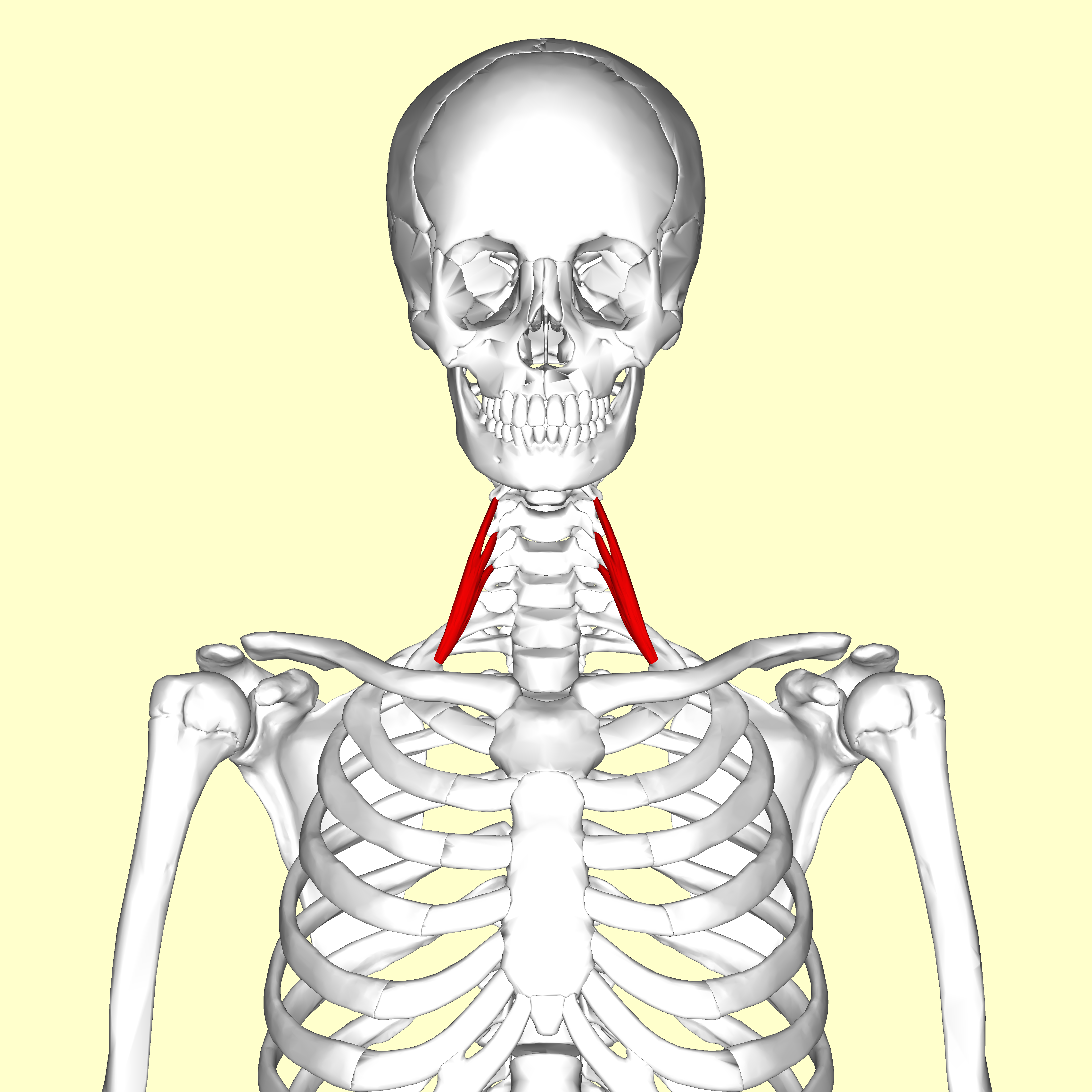
The scalene muscles are a group of three pairs of muscles in the lateral neck, namely the anterior scalene, middle scalene, and posterior scalene. They are innervated by the third to the eight cervical spinal nerves (C3-C8). The anterior and middle scalene muscles lift the first rib and bend the neck to the same side; the posterior scalene lifts the second rib and tilts the neck to the same side. The muscles are named . Structure The scalene muscles originate from the transverse processes from the cervical vertebrae of C2 to C7 and insert onto the first and second ribs. Anterior scalene The anterior scalene muscle ( la, scalenus anterior), lies deeply at the side of the neck, behind the sternocleidomastoid muscle. It arises from the anterior tubercles of the transverse processes of the third, fourth, fifth, and sixth cervical vertebrae, and descending, almost vertically, is inserted by a narrow, flat tendon into the scalene tubercle on the inner border of the first rib, and ... [...More Info...] [...Related Items...] OR: [Wikipedia] [Google] [Baidu] |
Deep Cervical Artery
The deep cervical artery (Profunda cervicalis) is an artery of the neck. Course It arises, in most cases, from the costocervical trunk, and is analogous to the posterior branch of an aortic intercostal artery: occasionally it is a separate branch from the subclavian artery. Passing backward, above the eighth cervical nerve and between the transverse process of the seventh cervical vertebra and the neck of the first rib, it runs up the back of the neck, between the semispinalis capitis and semispinalis cervicis, as high as the axis vertebra, supplying these and adjacent muscles, and anastomosing with the deep division of the descending branch of the occipital, and with branches of the vertebral. It gives off a spinal twig which enters the canal through the intervertebral foramen between the seventh cervical and first thoracic vertebrae In vertebrates, thoracic vertebrae compose the middle segment of the vertebral column, between the cervical vertebrae and the lumbar vert ... [...More Info...] [...Related Items...] OR: [Wikipedia] [Google] [Baidu] |
Supreme Intercostal Artery
The intercostal arteries are a group of arteries that supply the area between the ribs ("costae"), called the intercostal space. The highest intercostal artery (supreme intercostal artery or superior intercostal artery) is an artery in the human body that usually gives rise to the first and second posterior intercostal arteries, which supply blood to their corresponding intercostal space. It usually arises from the costocervical trunk, which is a branch of the subclavian artery. Some anatomists may contend that there is no supreme intercostal artery, only a supreme intercostal vein. The anterior intercostal branches of internal thoracic artery supply the upper five or six intercostal spaces. The internal thoracic artery (previously called as internal mammary artery) then divides into the superior epigastric artery and musculophrenic artery. The latter gives out the remaining anterior intercostal branches. Two in number in each space, these small vessels pass lateralward, one l ... [...More Info...] [...Related Items...] OR: [Wikipedia] [Google] [Baidu] |
Pleura
The pulmonary pleurae (''sing.'' pleura) are the two opposing layers of serous membrane overlying the lungs and the inside of the surrounding chest walls. The inner pleura, called the visceral pleura, covers the surface of each lung and dips between the lobes of the lung as ''fissures'', and is formed by the invagination of lung buds into each thoracic sac during embryonic development. The outer layer, called the parietal pleura, lines the inner surfaces of the thoracic cavity on each side of the mediastinum, and can be subdivided into ''mediastinal'' (covering the side surfaces of the fibrous pericardium, oesophagus and thoracic aorta), ''diaphragmatic'' (covering the upper surface of the diaphragm), ''costal'' (covering the inside of rib cage) and cervical (covering the underside of the suprapleural membrane) pleurae. The visceral and the mediastinal parietal pleurae are connected at the root of the lung ("hilum") through a smooth fold known as ''pleural reflections'', and ... [...More Info...] [...Related Items...] OR: [Wikipedia] [Google] [Baidu] |
Ribs
The rib cage, as an enclosure that comprises the ribs, vertebral column and sternum in the thorax of most vertebrates, protects vital organs such as the heart, lungs and great vessels. The sternum, together known as the thoracic cage, is a semi-rigid bony and cartilaginous structure which surrounds the thoracic cavity and supports the shoulder girdle to form the core part of the human skeleton. A typical human thoracic cage consists of 12 pairs of ribs and the adjoining costal cartilages, the sternum (along with the manubrium and xiphoid process), and the 12 thoracic vertebrae articulating with the ribs. Together with the skin and associated fascia and muscles, the thoracic cage makes up the thoracic wall and provides attachments for extrinsic skeletal muscles of the neck, upper limbs, upper abdomen and back. The rib cage intrinsically holds the muscles of respiration ( diaphragm, intercostal muscles, etc.) that are crucial for active inhalation and forced exhalation, and t ... [...More Info...] [...Related Items...] OR: [Wikipedia] [Google] [Baidu] |
Anastomoses
An anastomosis (, plural anastomoses) is a connection or opening between two things (especially cavities or passages) that are normally diverging or branching, such as between blood vessels, leaf veins, or streams. Such a connection may be normal (such as the foramen ovale in a fetus's heart) or abnormal (such as the patent foramen ovale in an adult's heart); it may be acquired (such as an arteriovenous fistula) or innate (such as the arteriovenous shunt of a metarteriole); and it may be natural (such as the aforementioned examples) or artificial (such as a surgical anastomosis). The reestablishment of an anastomosis that had become blocked is called a reanastomosis. Anastomoses that are abnormal, whether congenital or acquired, are often called fistulas. The term is used in medicine, biology, mycology, geology, and geography. Etymology Anastomosis: medical or Modern Latin, from Greek ἀναστόμωσις, anastomosis, "outlet, opening", Gr ana- "up, on, upon", stoma "mouth", ... [...More Info...] [...Related Items...] OR: [Wikipedia] [Google] [Baidu] |
Aortic Intercostal
The intercostal arteries are a group of arteries that supply the area between the ribs ("costae"), called the intercostal space. The highest intercostal artery (supreme intercostal artery or superior intercostal artery) is an artery in the human body that usually gives rise to the first and second posterior intercostal arteries, which supply blood to their corresponding intercostal space. It usually arises from the costocervical trunk, which is a branch of the subclavian artery. Some anatomists may contend that there is no supreme intercostal artery, only a supreme intercostal vein. The anterior intercostal branches of internal thoracic artery supply the upper five or six intercostal spaces. The internal thoracic artery (previously called as internal mammary artery) then divides into the superior epigastric artery and musculophrenic artery. The latter gives out the remaining anterior intercostal branches. Two in number in each space, these small vessels pass lateralward, one ly ... [...More Info...] [...Related Items...] OR: [Wikipedia] [Google] [Baidu] |
Thoracic Nerve
A spinal nerve is a mixed nerve, which carries motor, sensory, and autonomic signals between the spinal cord and the body. In the human body there are 31 pairs of spinal nerves, one on each side of the vertebral column. These are grouped into the corresponding cervical, thoracic, lumbar, sacral and coccygeal regions of the spine. There are eight pairs of cervical nerves, twelve pairs of thoracic nerves, five pairs of lumbar nerves, five pairs of sacral nerves, and one pair of coccygeal nerves. The spinal nerves are part of the peripheral nervous system. Structure Each spinal nerve is a mixed nerve, formed from the combination of nerve fibers from its dorsal and ventral roots. The dorsal root is the afferent sensory root and carries sensory information to the brain. The ventral root is the efferent motor root and carries motor information from the brain. The spinal nerve emerges from the spinal column through an opening (intervertebral foramen) between adjacent vertebrae. ... [...More Info...] [...Related Items...] OR: [Wikipedia] [Google] [Baidu] |


