|
Coronectomy
When extracting lower wisdom teeth, coronectomy is a treatment option involving removing the Crown_(tooth), crown of the lower wisdom tooth, whilst keeping the roots in place in healthy patients. This option is given to patients as an alternative to Dental extraction, extraction when the wisdom teeth are in close association with the inferior alveolar nerve, and so used to prevent damage to the nerve which may occur during extraction. Advantages Prevents potential neuropathy The risk of altered sensation is significantly lower than convention surgical removal of mandibular third molars with 8% of the cases affected temporarily and 3.6% of the cases got permanently affected. 30% of the roots will migrate post-coronectomy, erupting away from the inferior alveolar canal. This makes extraction of the remaining roots safer. Disadvantages There is a 5% chance of failure of coronectomy, the root will become mobilized during transection. In 5% of the cases, Dental follicle, follicl ... [...More Info...] [...Related Items...] OR: [Wikipedia] [Google] [Baidu] |
Dental Extraction
A dental extraction (also referred to as tooth extraction, exodontia, exodontics, or informally, tooth pulling) is the removal of teeth from the dental alveolus (socket) in the alveolar bone. Extractions are performed for a wide variety of reasons, but most commonly to remove teeth which have become unrestorable through tooth decay, periodontal disease, or dental trauma, especially when they are associated with toothache. Sometimes impacted wisdom teeth (wisdom teeth that are stuck and unable to grow normally into the mouth) cause recurrent infections of the gum (pericoronitis), and may be removed when other conservative treatments have failed (cleaning, antibiotics and operculectomy). In orthodontics, if the teeth are crowded, healthy teeth may be extracted (often bicuspids) to create space so the rest of the teeth can be straightened. Procedure Extractions could be categorized into non-surgical (simple) and surgical, depending on the type of tooth to be removed and other fac ... [...More Info...] [...Related Items...] OR: [Wikipedia] [Google] [Baidu] |
Inferior Alveolar Nerve
The inferior alveolar nerve (IAN) (also the inferior dental nerve) is a branch of the mandibular nerve, which is itself the third branch of the trigeminal nerve. The inferior alveolar nerves supply sensation to the lower teeth. Structure The inferior alveolar nerve is a branch of the mandibular nerve. After branching from the mandibular nerve, the inferior alveolar nerve travels behind the lateral pterygoid muscle. It gives off a branch, the mylohyoid nerve, and then enters the mandibular foramen. While in the mandibular canal within the mandible, it supplies the lower teeth (molars and second premolar) with sensory branches that form into the inferior dental plexus and give off small gingival and dental nerves to the teeth. Anteriorly, the nerve gives off the mental nerve at about the level of the mandibular 2nd premolars, which exits the mandible via the mental foramen and supplies sensory branches to the chin and lower lip. The inferior alveolar nerve continues anteriorl ... [...More Info...] [...Related Items...] OR: [Wikipedia] [Google] [Baidu] |
Wisdom Teeth
A third molar, commonly called wisdom tooth, is one of the three molars per quadrant of the human dentition. It is the most posterior of the three. The age at which wisdom teeth come through ( erupt) is variable, but this generally occurs between late teens and early twenties. Most adults have four wisdom teeth, one in each of the four quadrants, but it is possible to have none, fewer, or more, in which case the extras are called supernumerary teeth. Wisdom teeth may get stuck ( impacted) against other teeth if there is not enough space for them to come through normally. Impacted wisdom teeth are still sometimes removed for orthodontic treatment, believing that they move the other teeth and cause crowding, though this is not held anymore as true. Impacted wisdom teeth may suffer from tooth decay if oral hygiene becomes more difficult. Wisdom teeth which are partially erupted through the gum may also cause inflammation and infection in the surrounding gum tissues, termed pericoron ... [...More Info...] [...Related Items...] OR: [Wikipedia] [Google] [Baidu] |
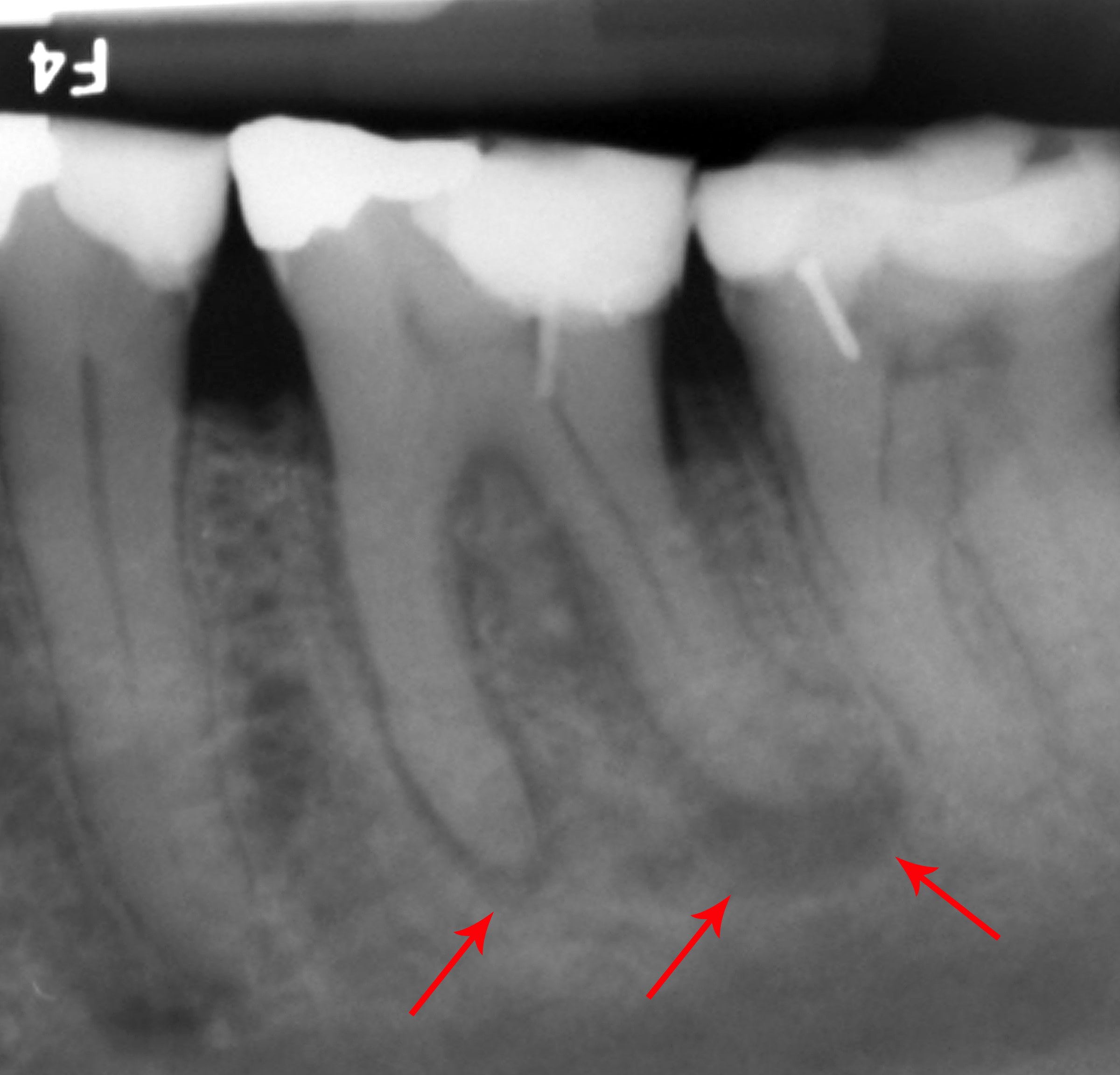
Radiograph Of Impacted Wisdom Tooth Near Nerve
Radiography is an imaging technique using X-rays, gamma rays, or similar ionizing radiation and non-ionizing radiation to view the internal form of an object. Applications of radiography include medical radiography ("diagnostic" and "therapeutic") and industrial radiography. Similar techniques are used in airport security (where "body scanners" generally use backscatter X-ray). To create an image in conventional radiography, a beam of X-rays is produced by an X-ray generator and is projected toward the object. A certain amount of the X-rays or other radiation is absorbed by the object, dependent on the object's density and structural composition. The X-rays that pass through the object are captured behind the object by a detector (either photographic film or a digital detector). The generation of flat two dimensional images by this technique is called projectional radiography. In computed tomography (CT scanning) an X-ray source and its associated detectors rotate around the su ... [...More Info...] [...Related Items...] OR: [Wikipedia] [Google] [Baidu] |
Radiograph
Radiography is an imaging technique using X-rays, gamma rays, or similar ionizing radiation and non-ionizing radiation to view the internal form of an object. Applications of radiography include medical radiography ("diagnostic" and "therapeutic") and industrial radiography. Similar techniques are used in airport security (where "body scanners" generally use backscatter X-ray). To create an image in conventional radiography, a beam of X-rays is produced by an X-ray generator and is projected toward the object. A certain amount of the X-rays or other radiation is absorbed by the object, dependent on the object's density and structural composition. The X-rays that pass through the object are captured behind the object by a detector (either photographic film or a digital detector). The generation of flat two dimensional images by this technique is called projectional radiography. In computed tomography (CT scanning) an X-ray source and its associated detectors rotate around the ... [...More Info...] [...Related Items...] OR: [Wikipedia] [Google] [Baidu] |
Articaine
Articaine is a dental amide-type local anesthetic. It is the most widely used local anesthetic in a number of European countriesOertel R, Ebert U, Rahn R, Kirch W. Clinical pharmacokinetics of articaine. Clin Pharmacokinet. 1997 Dec;33(6):418. and is available in many countries. It is the only local anaesthetic to contain a thiophene ring, meaning it can be described as 'thiophenic'; this conveys lipid solubility. History This drug was synthesized by pharmacologist Roman Muschaweck and chemist Robert Rippel. Muschaweck received a "O. Schmiedeberg" medal by the German Society for Experimental and Clinical Pharmacology and Toxicology for his work in 2002. It was brought to the German market in 1976 by Hoechst AG, a life-sciences German company (now Sanofi-Aventis), under the brand name Ultracain. This drug was also referred to as "carticaine" until 1984.Malamed SF. Handbook of local anaesthesia, p. 71, 5th ed. St. Louis, Mosby; 2004. In 1983 it was brought into the North Americ ... [...More Info...] [...Related Items...] OR: [Wikipedia] [Google] [Baidu] |
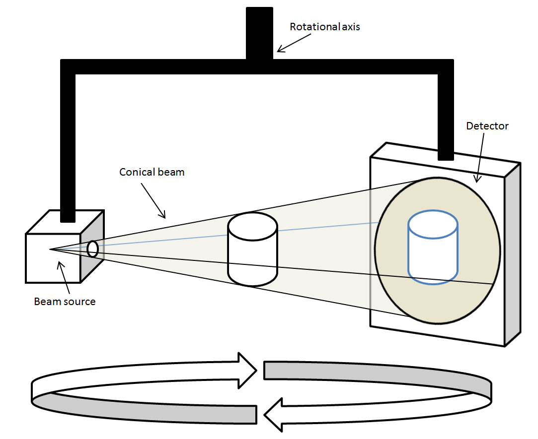
Cone Beam Computed Tomography
Cone beam computed tomography (or CBCT, also referred to as C-arm CT, cone beam volume CT, flat panel CT or Digital Volume Tomography (DVT)) is a medical imaging technique consisting of X-ray computed tomography where the X-rays are divergent, forming a cone. CBCT has become increasingly important in treatment planning and diagnosis in implant dentistry, ENT, orthopedics, and interventional radiology (IR), among other things. Perhaps because of the increased access to such technology, CBCT scanners are now finding many uses in dentistry, such as in the fields of oral surgery, endodontics and orthodontics. Integrated CBCT is also an important tool for patient positioning and verification in image-guided radiation therapy (IGRT). During dental/orthodontic imaging, the CBCT scanner rotates around the patient's head, obtaining up to nearly 600 distinct images. For interventional radiology, the patient is positioned offset to the table so that the region of interest is centered in ... [...More Info...] [...Related Items...] OR: [Wikipedia] [Google] [Baidu] |
Alveolar Osteitis
Alveolar osteitis, also known as dry socket, is inflammation of the alveolar bone (i.e., the alveolar process of the maxilla or mandible). Classically, this occurs as a postoperative complication of tooth extraction. Alveolar osteitis usually occurs where the blood clot fails to form or is lost from the socket (i.e., the defect left in the gum when a tooth is taken out). This leaves an empty socket where bone is exposed to the oral cavity, causing a localized alveolar osteitis limited to the lamina dura (i.e., the bone which lines the socket). This specific type is known as dry socket and is associated with increased pain and delayed healing time. Dry socket occurs in about 0.5–5% of routine dental extractions, and in about 25–30% of extractions of impacted mandibular third molars (wisdom teeth which are buried in the bone of the lower jaw and which erupt during adulthood). If it is going to occur, the pain of dry socket may appear as early as three days following surgery; ... [...More Info...] [...Related Items...] OR: [Wikipedia] [Google] [Baidu] |
Dental Follicle
The dental follicle, also known as dental sac, is made up of mesenchymal cells and fibres surrounding the enamel organ and dental papilla of a developing tooth. It is a vascular fibrous sac containing the developing tooth and its odontogenic organ. The dental follicle (DF) differentiates into the periodontal ligament. In addition, it may be the precursor of other cells of the periodontium, including osteoblasts, cementoblasts and fibroblasts. They develop into the alveolar bone, the cementum with Sharpey's fibers and the periodontal ligament fibers respectively. Similar to dental papilla, the dental follicle provides nutrition to the enamel organ and dental papilla and also have an extremely rich blood supply. Role in tooth eruption The formative role of the dental follicle starts when the crown of the tooth is fully developed and just before tooth eruption into the oral cavity. Although tooth eruption mechanisms have yet to be understood entirely, generally it can be agreed t ... [...More Info...] [...Related Items...] OR: [Wikipedia] [Google] [Baidu] |
Lamina Dura
Lamina dura is compact bone that lies adjacent to the periodontal ligament, in the tooth socket. The lamina dura surrounds the tooth socket and provides the attachment surface with which the Sharpey's fibers of the periodontal ligament perforate. On an x-ray a lamina dura will appear as a radiopaque line surrounding the tooth root. An intact lamina dura is seen as a sign of healthy periodontium. Lamina dura, along with the periodontal ligament, plays an important role in bone remodeling and thus in orthodontic tooth movement. Under the lamina dura is the less bright cancellous bone. Trabeculae are the tiny spicules of bone crisscrossing the cancellous bone that make it look spongy. These trabeculae separate the cancellous bone into tiny compartments which contain the blood producing marrow. See also *Alveolar bone The alveolar process () or alveolar bone is the thickened ridge of bone that contains the tooth sockets on the jaw bones (in humans, the maxilla and the mandible). ... [...More Info...] [...Related Items...] OR: [Wikipedia] [Google] [Baidu] |
Periodontal
Periodontology or periodontics (from Ancient Greek , – 'around'; and , – 'tooth', genitive , ) is the specialty of dentistry that studies supporting structures of teeth, as well as diseases and conditions that affect them. The supporting tissues are known as the periodontium, which includes the gingiva (gums), alveolar bone, cementum, and the periodontal ligament. A periodontist is a dentist that specializes in the prevention, diagnosis and treatment of periodontal disease and in the placement of dental implants. The periodontium The term ''periodontium'' is used to describe the group of structures that directly surround, support and protect the teeth. The periodontium is composed largely of the gingival tissue and the supporting bone. Gingivae Normal gingiva may range in color from light coral pink to heavily pigmented. The soft tissues and connective fibres that cover and protect the underlying cementum, periodontal ligament and alveolar bone are known as the gingivae ... [...More Info...] [...Related Items...] OR: [Wikipedia] [Google] [Baidu] |


