|
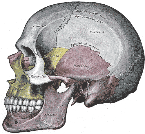
Coronal Sutures
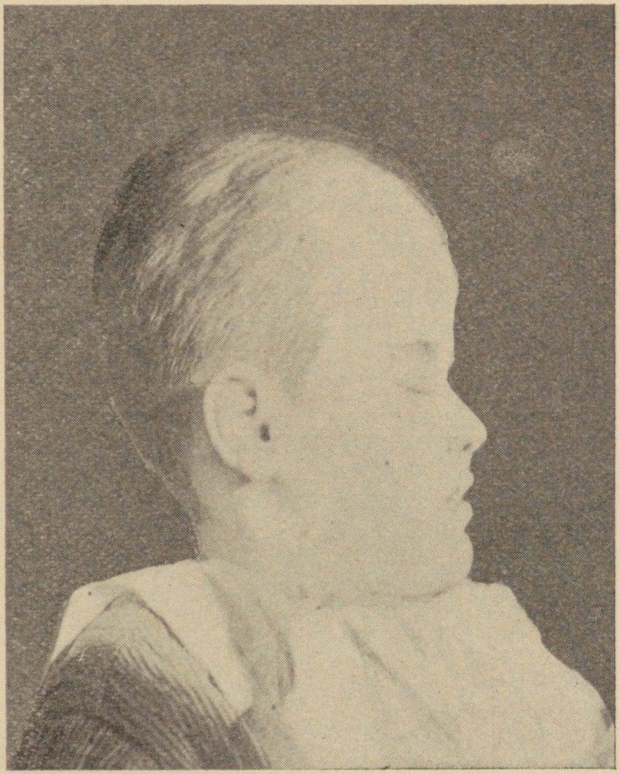
The coronal suture is a dense, fibrous connective tissue joint that separates the two parietal bones from the frontal bone of the skull. Structure The coronal suture lies between the paired parietal bones and the frontal bone of the skull. It runs from the pterion on each side. Nerve supply The coronal suture is likely supplied by a branch of the trigeminal nerve. Development The coronal suture is derived from the paraxial mesoderm. Clinical significance If certain bones of the skull grow too fast then premature fusion of the sutures may occur. This can result in skull deformities. There are two possible deformities that can be caused by the premature closure of the coronal suture: * a high, tower-like skull called "oxycephaly" or "turret skull". * a twisted and asymmetrical skull called "plagiocephaly". References * "Sagittal suture." ''Stedman's Medical Dictionary, 27th ed.'' (2000). * Moore, Keith L., and T.V.N. Persaud. ''The Developing Human: Clinically Orien ... [...More Info...] [...Related Items...] OR: [Wikipedia] [Google] [Baidu] |
Trigeminal Nerve
In neuroanatomy, the trigeminal nerve ( lit. ''triplet'' nerve), also known as the fifth cranial nerve, cranial nerve V, or simply CN V, is a cranial nerve responsible for sensation in the face and motor functions such as biting and chewing; it is the most complex of the cranial nerves. Its name ("trigeminal", ) derives from each of the two nerves (one on each side of the pons) having three major branches: the ophthalmic nerve (V), the maxillary nerve (V), and the mandibular nerve (V). The ophthalmic and maxillary nerves are purely sensory, whereas the mandibular nerve supplies motor as well as sensory (or "cutaneous") functions. Adding to the complexity of this nerve is that autonomic nerve fibers as well as special sensory fibers (taste) are contained within it. The motor division of the trigeminal nerve derives from the basal plate of the embryonic pons, and the sensory division originates in the cranial neural crest. Sensory information from the face and body is proc ... [...More Info...] [...Related Items...] OR: [Wikipedia] [Google] [Baidu] |
Churchill Livingstone
Churchill Livingstone is an academic publisher. It was formed in 1971 from the merger of Longman's medical list, E & S Livingstone (Edinburgh, Scotland) and J & A Churchill (London, England) and was owned by Pearson. Harcourt acquired Churchill Livingstone in 1997. It is now integrated as an imprint in Elsevier's health science division after Elsevier acquired Harcourt in 2001. In the past it published a number of classic medical texts, including Sir William Osler's textbook '' The Principles and Practice of Medicine, Gray's Anatomy,'' and ''Myles In Greek mythology, Myles (; Ancient Greek: Μύλης means 'mill-man') was an ancient king of Laconia. He was the son of the King Lelex and possibly the naiad Queen Cleocharia, and brother of Polycaon. Myles was the father of Eurotas who begott ...' Textbook for Midwives.'' In the 1980s, in addition to new texts in all areas of clinical medicine, it published an extensive list of medical and nursing textbooks in low-cost editions ... [...More Info...] [...Related Items...] OR: [Wikipedia] [Google] [Baidu] |
Joints
A joint or articulation (or articular surface) is the connection made between bones, ossicles, or other hard structures in the body which link an animal's skeletal system into a functional whole.Saladin, Ken. Anatomy & Physiology. 7th ed. McGraw-Hill Connect. Webp.274/ref> They are constructed to allow for different degrees and types of movement. Some joints, such as the knee, elbow, and shoulder, are self-lubricating, almost frictionless, and are able to withstand compression and maintain heavy loads while still executing smooth and precise movements. Other joints such as suture (joint), sutures between the bones of the skull permit very little movement (only during birth) in order to protect the brain and the sense organs. The connection between a tooth and the jawbone is also called a joint, and is described as a fibrous joint known as a gomphosis. Joints are classified both structurally and functionally. Classification The number of joints depends on if Sesamoid bone, sesamoi ... [...More Info...] [...Related Items...] OR: [Wikipedia] [Google] [Baidu] |
Human Head And Neck
Humans (''Homo sapiens'') are the most abundant and widespread species of primate, characterized by bipedalism and exceptional cognitive skills due to a large and complex brain. This has enabled the development of advanced tools, culture, and language. Humans are highly social and tend to live in complex social structures composed of many cooperating and competing groups, from families and kinship networks to political states. Social interactions between humans have established a wide variety of values, social norms, and rituals, which bolster human society. Its intelligence and its desire to understand and influence the environment and to explain and manipulate phenomena have motivated humanity's development of science, philosophy, mythology, religion, and other fields of study. Although some scientists equate the term ''humans'' with all members of the genus ''Homo'', in common usage, it generally refers to ''Homo sapiens'', the only extant member. Anatomically mode ... [...More Info...] [...Related Items...] OR: [Wikipedia] [Google] [Baidu] |
Cranial Sutures
In anatomy, fibrous joints are joints connected by fibrous tissue, consisting mainly of collagen Collagen () is the main structural protein in the extracellular matrix found in the body's various connective tissues. As the main component of connective tissue, it is the most abundant protein in mammals, making up from 25% to 35% of the whole .... These are fixed joints where bones are united by a layer of white fibrous tissue of varying thickness. In the skull the joints between the bones are called Suture (anatomy), sutures. Such immovable joints are also referred to as synarthrosis, synarthroses. Types Most fibrous joints are also called "fixed" or "immovable". These joints have no joint cavity and are connected via fibrous connective tissue. The skull bones are connected by fibrous joints called ''#Sutures, sutures''. In fetus, fetal skulls the sutures are wide to allow slight movement during birth. They later become rigid (synarthrosis, synarthrodial). Some of the long bo ... [...More Info...] [...Related Items...] OR: [Wikipedia] [Google] [Baidu] |
Frontal Bone
The frontal bone is a bone in the human skull. The bone consists of two portions.''Gray's Anatomy'' (1918) These are the vertically oriented squamous part, and the horizontally oriented orbital part, making up the bony part of the forehead, part of the bony orbital cavity holding the eye, and part of the bony part of the nose respectively. The name comes from the Latin word ''frons'' (meaning " forehead"). Structure of the frontal bone The frontal bone is made up of two main parts. These are the squamous part, and the orbital part. The squamous part marks the vertical, flat, and also the biggest part, and the main region of the forehead. The orbital part is the horizontal and second biggest region of the frontal bone. It enters into the formation of the roofs of the orbital and nasal cavities. Sometimes a third part is included as the nasal part of the frontal bone, and sometimes this is included with the squamous part. The nasal part is between the brow ridges, and ends in ... [...More Info...] [...Related Items...] OR: [Wikipedia] [Google] [Baidu] |
Plagiocephaly
Plagiocephaly, also known as flat head syndrome, is a condition characterized by an asymmetrical distortion (flattening of one side) of the skull. A mild and widespread form is characterized by a flat spot on the back or one side of the head caused by remaining in a supine position for prolonged periods. Plagiocephaly is a diagonal asymmetry across the head shape. Often it is a flattening which is to one side at the back of the head and there is often some facial asymmetry. Depending on whether synostosis is involved, plagiocephaly divides into two groups: synostotic, with one or more fused cranial sutures, and non-synostotic (deformational). Surgical treatment of these groups includes the deference method; however, the treatment of deformational plagiocephaly is controversial. Brachycephaly describes a very wide head shape with a flattening across the whole back of the head. Causes Slight plagiocephaly is routinely diagnosed at birth and may be the result of a restrictive intrau ... [...More Info...] [...Related Items...] OR: [Wikipedia] [Google] [Baidu] |
Oxycephaly
Turricephaly is a type of cephalic disorder where the head appears tall with a small length and width. It is due to premature closure of the coronal suture plus any other suture, like the lambdoid, or it may be used to describe the premature fusion of all sutures. It should be differentiated from Crouzon syndrome. Oxycephaly (or acrocephaly) is a form of turricephaly where the head is cone-shaped, and is the most severe of the craniosynostoses. Presentation Common associations It may be associated with: * 8th cranial nerve lesion * Optic nerve compression * Intellectual disability * Syndactyly Syndactyly is a condition wherein two or more digits are fused together. It occurs normally in some mammals, such as the siamang and diprotodontia, but is an unusual condition in humans. The term is from Greek σύν, ''syn'' 'together' and δάκ� ... Diagnosis Treatment See also * Acrocephalosyndactylia References Further reading NINDS Overview* External links Congeni ... [...More Info...] [...Related Items...] OR: [Wikipedia] [Google] [Baidu] |
Paraxial Mesoderm
Paraxial mesoderm, also known as presomitic or somitic mesoderm is the area of mesoderm in the neurulating embryo that flanks and forms simultaneously with the neural tube. The cells of this region give rise to somites, blocks of tissue running along both sides of the neural tube, which form muscle and the tissues of the back, including connective tissue and the dermis. Formation and somitogenesis The paraxial and other regions of the mesoderm are thought to be specified by bone morphogenetic proteins, or BMPs, along an axis spanning from the center to the sides of the body. Members of the FGF family also play an important role, as does the WNT pathway. In particular, Noggin, a downstream target of the Wnt pathway, antagonizes BMP signaling, forming boundaries where antagonists meet and limiting this signaling to a particular region of the mesoderm. Together, these pathways provide the initial specification of the paraxial mesoderm and maintain this identity. This specific ... [...More Info...] [...Related Items...] OR: [Wikipedia] [Google] [Baidu] |
Pterion
The pterion is the region where the frontal, parietal, temporal, and sphenoid bones join. It is located on the side of the skull, just behind the temple. Structure The pterion is located in the temporal fossa, approximately 2.6 cm behind and 1.3 cm above the posterolateral margin of the frontozygomatic suture. It is the junction between four bones: * the parietal bone. * the squamous part of temporal bone. * the greater wing of sphenoid bone. * the frontal bone. These bones are typically joined by five cranial sutures: * the sphenoparietal suture joins the sphenoid and parietal bones. * the coronal suture joins the frontal bone to the sphenoid and parietal bones. * the squamous suture joins the temporal bone to the sphenoid and parietal bones. * the sphenofrontal suture joins the sphenoid and frontal bones. * the sphenosquamosal suture joins the sphenoid and temporal bones. Clinical significance Haematoma The pterion is known as the weakest part of the sk ... [...More Info...] [...Related Items...] OR: [Wikipedia] [Google] [Baidu] |
Skull
The skull is a bone protective cavity for the brain. The skull is composed of four types of bone i.e., cranial bones, facial bones, ear ossicles and hyoid bone. However two parts are more prominent: the cranium and the mandible. In humans, these two parts are the neurocranium and the viscerocranium ( facial skeleton) that includes the mandible as its largest bone. The skull forms the anterior-most portion of the skeleton and is a product of cephalisation—housing the brain, and several sensory structures such as the eyes, ears, nose, and mouth. In humans these sensory structures are part of the facial skeleton. Functions of the skull include protection of the brain, fixing the distance between the eyes to allow stereoscopic vision, and fixing the position of the ears to enable sound localisation of the direction and distance of sounds. In some animals, such as horned ungulates (mammals with hooves), the skull also has a defensive function by providing the mount (on the front ... [...More Info...] [...Related Items...] OR: [Wikipedia] [Google] [Baidu] |
Saunders (imprint)
Saunders is an American academic publisher based in the United States. It is currently an imprint of Elsevier. Formerly independent, the W. B. Saunders company was acquired by CBS in 1968, who added it to their publishing division Holt, Rinehart & Winston. When CBS left the publishing field in 1986, it sold the academic publishing units to Harcourt Brace Jovanovich. Harcourt was acquired by Reed Elsevier in 2001. . . Retrieved May 2, 2015. W. B. Saunders published the Kinsey Reports and |


.jpg)

