|
Common Iliac Vein
In human anatomy, the common iliac veins are formed by the external iliac veins and internal iliac veins. The left and right common iliac veins come together in the abdomen at the level of the fifth lumbar vertebra, forming the inferior vena cava. They drain blood from the pelvis and lower limbs. Both common iliac veins are accompanied along their course by common iliac arteries. Structure The external iliac vein and internal iliac vein unite in front of the sacroiliac joint to form the common iliac veins. Both common iliac veins ascend to form the inferior vena cava behind the right common iliac artery at the level of the fifth lumbar vertebra. The vena cava is to the right of the midline and therefore the left common iliac vein is longer than the right. The left common iliac vein occasionally travels upwards to the left of the aorta to the level of the kidney, where it receives the left renal vein and crosses in front of the aorta to join the inferior vena cava. The right ... [...More Info...] [...Related Items...] OR: [Wikipedia] [Google] [Baidu] |
Abdomen
The abdomen (colloquially called the belly, tummy, midriff, tucky or stomach) is the part of the body between the thorax (chest) and pelvis, in humans and in other vertebrates. The abdomen is the front part of the abdominal segment of the torso. The area occupied by the abdomen is called the abdominal cavity. In arthropods it is the posterior tagma of the body; it follows the thorax or cephalothorax. In humans, the abdomen stretches from the thorax at the thoracic diaphragm to the pelvis at the pelvic brim. The pelvic brim stretches from the lumbosacral joint (the intervertebral disc between L5 and S1) to the pubic symphysis and is the edge of the pelvic inlet. The space above this inlet and under the thoracic diaphragm is termed the abdominal cavity. The boundary of the abdominal cavity is the abdominal wall in the front and the peritoneal surface at the rear. In vertebrates, the abdomen is a large body cavity enclosed by the abdominal muscles, at front and to ... [...More Info...] [...Related Items...] OR: [Wikipedia] [Google] [Baidu] |
Kidney
The kidneys are two reddish-brown bean-shaped organs found in vertebrates. They are located on the left and right in the retroperitoneal space, and in adult humans are about in length. They receive blood from the paired renal arteries; blood exits into the paired renal veins. Each kidney is attached to a ureter, a tube that carries excreted urine to the bladder. The kidney participates in the control of the volume of various body fluids, fluid osmolality, acid–base balance, various electrolyte concentrations, and removal of toxins. Filtration occurs in the glomerulus: one-fifth of the blood volume that enters the kidneys is filtered. Examples of substances reabsorbed are solute-free water, sodium, bicarbonate, glucose, and amino acids. Examples of substances secreted are hydrogen, ammonium, potassium and uric acid. The nephron is the structural and functional unit of the kidney. Each adult human kidney contains around 1 million nephrons, while a mouse kidney con ... [...More Info...] [...Related Items...] OR: [Wikipedia] [Google] [Baidu] |
Angiology
Angiology (from Greek , ''angeīon'', "vessel"; and , ''-logia'') is the medical specialty dedicated to studying the circulatory system and of the lymphatic system, i.e., arteries, veins and lymphatic vessels. In the UK, this field is more often termed ''angiology'', and in the United States the term vascular medicine is more frequent. The field of vascular medicine (angiology) is the field that deals with preventing, diagnosing and treating vascular and blood vessel related diseases. Overview Arterial diseases include the aorta (aneurysms/dissection) and arteries supplying the legs, hands, kidneys, brain, intestines. It also covers arterial thrombosis and embolism; vasculitides; and vasospastic disorders. Naturally, it deals with preventing cardiovascular diseases such as heart attack and stroke. Venous diseases include venous thrombosis, chronic venous insufficiency, and varicose veins. Lymphatic diseases include primary and secondary forms of lymphedema. It also inv ... [...More Info...] [...Related Items...] OR: [Wikipedia] [Google] [Baidu] |
Veins Of The Torso
Veins are blood vessels in humans and most other animals that carry blood towards the heart. Most veins carry deoxygenated blood from the tissues back to the heart; exceptions are the pulmonary and umbilical veins, both of which carry oxygenated blood to the heart. In contrast to veins, arteries carry blood away from the heart. Veins are less muscular than arteries and are often closer to the skin. There are valves (called ''pocket valves'') in most veins to prevent backflow. Structure Veins are present throughout the body as tubes that carry blood back to the heart. Veins are classified in a number of ways, including superficial vs. deep, pulmonary vs. systemic, and large vs. small. *Superficial veins are those closer to the surface of the body, and have no corresponding arteries. *Deep veins are deeper in the body and have corresponding arteries. * Perforator veins drain from the superficial to the deep veins. These are usually referred to in the lower limbs and feet. *Communic ... [...More Info...] [...Related Items...] OR: [Wikipedia] [Google] [Baidu] |
Venous Thromboembolism
Venous thrombosis is blockage of a vein caused by a thrombus (blood clot). A common form of venous thrombosis is deep vein thrombosis (DVT), when a blood clot forms in the deep veins. If a thrombus breaks off (embolizes) and flows to the lungs to lodge there, it becomes a pulmonary embolism (PE), a blood clot in the lungs. The conditions of DVT only, DVT with PE, and PE only, are all captured by the term venous thromboembolism (VTE). The initial treatment for VTE is typically either low-molecular-weight heparin (LMWH) or unfractionated heparin, or increasingly with direct acting oral anticoagulants (DOAC). Those initially treated with heparins can be switched to other anticoagulants (warfarin, DOACs), although pregnant women and some people with cancer receive ongoing heparin treatment. Superficial venous thrombosis or phlebitis affects the superficial veins of the upper or lower extremity and only require anticoagulation in specific situations, and may be treated with anti-infla ... [...More Info...] [...Related Items...] OR: [Wikipedia] [Google] [Baidu] |
Fibrosis
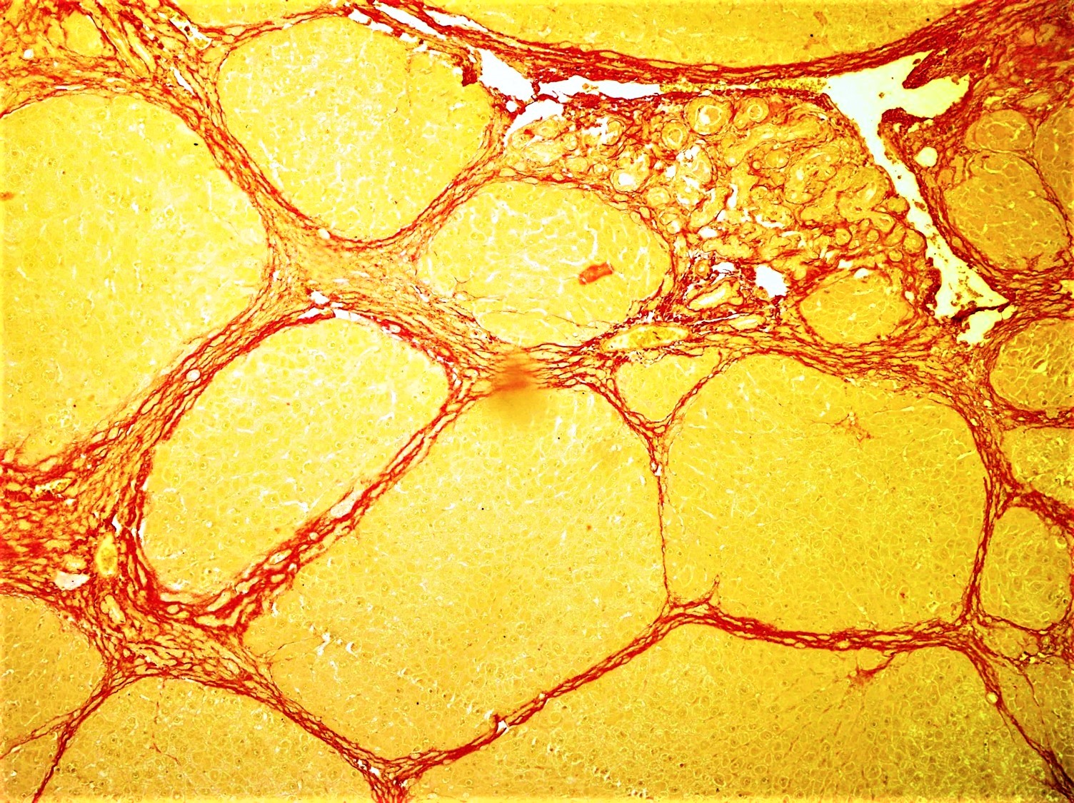
Fibrosis, also known as fibrotic scarring, is a pathological wound healing in which connective tissue replaces normal parenchymal tissue to the extent that it goes unchecked, leading to considerable tissue remodelling and the formation of permanent scar tissue. Repeated injuries, chronic inflammation and repair are susceptible to fibrosis where an accidental excessive accumulation of extracellular matrix components, such as the collagen is produced by fibroblasts, leading to the formation of a permanent fibrotic scar. In response to injury, this is called scarring, and if fibrosis arises from a single cell line, this is called a fibroma. Physiologically, fibrosis acts to deposit connective tissue, which can interfere with or totally inhibit the normal architecture and function of the underlying organ or tissue. Fibrosis can be used to describe the pathological state of excess deposition of fibrous tissue, as well as the process of connective tissue deposition in healing. Defin ... [...More Info...] [...Related Items...] OR: [Wikipedia] [Google] [Baidu] |
Collagen
Collagen () is the main structural protein in the extracellular matrix found in the body's various connective tissues. As the main component of connective tissue, it is the most abundant protein in mammals, making up from 25% to 35% of the whole-body protein content. Collagen consists of amino acids bound together to form a triple helix of elongated fibril known as a collagen helix. It is mostly found in connective tissue such as cartilage, bones, tendons, ligaments, and skin. Depending upon the degree of mineralization, collagen tissues may be rigid (bone) or compliant (tendon) or have a gradient from rigid to compliant (cartilage). Collagen is also abundant in corneas, blood vessels, the gut, intervertebral discs, and the dentin in teeth. In muscle tissue, it serves as a major component of the endomysium. Collagen constitutes one to two percent of muscle tissue and accounts for 6% of the weight of the skeletal muscle tissue. The fibroblast is the most common ... [...More Info...] [...Related Items...] OR: [Wikipedia] [Google] [Baidu] |
Elastin
Elastin is a protein that in humans is encoded by the ''ELN'' gene. Elastin is a key component of the extracellular matrix in gnathostomes (jawed vertebrates). It is highly elastic and present in connective tissue allowing many tissues in the body to resume their shape after stretching or contracting. Elastin helps skin to return to its original position when it is poked or pinched. Elastin is also an important load-bearing tissue in the bodies of vertebrates and used in places where mechanical energy is required to be stored. Function The ''ELN'' gene encodes a protein that is one of the two components of elastic fibers. The encoded protein is rich in hydrophobic amino acids such as glycine and proline, which form mobile hydrophobic regions bounded by crosslinks between lysine residues. Multiple transcript variants encoding different isoforms have been found for this gene. Elastin's soluble precursor is tropoelastin. The characterization of disorder is consistent with an ... [...More Info...] [...Related Items...] OR: [Wikipedia] [Google] [Baidu] |
Inflammatory Response
Inflammation (from la, inflammatio) is part of the complex biological response of body tissues to harmful stimuli, such as pathogens, damaged cells, or irritants, and is a protective response involving immune cells, blood vessels, and molecular mediators. The function of inflammation is to eliminate the initial cause of cell injury, clear out necrotic cells and tissues damaged from the original insult and the inflammatory process, and initiate tissue repair. The five cardinal signs are heat, pain, redness, swelling, and loss of function (Latin ''calor'', ''dolor'', ''rubor'', ''tumor'', and ''functio laesa''). Inflammation is a generic response, and therefore it is considered as a mechanism of innate immunity, as compared to adaptive immunity, which is specific for each pathogen. Too little inflammation could lead to progressive tissue destruction by the harmful stimulus (e.g. bacteria) and compromise the survival of the organism. In contrast, too much inflammation, in the ... [...More Info...] [...Related Items...] OR: [Wikipedia] [Google] [Baidu] |
Pulse
In medicine, a pulse represents the tactile arterial palpation of the cardiac cycle (heartbeat) by trained fingertips. The pulse may be palpated in any place that allows an artery to be compressed near the surface of the body, such as at the neck (carotid artery), wrist (radial artery), at the groin (femoral artery), behind the knee ( popliteal artery), near the ankle joint (posterior tibial artery), and on foot ( dorsalis pedis artery). Pulse (or the count of arterial pulse per minute) is equivalent to measuring the heart rate. The heart rate can also be measured by listening to the heart beat by auscultation, traditionally using a stethoscope and counting it for a minute. The radial pulse is commonly measured using three fingers. This has a reason: the finger closest to the heart is used to occlude the pulse pressure, the middle finger is used get a crude estimate of the blood pressure, and the finger most distal to the heart (usually the ring finger) is used to nullify the ... [...More Info...] [...Related Items...] OR: [Wikipedia] [Google] [Baidu] |
May–Thurner Syndrome
May–Thurner syndrome (MTS), also known as the iliac vein compression syndrome, is a condition in which compression of the common venous outflow tract of the left lower extremity may cause discomfort, swelling, pain or clots (deep venous thrombosis) in the iliofemoral veins. Specifically, the problem is due to left common iliac vein compression by the overlying right common iliac artery. This leads to stasis of blood, which predisposes to the formation of blood clots. Uncommon variations of MTS have been described, such as the right common iliac vein getting compressed by the right common iliac artery. In the 21st century, the May–Thurner syndrome definition has been expanded to a broader disease profile known as nonthrombotic iliac vein lesions (NIVL) which can involve both the right and left iliac veins as well as multiple other named venous segments. This syndrome frequently manifests as pain when the limb is dependent (hanging down the edge of a bed/chair) and/or signif ... [...More Info...] [...Related Items...] OR: [Wikipedia] [Google] [Baidu] |
Median Sacral Vein
The median sacral vein (or middle sacral veins) is a vein of the abdomen. It accompanies the median sacral artery along the front of the sacrum. It ends in the left common iliac vein. Sometimes, it ends in the angle of junction of the two common iliac veins In human anatomy, the common iliac veins are formed by the external iliac veins and internal iliac veins. The left and right common iliac veins come together in the abdomen at the level of the fifth lumbar vertebra, forming the inferior vena ca .... References External links {{circulatory-stub Veins of the torso ... [...More Info...] [...Related Items...] OR: [Wikipedia] [Google] [Baidu] |


