|
Cochlear Amplifier
The cochlear amplifier is a positive feedback mechanism within the cochlea that provides acute sensitivity in the mammalian auditory system. The main component of the cochlear amplifier is the outer hair cell (OHC) which increases the amplitude and frequency selectivity of sound vibrations using electromechanical feedback. Discovery The cochlear amplifier was first proposed in 1948 by Gold. This was around the time when Georg von Békésy was publishing articles observing the propagation of passive travelling waves in the dead cochlea. Thirty years later the first recordings of emissions from the ear were captured by Kemp. This was confirmation that such an active mechanism was present in the ear. These emissions are now termed otoacoustic emissions and are produced by the cochlear amplifier. The first modeling effort to define the cochlear amplifier was a simple augmentation of Georg von Békésy's passive traveling wave with an active component. In such a model, a lopsided pr ... [...More Info...] [...Related Items...] OR: [Wikipedia] [Google] [Baidu] |
Cochlea
The cochlea is the part of the inner ear involved in hearing. It is a spiral-shaped cavity in the bony labyrinth, in humans making 2.75 turns around its axis, the modiolus. A core component of the cochlea is the Organ of Corti, the sensory organ of hearing, which is distributed along the partition separating the fluid chambers in the coiled tapered tube of the cochlea. The name cochlea derives . Structure The cochlea (plural is cochleae) is a spiraled, hollow, conical chamber of bone, in which waves propagate from the base (near the middle ear and the oval window) to the apex (the top or center of the spiral). The spiral canal of the cochlea is a section of the bony labyrinth of the inner ear that is approximately 30 mm long and makes 2 turns about the modiolus. The cochlear structures include: * Three ''scalae'' or chambers: ** the vestibular duct or ''scala vestibuli'' (containing perilymph), which lies superior to the cochlear duct and abuts the oval window ** the ty ... [...More Info...] [...Related Items...] OR: [Wikipedia] [Google] [Baidu] |
Rarefaction Phase
Rarefaction is the reduction of an item's density, the opposite of compression. Like compression, which can travel in waves (sound waves, for instance), rarefaction waves also exist in nature. A common rarefaction wave is the area of low relative pressure following a shock wave (see picture). Rarefaction waves expand with time (much like sea waves spread out as they reach a beach); in most cases rarefaction waves keep the same overall profile ('shape') at all times throughout the wave's movement: it is a ''self-similar expansion''. Each part of the wave travels at the local speed of sound, in the local medium. This expansion behaviour contrasts with that of pressure increases, which gets narrower with time until they steepen into shock waves. When angle of incidence is greater than angle of refraction, then light travels from denser to rarer medium. When angle of incidence is smaller than angle of refraction then light travels from rarer to denser medium Physical examples A nat ... [...More Info...] [...Related Items...] OR: [Wikipedia] [Google] [Baidu] |
Actin
Actin is a family of globular multi-functional proteins that form microfilaments in the cytoskeleton, and the thin filaments in muscle fibrils. It is found in essentially all eukaryotic cells, where it may be present at a concentration of over 100 μM; its mass is roughly 42 kDa, with a diameter of 4 to 7 nm. An actin protein is the monomeric subunit of two types of filaments in cells: microfilaments, one of the three major components of the cytoskeleton, and thin filaments, part of the contractile apparatus in muscle cells. It can be present as either a free monomer called G-actin (globular) or as part of a linear polymer microfilament called F-actin (filamentous), both of which are essential for such important cellular functions as the mobility and contraction of cells during cell division. Actin participates in many important cellular processes, including muscle contraction, cell motility, cell division and cytokinesis, vesicle and organelle movement, cell sign ... [...More Info...] [...Related Items...] OR: [Wikipedia] [Google] [Baidu] |
Myosin
Myosins () are a superfamily of motor proteins best known for their roles in muscle contraction and in a wide range of other motility processes in eukaryotes. They are ATP-dependent and responsible for actin-based motility. The first myosin (M2) to be discovered was in 1864 by Wilhelm Kühne. Kühne had extracted a viscous protein from skeletal muscle that he held responsible for keeping the tension state in muscle. He called this protein ''myosin''. The term has been extended to include a group of similar ATPases found in the cells of both striated muscle tissue and smooth muscle tissue. Following the discovery in 1973 of enzymes with myosin-like function in '' Acanthamoeba castellanii'', a global range of divergent myosin genes have been discovered throughout the realm of eukaryotes. Although myosin was originally thought to be restricted to muscle cells (hence '' myo-''(s) + '' -in''), there is no single "myosin"; rather it is a very large superfamily of genes whose p ... [...More Info...] [...Related Items...] OR: [Wikipedia] [Google] [Baidu] |
Nonlinear Capacitance
In mathematics and science, a nonlinear system is a system in which the change of the output is not proportional to the change of the input. Nonlinear problems are of interest to engineers, biologists, physicists, mathematicians, and many other scientists because most systems are inherently nonlinear in nature. Nonlinear dynamical systems, describing changes in variables over time, may appear chaotic, unpredictable, or counterintuitive, contrasting with much simpler linear systems. Typically, the behavior of a nonlinear system is described in mathematics by a nonlinear system of equations, which is a set of simultaneous equations in which the unknowns (or the unknown functions in the case of differential equations) appear as variables of a polynomial of degree higher than one or in the argument of a function (mathematics), function which is not a polynomial of degree one. In other words, in a nonlinear system of equations, the equation(s) to be solved cannot be written as a linea ... [...More Info...] [...Related Items...] OR: [Wikipedia] [Google] [Baidu] |
Prestin
Prestin is a protein that is critical to sensitive hearing in mammals. It is encoded by the ''SLC26A5'' (solute carrier anion transporter family 26, member 5) gene. Prestin is the motor protein of the outer hair cells of the inner ear of the mammalian cochlea. It is highly expressed in the outer hair cells, and is not expressed in the nonmotile inner hair cells. Immunolocalization shows prestin is expressed in the lateral plasma membrane of the outer hair cells, the region wherelectromotilityoccurs. The expression pattern correlates with the appearance of outer hair cell electromotility. Function Prestin is essential in auditory processing. It is specifically expressed in the lateral membrane of outer hair cells (OHCs) of the cochlea. There is no significant difference between prestin density in high-frequency and low-frequency regions of the cochlea in fully developed mammals. There is good evidence that prestin has undergone adaptive evolution in mammals associated with acq ... [...More Info...] [...Related Items...] OR: [Wikipedia] [Google] [Baidu] |
Membrane Potential
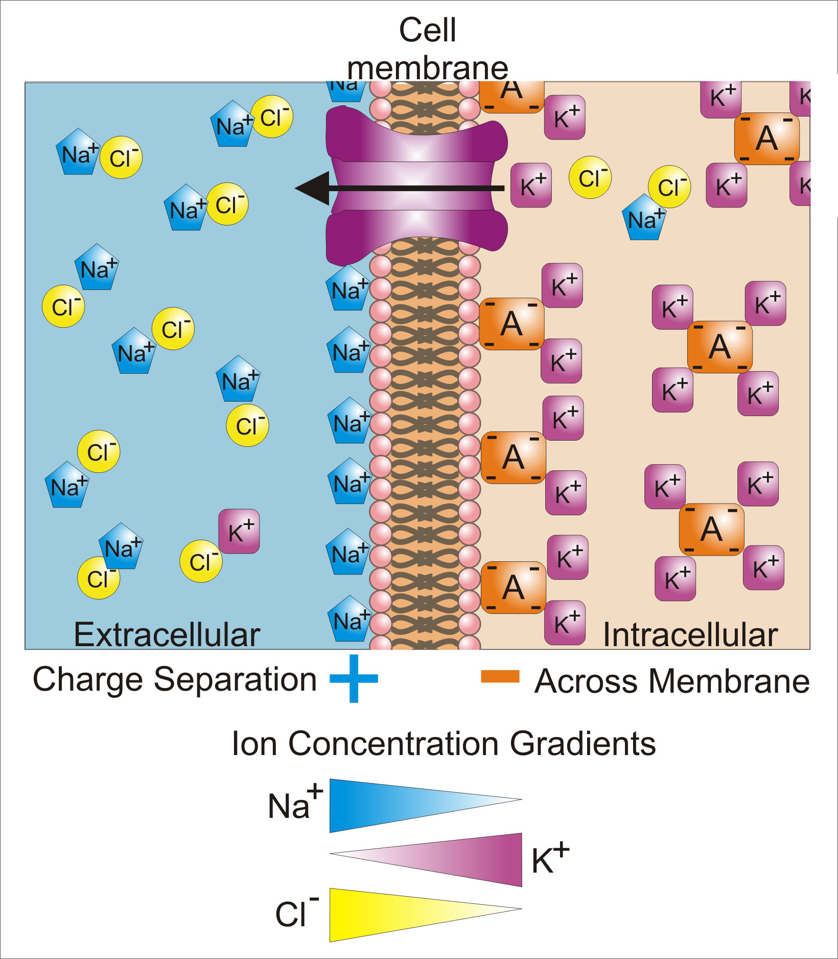
Membrane potential (also transmembrane potential or membrane voltage) is the difference in electric potential between the interior and the exterior of a biological cell. That is, there is a difference in the energy required for electric charges to move from the internal to exterior cellular environments and vice versa, as long as there is no acquisition of kinetic energy or the production of radiation. The concentration gradients of the charges directly determine this energy requirement. For the exterior of the cell, typical values of membrane potential, normally given in units of milli volts and denoted as mV, range from –80 mV to –40 mV. All animal cells are surrounded by a membrane composed of a lipid bilayer with proteins embedded in it. The membrane serves as both an insulator and a diffusion barrier to the movement of ions. Transmembrane proteins, also known as ion transporter or ion pump proteins, actively push ions across the membrane and establish concentration gradi ... [...More Info...] [...Related Items...] OR: [Wikipedia] [Google] [Baidu] |
Stereocilia (inner Ear)
In the inner ear, stereocilia are the mechanosensing organelles of hair cells, which respond to fluid motion in numerous types of animals for various functions, including hearing and balance. They are about 10–50 micrometers in length and share some similar features of microvilli.Caceci, T. VM8054 Veterinary Histology: Male Reproductive System. http://education.vetmed.vt.edu/Curriculum/VM8054/Labs/Lab27/Lab27.htm (accessed 2/16/06). The hair cells turn the fluid pressure and other mechanical stimuli into electric stimuli via the many microvilli that make up stereocilia rods.Alberts, B., Johnson, A., Lewis, J., Raff, M., Roberts, K. and Walter, P. (2002) The Molecular Biology of the Cell. Garland Science Textbooks. Stereocilia exist in the auditory and vestibular systems. Morphology Resembling hair-like projections, the stereocilia are arranged in bundles of 30-300. Within the bundles the stereocilia are often lined up in several rows of increasing height, similar to a stairca ... [...More Info...] [...Related Items...] OR: [Wikipedia] [Google] [Baidu] |
Basilar Membrane
The basilar membrane is a stiff structural element within the cochlea of the inner ear which separates two liquid-filled tubes that run along the coil of the cochlea, the scala media and the scala tympani. The basilar membrane moves up and down in response to incoming sound waves, which are converted to traveling waves on the basilar membrane. Structure The basilar membrane is a pseudo-resonant structure that, like the strings on an instrument, varies in width and stiffness. But unlike the parallel strings of a guitar, the basilar membrane is not a discrete set of resonant structures, but a single structure with varying width, stiffness, mass, damping, and duct dimensions along its length. The motion of the basilar membrane is generally described as a traveling wave. The properties of the membrane at a given point along its length determine its characteristic frequency (CF), the frequency at which it is most sensitive to sound vibrations. The basilar membrane is widest (0.42– ... [...More Info...] [...Related Items...] OR: [Wikipedia] [Google] [Baidu] |
Hair Cell
Hair cells are the sensory receptors of both the auditory system and the vestibular system in the ears of all vertebrates, and in the lateral line organ of fishes. Through mechanotransduction, hair cells detect movement in their environment. In mammals, the auditory hair cells are located within the spiral organ of Corti on the thin basilar membrane in the cochlea of the inner ear. They derive their name from the tufts of stereocilia called ''hair bundles'' that protrude from the apical surface of the cell into the fluid-filled cochlear duct. The stereocilia number from 50-100 in each cell while being tightly packed together and decrease in size the further away they are located from the kinocilium. The hair bundles are arranged as stiff columns that move at their base in response to stimuli applied to the tips. Mammalian cochlear hair cells are of two anatomically and functionally distinct types, known as outer, and inner hair cells. Damage to these hair cells results in ... [...More Info...] [...Related Items...] OR: [Wikipedia] [Google] [Baidu] |
Outer Hair Cell
Hair cells are the sensory receptors of both the auditory system and the vestibular system in the ears of all vertebrates, and in the lateral line organ of fishes. Through mechanotransduction, hair cells detect movement in their environment. In mammals, the auditory hair cells are located within the spiral organ of Corti on the thin basilar membrane in the cochlea of the inner ear. They derive their name from the tufts of stereocilia called ''hair bundles'' that protrude from the apical surface of the cell into the fluid-filled cochlear duct. The stereocilia number from 50-100 in each cell while being tightly packed together and decrease in size the further away they are located from the kinocilium. The hair bundles are arranged as stiff columns that move at their base in response to stimuli applied to the tips. Mammalian cochlear hair cells are of two anatomically and functionally distinct types, known as outer, and inner hair cells. Damage to these hair cells results in d ... [...More Info...] [...Related Items...] OR: [Wikipedia] [Google] [Baidu] |
Organ Of Corti
The organ of Corti, or spiral organ, is the receptor organ for hearing and is located in the mammalian cochlea. This highly varied strip of epithelial cells allows for transduction of auditory signals into nerve impulses' action potential. Transduction occurs through vibrations of structures in the inner ear causing displacement of cochlear fluid and movement of hair cells at the organ of Corti to produce electrochemical signals.The Ear Pujol, R., Irving, S., 2013 Italian anatomist (1822–1876) discovered the organ of Corti in 1851. The structure evolved from the [...More Info...] [...Related Items...] OR: [Wikipedia] [Google] [Baidu] |


