|
Coccygeus
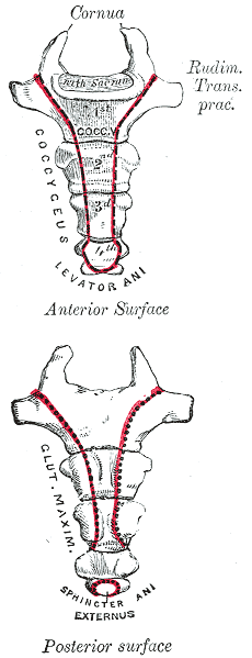
The coccygeus muscle or ischiococcygeus is a muscle of the pelvic floor, located posterior to levator ani and anterior to the sacrospinous ligament. Structure The coccygeus muscle is posterior to levator ani and anterior to the sacrospinous ligament in the pelvic floor. It is a triangular plane of muscular and tendinous fibers. It arises by its apex from the spine of the ischium and sacrospinous ligament. It is inserted by its base into the margin of the coccyx and into the side of the lowest piece of the sacrum. In combination with the levator ani, it forms the pelvic diaphragm. The pudendal nerve runs between the coccygeus muscle and the piriformis muscle, superficial to the coccygeus muscle. Nerve supply The coccygeus muscle is innervated by the pudendal nerve, which runs between it and the piriformis muscle. Function The coccygeus muscle assists the levator ani and piriformis muscle in closing in the back part of the outlet of the pelvis. This helps to support t ... [...More Info...] [...Related Items...] OR: [Wikipedia] [Google] [Baidu] |
Pubococcygeus Muscle
The levator ani is a broad, thin muscle group, situated on either side of the pelvis. It is formed from three muscle components: the pubococcygeus, the iliococcygeus, and the puborectalis. It is attached to the inner surface of each side of the lesser pelvis, and these unite to form the greater part of the pelvic floor. The coccygeus muscle completes the pelvic floor, which is also called the ''pelvic diaphragm''. It supports the viscera in the pelvic cavity, and surrounds the various structures that pass through it. The levator ani is the main pelvic floor muscle and painfully contracts during vaginismus. It also contracts rhythmically during orgasm. Structure The levator ani is made up of 3 parts: * Iliococcygeus muscle * Pubococcygeus muscle * Puborectalis muscle The iliococcygeus arises from the inner side of the ischium (the lower and back part of the hip bone) and from the posterior part of the tendinous arch of the obturator fascia, and is attached to the coccyx and a ... [...More Info...] [...Related Items...] OR: [Wikipedia] [Google] [Baidu] |
Levator Ani
The levator ani is a broad, thin muscle group, situated on either side of the pelvis. It is formed from three muscle components: the pubococcygeus, the iliococcygeus, and the puborectalis. It is attached to the inner surface of each side of the lesser pelvis, and these unite to form the greater part of the pelvic floor. The coccygeus muscle completes the pelvic floor, which is also called the ''pelvic diaphragm''. It supports the viscera in the pelvic cavity, and surrounds the various structures that pass through it. The levator ani is the main pelvic floor muscle and painfully contracts during vaginismus. It also contracts rhythmically during orgasm. Structure The levator ani is made up of 3 parts: * Iliococcygeus muscle * Pubococcygeus muscle * Puborectalis muscle The iliococcygeus arises from the inner side of the ischium (the lower and back part of the hip bone) and from the posterior part of the tendinous arch of the obturator fascia, and is attached to the coccyx and a ... [...More Info...] [...Related Items...] OR: [Wikipedia] [Google] [Baidu] |
Pelvic Floor
The pelvic floor or pelvic diaphragm is composed of muscle fibers of the levator ani, the coccygeus muscle, and associated connective tissue which span the area underneath the pelvis. The pelvic diaphragm is a muscular partition formed by the levatores ani and coccygei, with which may be included the parietal pelvic fascia on their upper and lower aspects. The pelvic floor separates the pelvic cavity above from the perineal region (including perineum) below. Both males and females have a pelvic floor. To accommodate the birth canal, a female's pelvic cavity is larger than a male's. Structure The right and left levator ani lie almost horizontally in the floor of the pelvis, separated by a narrow gap that transmits the urethra, vagina, and anal canal. The levator ani is usually considered in three parts: pubococcygeus, puborectalis, and iliococcygeus. The pubococcygeus, the main part of the levator, runs backward from the body of the pubis toward the coccyx and may be damaged during ... [...More Info...] [...Related Items...] OR: [Wikipedia] [Google] [Baidu] |
Pelvic Diaphragm
The pelvic floor or pelvic diaphragm is composed of muscle fibers of the levator ani, the coccygeus muscle, and associated connective tissue which span the area underneath the pelvis. The pelvic diaphragm is a muscular partition formed by the levatores ani and coccygei, with which may be included the parietal pelvic fascia on their upper and lower aspects. The pelvic floor separates the pelvic cavity above from the perineal region (including perineum) below. Both males and females have a pelvic floor. To accommodate the birth canal, a female's pelvic cavity is larger than a male's. Structure The right and left levator ani lie almost horizontally in the floor of the pelvis, separated by a narrow gap that transmits the urethra, vagina, and anal canal. The levator ani is usually considered in three parts: pubococcygeus, puborectalis, and iliococcygeus. The pubococcygeus, the main part of the levator, runs backward from the body of the pubis toward the coccyx and may be damaged during ... [...More Info...] [...Related Items...] OR: [Wikipedia] [Google] [Baidu] |
Coccyx
The coccyx ( : coccyges or coccyxes), commonly referred to as the tailbone, is the final segment of the vertebral column in all apes, and analogous structures in certain other mammals such as horses. In tailless primates (e.g. humans and other great apes) since ''Nacholapithecus'' (a Miocene hominoid),Nakatsukasa 2004, ''Acquisition of bipedalism'' (SeFig. 5entitled ''First coccygeal/caudal vertebra in short-tailed or tailless primates.''.) the coccyx is the remnant of a vestigial tail. In animals with bony tails, it is known as ''tailhead'' or ''dock'', in bird anatomy as ''tailfan''. It comprises three to five separate or fused coccygeal vertebrae below the sacrum, attached to the sacrum by a fibrocartilaginous joint, the sacrococcygeal symphysis, which permits limited movement between the sacrum and the coccyx. Structure The coccyx is formed of three, four or five rudimentary vertebrae. It articulates superiorly with the sacrum. In each of the first three segments may ... [...More Info...] [...Related Items...] OR: [Wikipedia] [Google] [Baidu] |
Extensor Coccygis
The coccyx ( : coccyges or coccyxes), commonly referred to as the tailbone, is the final segment of the vertebral column in all apes, and analogous structures in certain other mammals such as horses. In tailless primates (e.g. humans and other great apes) since ''Nacholapithecus'' (a Miocene hominoid),Nakatsukasa 2004, ''Acquisition of bipedalism'' (SeFig. 5entitled ''First coccygeal/caudal vertebra in short-tailed or tailless primates.''.) the coccyx is the remnant of a vestigial tail. In animals with bony tails, it is known as ''tailhead'' or ''dock'', in bird anatomy as ''tailfan''. It comprises three to five separate or fused coccygeal vertebrae below the sacrum, attached to the sacrum by a fibrocartilaginous joint, the sacrococcygeal symphysis, which permits limited movement between the sacrum and the coccyx. Structure The coccyx is formed of three, four or five rudimentary vertebrae. It articulates superiorly with the sacrum. In each of the first three segments may be ... [...More Info...] [...Related Items...] OR: [Wikipedia] [Google] [Baidu] |
Coccyx
The coccyx ( : coccyges or coccyxes), commonly referred to as the tailbone, is the final segment of the vertebral column in all apes, and analogous structures in certain other mammals such as horses. In tailless primates (e.g. humans and other great apes) since ''Nacholapithecus'' (a Miocene hominoid),Nakatsukasa 2004, ''Acquisition of bipedalism'' (SeFig. 5entitled ''First coccygeal/caudal vertebra in short-tailed or tailless primates.''.) the coccyx is the remnant of a vestigial tail. In animals with bony tails, it is known as ''tailhead'' or ''dock'', in bird anatomy as ''tailfan''. It comprises three to five separate or fused coccygeal vertebrae below the sacrum, attached to the sacrum by a fibrocartilaginous joint, the sacrococcygeal symphysis, which permits limited movement between the sacrum and the coccyx. Structure The coccyx is formed of three, four or five rudimentary vertebrae. It articulates superiorly with the sacrum. In each of the first three segments may ... [...More Info...] [...Related Items...] OR: [Wikipedia] [Google] [Baidu] |
Pudendal Nerve
The pudendal nerve is the main nerve of the perineum. It carries sensation from the external genitalia of both sexes and the skin around the anus and perineum, as well as the motor supply to various pelvic muscles, including the male or female external urethral sphincter and the external anal sphincter. If damaged, most commonly by childbirth, lesions may cause sensory loss or fecal incontinence. The nerve may be temporarily blocked as part of an anaesthetic procedure. The pudendal canal that carries the pudendal nerve is also known by the eponymous term "Alcock's canal", after Benjamin Alcock, an Irish anatomist who documented the canal in 1836. Structure The pudendal nerve is paired, meaning there are two nerves, one on the left and one on the right side of the body. Each is formed as three roots immediately converge above the upper border of the sacrotuberous ligament and the coccygeus muscle. The three roots become two cords when the middle and lower root join to fo ... [...More Info...] [...Related Items...] OR: [Wikipedia] [Google] [Baidu] |
Sacral Spinal Nerve 4
The sacral spinal nerve 4 (S4) is a spinal nerve of the sacral segment. Nervous System -- Groups of Nerves It originates from the from below the 4th body of the 
Muscles S4 supplies many muscles, either directly or through nerves originating from S4. They are not innervated with S4 as single origin, but partly by S4 and partly by other spinal nerves. The muscles ar ...[...More Info...] [...Related Items...] OR: [Wikipedia] [Google] [Baidu] |
Sacral Spinal Nerve 3
The sacral spinal nerve 3 (S3) is a spinal nerve of the sacral segment. Nervous System -- Groups of Nerves It originates from the spinal column from below the 3rd body of the sacrum. 
Muscles S3 supplies many muscles, either directly or through nerves originating from S3. They are not innervated with S3 as single origin, but partly by S3 and partly by other spinal nerves. The muscles are: *[...More Info...] [...Related Items...] OR: [Wikipedia] [Google] [Baidu] |
Sacrum
The sacrum (plural: ''sacra'' or ''sacrums''), in human anatomy, is a large, triangular bone at the base of the spine that forms by the fusing of the sacral vertebrae (S1S5) between ages 18 and 30. The sacrum situates at the upper, back part of the pelvic cavity, between the two wings of the pelvis. It forms joints with four other bones. The two projections at the sides of the sacrum are called the alae (wings), and articulate with the ilium at the L-shaped sacroiliac joints. The upper part of the sacrum connects with the last lumbar vertebra (L5), and its lower part with the coccyx (tailbone) via the sacral and coccygeal cornua. The sacrum has three different surfaces which are shaped to accommodate surrounding pelvic structures. Overall it is concave (curved upon itself). The base of the sacrum, the broadest and uppermost part, is tilted forward as the sacral promontory internally. The central part is curved outward toward the posterior, allowing greater room for the pel ... [...More Info...] [...Related Items...] OR: [Wikipedia] [Google] [Baidu] |

.png)


