|
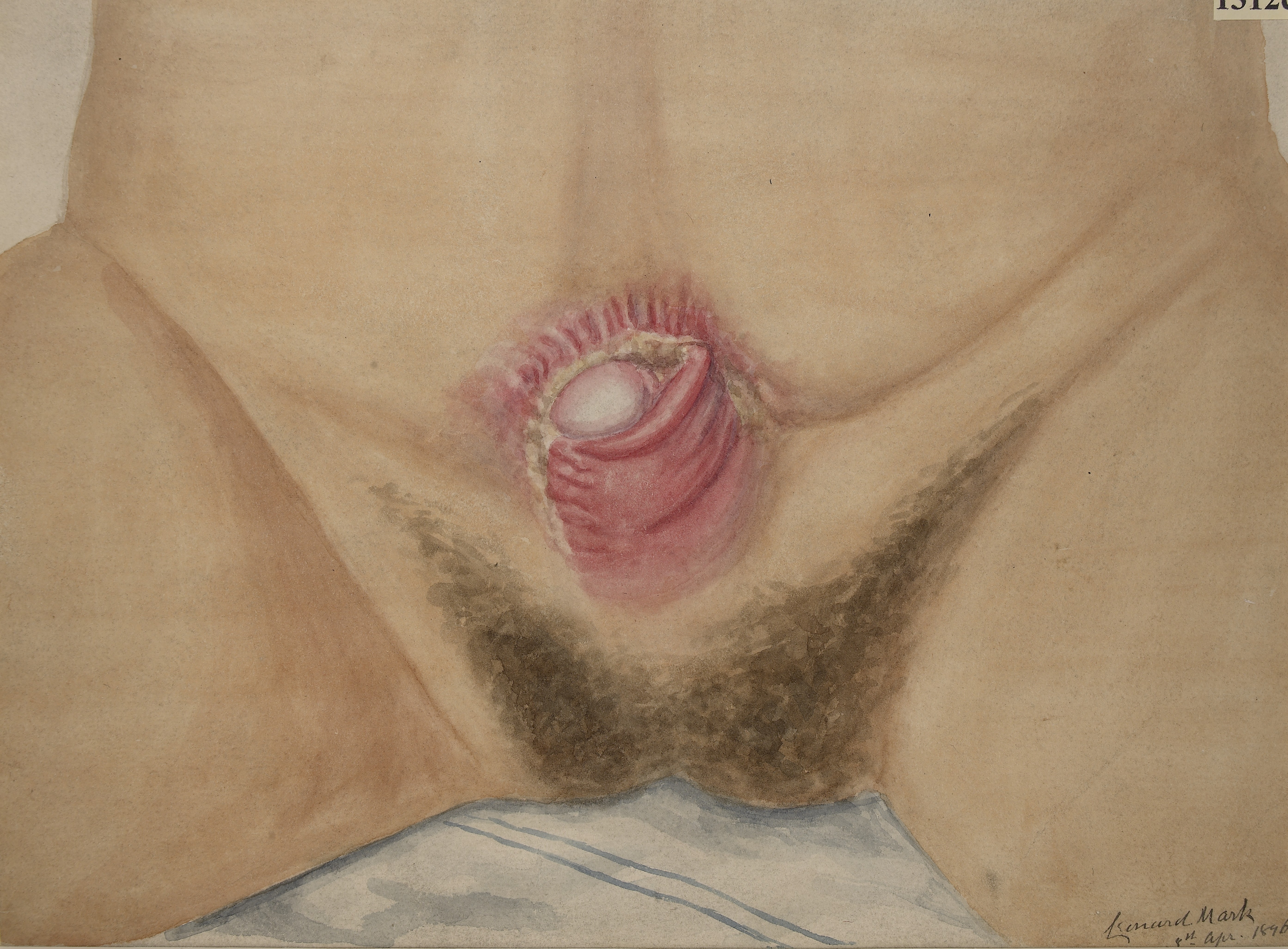
Cloacal Exstrophy
Cloacal exstrophy (EC) is a severe birth defect wherein much of the abdominal organs (the bladder and intestines) are exposed. It often causes the splitting of the bladder, genitalia, and the anus. It is sometimes called OEIS complex. Diagnostic tests can include ultrasound, voiding cystourethrogram (VCUG), intravenous pyelogram (IVP), nuclear renogram, computerized axial tomography (CT scan), and magnetic resonance imaging (MRI). Cloacal exstrophy is a rare birth defect, present in 1/200,000 pregnancies and 1/400,000 live births. It is associated with a defect of the ventral body wall and can be caused by inhibited mesodermal migration. The defect can often be comorbid with spinal bifida Spina bifida (Latin for 'split spine'; SB) is a birth defect in which there is incomplete closing of the spine and the membranes around the spinal cord during early development in pregnancy. There are three main types: spina bifida occulta, ... and kidney abnormalities. See also * Bl ... [...More Info...] [...Related Items...] OR: [Wikipedia] [Google] [Baidu] |
Omphalocele
Omphalocele or omphalocoele also called exomphalos, is a rare abdominal wall defect. Beginning at the 6th week of development, rapid elongation of the gut and increased liver size reduces intra abdominal space, which pushes intestinal loops out of the abdominal cavity. Around 10th week, the intestine returns to the abdominal cavity and the process is completed by the 12th week. Persistence of intestine or the presence of other abdominal viscera (e.g. stomach, liver) in the umbilical cord results in an omphalocele. Omphalocele occurs in 1 in 4,000 births and is associated with a high rate of mortality (25%) and severe malformations, such as cardiac anomalies (50%), neural tube defect (40%), exstrophy of the bladder and Beckwith–Wiedemann syndrome. Approximately 15% of live-born infants with omphalocele have chromosomal abnormalities. About 30% of infants with an omphalocele have other congenital abnormalities. Signs and symptoms The sac, which is formed from an outpouching of ... [...More Info...] [...Related Items...] OR: [Wikipedia] [Google] [Baidu] |
CT Scan
A computed tomography scan (CT scan; formerly called computed axial tomography scan or CAT scan) is a medical imaging technique used to obtain detailed internal images of the body. The personnel that perform CT scans are called radiographers or radiology technologists. CT scanners use a rotating X-ray tube and a row of detectors placed in a gantry to measure X-ray attenuations by different tissues inside the body. The multiple X-ray measurements taken from different angles are then processed on a computer using tomographic reconstruction algorithms to produce tomographic (cross-sectional) images (virtual "slices") of a body. CT scans can be used in patients with metallic implants or pacemakers, for whom magnetic resonance imaging (MRI) is contraindicated. Since its development in the 1970s, CT scanning has proven to be a versatile imaging technique. While CT is most prominently used in medical diagnosis, it can also be used to form images of non-living objects. The 1979 N ... [...More Info...] [...Related Items...] OR: [Wikipedia] [Google] [Baidu] |
Congenital Disorders Of Urinary System
A birth defect, also known as a congenital disorder, is an abnormal condition that is present at birth regardless of its cause. Birth defects may result in disabilities that may be physical, intellectual, or developmental. The disabilities can range from mild to severe. Birth defects are divided into two main types: structural disorders in which problems are seen with the shape of a body part and functional disorders in which problems exist with how a body part works. Functional disorders include metabolic and degenerative disorders. Some birth defects include both structural and functional disorders. Birth defects may result from genetic or chromosomal disorders, exposure to certain medications or chemicals, or certain infections during pregnancy. Risk factors include folate deficiency, drinking alcohol or smoking during pregnancy, poorly controlled diabetes, and a mother over the age of 35 years old. Many are believed to involve multiple factors. Birth defects may be visib ... [...More Info...] [...Related Items...] OR: [Wikipedia] [Google] [Baidu] |
Bladder Exstrophy
Bladder exstrophy is a congenital anomaly that exists along the spectrum of the exstrophy-epispadias complex, and most notably involves protrusion of the urinary bladder through a defect in the abdominal wall. Its presentation is variable, often including abnormalities of the bony pelvis, pelvic floor, and genitalia. The underlying embryologic mechanism leading to bladder exstrophy is unknown, though it is thought to be in part due to failed reinforcement of the cloacal membrane by underlying mesoderm. Exstrophy means the inversion of a hollow organ. Signs and symptoms The classic manifestation of bladder exstrophy presents with: * A defect in the abdominal wall occupied by both the exstrophied bladder as well as a portion of the urethra * A flattened puborectal sling * Separation of the pubic symphysis * Shortening of a pubic rami * External rotation of the pelvis. Females frequently have a displaced and narrowed vaginal orifice, a bifid clitoris, and divergent labia. Cau ... [...More Info...] [...Related Items...] OR: [Wikipedia] [Google] [Baidu] |
Spinal Bifida
Spina bifida (Latin for 'split spine'; SB) is a birth defect in which there is incomplete closing of the spine and the membranes around the spinal cord during early development in pregnancy. There are three main types: spina bifida occulta, meningocele and myelomeningocele. Meningocele and myelomeningocele may be grouped as spina bifida cystica. The most common location is the lower back, but in rare cases it may be in the middle back or neck. Occulta has no or only mild signs, which may include a hairy patch, dimple, dark spot or swelling on the back at the site of the gap in the spine. Meningocele typically causes mild problems, with a sac of fluid present at the gap in the spine. Myelomeningocele, also known as open spina bifida, is the most severe form. Problems associated with this form include poor ability to walk, impaired bladder or bowel control, accumulation of fluid in the brain (hydrocephalus), a tethered spinal cord and latex allergy. Learning problems are r ... [...More Info...] [...Related Items...] OR: [Wikipedia] [Google] [Baidu] |
Mesoderm
The mesoderm is the middle layer of the three germ layers that develops during gastrulation in the very early development of the embryo of most animals. The outer layer is the ectoderm, and the inner layer is the endoderm.Langman's Medical Embryology, 11th edition. 2010. The mesoderm forms mesenchyme, mesothelium, non-epithelial blood cells and coelomocytes. Mesothelium lines coeloms. Mesoderm forms the muscles in a process known as myogenesis, septa (cross-wise partitions) and mesenteries (length-wise partitions); and forms part of the gonads (the rest being the gametes). Myogenesis is specifically a function of mesenchyme. The mesoderm differentiates from the rest of the embryo through intercellular signaling, after which the mesoderm is polarized by an organizing center. The position of the organizing center is in turn determined by the regions in which beta-catenin is protected from degradation by GSK-3. Beta-catenin acts as a co-factor that alters the activity of ... [...More Info...] [...Related Items...] OR: [Wikipedia] [Google] [Baidu] |
Ventral
Standard anatomical terms of location are used to unambiguously describe the anatomy of animals, including humans. The terms, typically derived from Latin or Greek roots, describe something in its standard anatomical position. This position provides a definition of what is at the front ("anterior"), behind ("posterior") and so on. As part of defining and describing terms, the body is described through the use of anatomical planes and anatomical axes. The meaning of terms that are used can change depending on whether an organism is bipedal or quadrupedal. Additionally, for some animals such as invertebrates, some terms may not have any meaning at all; for example, an animal that is radially symmetrical will have no anterior surface, but can still have a description that a part is close to the middle ("proximal") or further from the middle ("distal"). International organisations have determined vocabularies that are often used as standard vocabularies for subdisciplines of anatomy ... [...More Info...] [...Related Items...] OR: [Wikipedia] [Google] [Baidu] |
Magnetic Resonance Imaging
Magnetic resonance imaging (MRI) is a medical imaging technique used in radiology to form pictures of the anatomy and the physiological processes inside the body. MRI scanners use strong magnetic fields, magnetic field gradients, and radio waves to generate images of the organs in the body. MRI does not involve X-rays or the use of ionizing radiation, which distinguishes it from computed tomography (CT) and positron emission tomography (PET) scans. MRI is a medical application of nuclear magnetic resonance (NMR) which can also be used for imaging in other NMR applications, such as NMR spectroscopy. MRI is widely used in hospitals and clinics for medical diagnosis, staging and follow-up of disease. Compared to CT, MRI provides better contrast in images of soft tissues, e.g. in the brain or abdomen. However, it may be perceived as less comfortable by patients, due to the usually longer and louder measurements with the subject in a long, confining tube, although "open ... [...More Info...] [...Related Items...] OR: [Wikipedia] [Google] [Baidu] |
Intravenous Pyelogram
Pyelogram (or pyelography or urography) is a form of imaging of the renal pelvis and ureter. Types include: * Intravenous pyelogram – In which a contrast solution is introduced through a vein into the circulatory system. * Retrograde pyelogram – Any pyelogram in which contrast medium is introduced from the lower urinary tract and flows toward the kidney (i.e. in a "retrograde" direction, against the normal flow of urine). * Anterograde pyelogram (also antegrade pyelogram) – A pyelogram where a contrast medium passes from the kidneys toward the bladder, mimicking the normal flow of urine. * Gas pyelogram – A pyelogram that uses a gaseous rather than liquid contrast medium. It may also form without the injection of a gas, when gas producing micro-organisms infect the most upper parts of urinary system. Intravenous pyelogram An intravenous pyelogram (IVP), also called an intravenous urogram (IVU), is a radiological procedure used to visualize abnormalities of the urinary s ... [...More Info...] [...Related Items...] OR: [Wikipedia] [Google] [Baidu] |
Imperforate Anus
An imperforate anus or anorectal malformations (ARMs) are birth defects in which the rectum is malformed. ARMs are a spectrum of different congenital anomalies which vary from fairly minor lesions to complex anomalies. The cause of ARMs is unknown; the genetic basis of these anomalies is very complex because of their anatomical variability. In 8% of patients, genetic factors are clearly associated with ARMs. Anorectal malformation in Currarino syndrome represents the only association for which the gene '' HLXB9'' has been identified. Types There are other forms of anorectal malformations though imperforate anus is most common. Other variants include anterior ectopic anus. This form is more commonly seen in females and presents with constipation. Presentation There are several forms of imperforate anus and anorectal malformations. The new classification is in relation of the type of associated fistula. The classical Wingspread classification was in low and high anomalies: * A ... [...More Info...] [...Related Items...] OR: [Wikipedia] [Google] [Baidu] |
Voiding Cystourethrography
In urology, voiding cystourethrography (VCUG) is a frequently performed technique for visualizing a person's urethra and urinary bladder while the person urinates (voids). It is used in the diagnosis of vesicoureteral reflux (kidney reflux), among other disorders. The technique consists of catheterizing the person in order to fill the bladder with a radiocontrast agent, typically diatrizoic acid. Under fluoroscopy (real time x-rays) the radiologist watches the contrast enter the bladder and looks at the anatomy of the patient. If the contrast moves into the ureters and back into the kidneys, the radiologist makes the diagnosis of vesicoureteral reflux, and gives the degree of severity a score. The exam ends when the person voids while the radiologist is watching under fluoroscopy. Consumption of fluid promotes excretion of contrast media after the procedure. It is important to watch the contrast during voiding, because this is when the bladder has the most pressure, and it is most ... [...More Info...] [...Related Items...] OR: [Wikipedia] [Google] [Baidu] |
Anus
The anus (Latin, 'ring' or 'circle') is an opening at the opposite end of an animal's digestive tract from the mouth. Its function is to control the expulsion of feces, the residual semi-solid waste that remains after food digestion, which, depending on the type of animal, includes: matter which the animal cannot digest, such as bones; Summary at food material after the nutrients have been extracted, for example cellulose or lignin; ingested matter which would be toxic if it remained in the digestive tract; and dead or excess gut bacteria and other endosymbionts. Amphibians, reptiles, and birds use the same orifice (known as the cloaca) for excreting liquid and solid wastes, for copulation and egg-laying. Monotreme mammals also have a cloaca, which is thought to be a feature inherited from the earliest amniotes via the therapsids. Marsupials have a single orifice for excreting both solids and liquids and, in females, a separate vagina for reproduction. Female placenta ... [...More Info...] [...Related Items...] OR: [Wikipedia] [Google] [Baidu] |


