|
Clavipectoral Triangle
The clavipectoral triangle (also known as the deltopectoral triangle) is an anatomical region found in humans and other animals. It is bordered by the following structures: * Clavicle (superiorly) * Lateral border of Pectoralis Major (medially) * Medial border of Deltoid muscle (laterally) It contains the cephalic vein, and deltopectoral fascia, which is a layer of deep fascia that invests the three structures that make up the border of the triangle. The deltoid branch of the thoracoacromial artery also passes through this triangle, giving branches to both the deltoid and pectoralis major muscles. The subclavian vein and the subclavian artery may be accessed via this triangle, as they are deep to it. Clinical significance * Palpation of coracoid process of scapula The coracoid process of the scapula is not subcutaneous; It is covered by the anterior border of the deltoid. However, the tip of the coracoid process can be felt on deep palpation on the lateral aspect of the ... [...More Info...] [...Related Items...] OR: [Wikipedia] [Google] [Baidu] |
Superficial Vein
Superficial veins are veins that are close to the surface of the body, as opposed to deep veins, which are far from the surface. Superficial veins are not paired with an artery, unlike the deep veins, which are typically associated with an artery of the same name. Superficial veins are important physiologically for cooling of the body. When the body is too hot, the body shunts blood from the deep veins to the superficial veins to facilitate heat transfer to the body's surroundings. Superficial veins are often visible underneath the skin. Those below the level of the heart tend to bulge out, which can be readily witnessed in the hand, where the veins bulge significantly less after the arm has been raised above the head for a short time. Veins become more visually prominent when lifting heavy weight, especially after a period of proper strength training. Physiologically, the superficial veins are not as important as the deep veins (as they carry less blood) and are sometimes r ... [...More Info...] [...Related Items...] OR: [Wikipedia] [Google] [Baidu] |
Pectoralis Major
The pectoralis major () is a thick, fan-shaped or triangular convergent muscle, situated at the chest of the human body. It makes up the bulk of the chest muscles and lies under the breast. Beneath the pectoralis major is the pectoralis minor, a thin, triangular muscle. The pectoralis major's primary functions are flexion, adduction, and internal rotation of the humerus. The pectoral major may colloquially be referred to as "pecs", "pectoral muscle", or "chest muscle", because it is the largest and most superficial muscle in the chest area. Structure It arises from the anterior surface of the sternal half of the clavicle from breadth of the half of the anterior surface of the sternum, as low down as the attachment of the cartilage of the sixth or seventh rib; from the cartilages of all the true ribs, with the exception, frequently, of the first or seventh, and from the aponeurosis of the abdominal external oblique muscle. From this extensive origin the fibers converge toward the ... [...More Info...] [...Related Items...] OR: [Wikipedia] [Google] [Baidu] |
Deltoid Muscle
The deltoid muscle is the muscle forming the rounded contour of the human shoulder. It is also known as the 'common shoulder muscle', particularly in other animals such as the domestic cat. Anatomically, the deltoid muscle appears to be made up of three distinct sets of muscle fibers, namely the # anterior or clavicular part (pars clavicularis) # posterior or scapular part (pars scapularis) # intermediate or acromial part (pars acromialis) However, electromyography suggests that it consists of at least seven groups that can be independently coordinated by the nervous system. It was previously called the deltoideus (plural ''deltoidei'') and the name is still used by some anatomists. It is called so because it is in the shape of the Greek capital letter delta (Δ). Deltoid is also further shortened in slang as "delt". A study of 30 shoulders revealed an average mass of in humans, ranging from to . Structure Previous studies showed that the insertions of the tendons of the delto ... [...More Info...] [...Related Items...] OR: [Wikipedia] [Google] [Baidu] |
Cephalic Vein
In human anatomy, the cephalic vein is a superficial vein in the arm. It originates from the radial end of the dorsal venous network of hand, and ascends along the radial (lateral) side of the arm before emptying into the axillary vein. At the elbow, it communicates with the basilic vein via the median cubital vein. Anatomy The cephalic vein is situated within the superficial fascia along the anterolateral surface of the biceps. Origin The cephalic vein forms over the anatomical snuffbox at the radial end of the dorsal venous network of hand. Course and relations From its origin, it ascends ascends up the lateral aspect of the radius. Near the shoulder, the cephalic vein passes between the deltoid and pectoralis major muscles (deltopectoral groove) and through the clavipectoral triangle, where it empties into the axillary vein. Anastomoses It communicates with the basilic vein via the median cubital vein at the elbow. Clinical significance The cephalic vein ... [...More Info...] [...Related Items...] OR: [Wikipedia] [Google] [Baidu] |
Dartmouth Medical School
The Geisel School of Medicine at Dartmouth is the graduate medical school of Dartmouth College in Hanover, New Hampshire. The fourth oldest medical school in the United States, it was founded in 1797 by New England physician Nathan Smith. It is one of the seven Ivy League medical schools. Several milestones in medical care and research have taken place at Dartmouth, including the introduction of stethoscopes to U.S. medical education (1838), the first clinical x-ray (1896), and the first intensive care unit (ICU) in the United States (1955). The Geisel School of Medicine grants the Doctor of Medicine (MD) and Doctor of Philosophy (PhD) degrees. The school has a student body of approximately 700 students and more than 2,300 faculty and researchers. Geisel organizes research through over a dozen research centers and institutes, attracting more than $140 million in grants annually, and is ranked as a top medical school by '' U.S. News & World Report'' for both primary care and biom ... [...More Info...] [...Related Items...] OR: [Wikipedia] [Google] [Baidu] |
Thoracoacromial Artery
The thoracoacromial artery (acromiothoracic artery; thoracic axis) is a short trunk that arises from the second part of the axillary artery, its origin being generally overlapped by the upper edge of the pectoralis minor. Structure Projecting forward to the upper border of the Pectoralis minor, it pierces the coracoclavicular fascia The clavipectoral fascia (costocoracoid membrane; coracoclavicular fascia) is a strong fascia situated under cover of the clavicular portion of the pectoralis major. It occupies the interval between the pectoralis minor and subclavius, and pro ... and divides into four branches—pectoral, acromial, clavicular, and deltoid. Additional images File:Gray523.png, The axillary artery and its branches. References External links * * * - "Pectoral Region: Thoracoacromial Artery and its Branches" * - "The axillary artery and its major branches shown in relation to major landmarks." {{Authority control Arteries of the upper limb ... [...More Info...] [...Related Items...] OR: [Wikipedia] [Google] [Baidu] |
Subclavian Vein
The subclavian vein is a paired large vein, one on either side of the body, that is responsible for draining blood from the upper extremities, allowing this blood to return to the heart. The left subclavian vein plays a key role in the absorption of lipids, by allowing products that have been carried by lymph in the thoracic duct to enter the bloodstream. The diameter of the subclavian veins is approximately 1–2 cm, depending on the individual. Structure Each subclavian vein is a continuation of the axillary vein and runs from the outer border of the first rib to the medial border of anterior scalene muscle. From here it joins with the internal jugular vein to form the brachiocephalic vein (also known as "innominate vein"). The angle of union is termed the venous angle. The subclavian vein follows the subclavian artery and is separated from the subclavian artery by the insertion of anterior scalene. Thus, the subclavian vein lies anterior to the anterior scalene while the su ... [...More Info...] [...Related Items...] OR: [Wikipedia] [Google] [Baidu] |
Subclavian Artery
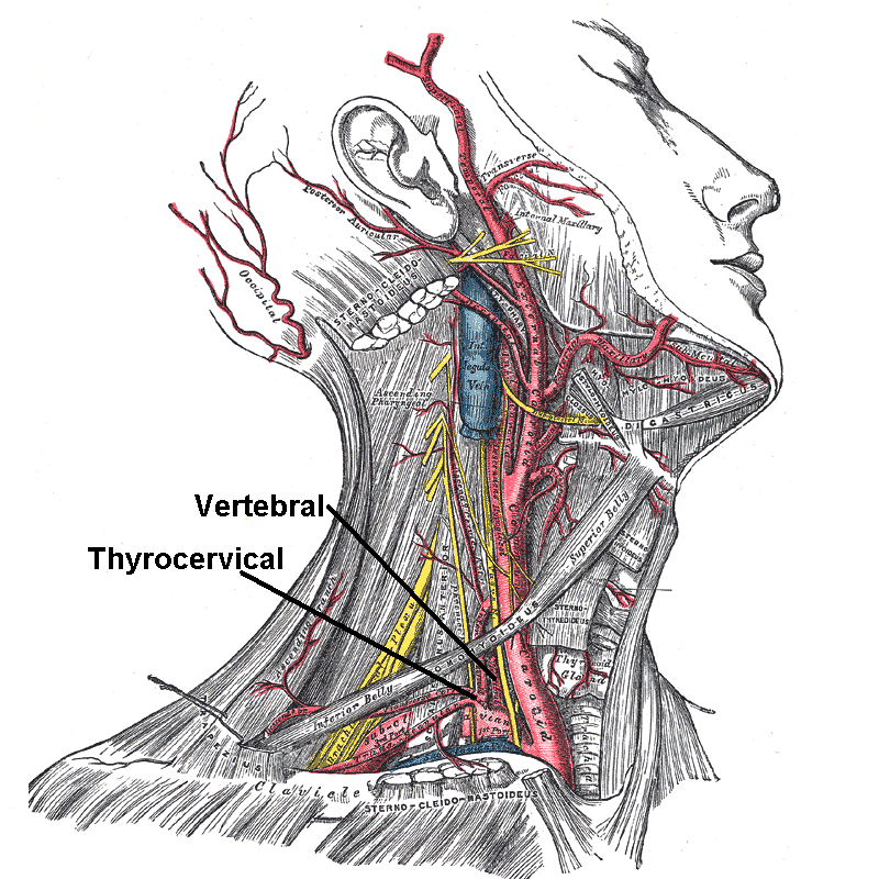
In human anatomy, the subclavian arteries are paired major arteries of the upper thorax, below the clavicle. They receive blood from the aortic arch. The left subclavian artery supplies blood to the left arm and the right subclavian artery supplies blood to the right arm, with some branches supplying the head and thorax. On the left side of the body, the subclavian comes directly off the aortic arch, while on the right side it arises from the relatively short brachiocephalic artery when it bifurcates into the subclavian and the right common carotid artery. The usual branches of the subclavian on both sides of the body are the vertebral artery, the internal thoracic artery, the thyrocervical trunk, the costocervical trunk and the dorsal scapular artery, which may branch off the transverse cervical artery, which is a branch of the thyrocervical trunk. The subclavian becomes the axillary artery at the lateral border of the first rib. Structure From its origin, the subclavian artery t ... [...More Info...] [...Related Items...] OR: [Wikipedia] [Google] [Baidu] |
Deltopectoral Groove
The deltopectoral groove is an indentation in the muscular structure between the deltoid muscle and pectoralis major. It is the location through which the cephalic vein passes and where the coracoid process is most easily palpable. See also * Deltopectoral triangle The clavipectoral triangle (also known as the deltopectoral triangle) is an anatomical region found in humans and other animals. It is bordered by the following structures: * Clavicle (superiorly) * Lateral border of Pectoralis Major (medially ... Additional images File:Gray410.png, Superficial muscles of the chest and front of the arm. File:Gray574.png, Superficial veins of the upper limb References External links Diagram at anatomyatlases.org(see #91) * http://ect.downstate.edu/courseware/haonline/labs/L06/020200.htm Upper limb anatomy {{musculoskeletal-stub ... [...More Info...] [...Related Items...] OR: [Wikipedia] [Google] [Baidu] |


