|
Ciliopathies
A ciliopathy is any genetic disorder that affects the cellular cilia or the cilia anchoring structures, the basal bodies, or ciliary function. Primary cilia are important in guiding the process of development, so abnormal ciliary function while an embryo is developing can lead to a set of malformations that can occur regardless of the particular genetic problem. The similarity of the clinical features of these developmental disorders means that they form a recognizable cluster of syndromes, loosely attributed to abnormal ciliary function and hence called ciliopathies. Regardless of the actual genetic cause, it is clustering of a set of characteristic physiological features which define whether a syndrome is a ciliopathy. Although ciliopathies are usually considered to involve proteins that localize to motile and/or immotile (primary) cilia or centrosomes, it is possible for ciliopathies to be associated with unexpected proteins such as XPNPEP3, which localizes to mitochondria but ... [...More Info...] [...Related Items...] OR: [Wikipedia] [Google] [Baidu] |
Cilium
The cilium, plural cilia (), is a membrane-bound organelle found on most types of eukaryotic cell, and certain microorganisms known as ciliates. Cilia are absent in bacteria and archaea. The cilium has the shape of a slender threadlike projection that extends from the surface of the much larger cell body. Eukaryotic flagella found on sperm cells and many protozoans have a similar structure to motile cilia that enables swimming through liquids; they are longer than cilia and have a different undulating motion. There are two major classes of cilia: ''motile'' and ''non-motile'' cilia, each with a subtype, giving four types in all. A cell will typically have one primary cilium or many motile cilia. The structure of the cilium core called the axoneme determines the cilium class. Most motile cilia have a central pair of single microtubules surrounded by nine pairs of double microtubules called a 9+2 axoneme. Most non-motile cilia have a 9+0 axoneme that lacks the central pair o ... [...More Info...] [...Related Items...] OR: [Wikipedia] [Google] [Baidu] |
Cilia
The cilium, plural cilia (), is a membrane-bound organelle found on most types of eukaryotic cell, and certain microorganisms known as ciliates. Cilia are absent in bacteria and archaea. The cilium has the shape of a slender threadlike projection that extends from the surface of the much larger cell body. Eukaryotic flagella found on sperm cells and many protozoans have a similar structure to motile cilia that enables swimming through liquids; they are longer than cilia and have a different undulating motion. There are two major classes of cilia: ''motile'' and ''non-motile'' cilia, each with a subtype, giving four types in all. A cell will typically have one primary cilium or many motile cilia. The structure of the cilium core called the axoneme determines the cilium class. Most motile cilia have a central pair of single microtubules surrounded by nine pairs of double microtubules called a 9+2 axoneme. Most non-motile cilia have a 9+0 axoneme that lacks the central pair of mi ... [...More Info...] [...Related Items...] OR: [Wikipedia] [Google] [Baidu] |
XPNPEP3
Xaa-Pro aminopeptidase 3, also known as aminopeptidase P3, is an enzyme that in humans is encoded by the ''XPNPEP3'' gene. XPNPEP3 localizes to mitochondria in renal cells and to kidney tubules in a cell type-specific pattern. Mutations in ''XPNPEP3'' gene have been identified as a cause of a nephronophthisis-like disease. Structure Gene The ''XPNPEP3'' gene is located at chromosome 22q13.2, consisting of 12 exons. Two splice variants of XPNPEP3, APP3m and APP3c, exist in mitochondria and cytosol, respectively. Protein APP3m has an N-terminal mitochondrial-targeting sequence (MTS) domain importing APP3m into mitochondria, where the domain is removed proteolytically and APP3m functions as a 51-kDa mature protein. By contrast, APP3c, lacks the MTS and is expressed in the cytosol. Arginine in MTS is required for mitochondrial transport. Function XPNPEP3 belongs to a family of X-pro-aminopeptidases (EC 3.4.11.9) that utilize a metal cofactor and remove the N-terminal amino acid f ... [...More Info...] [...Related Items...] OR: [Wikipedia] [Google] [Baidu] |
Anencephaly
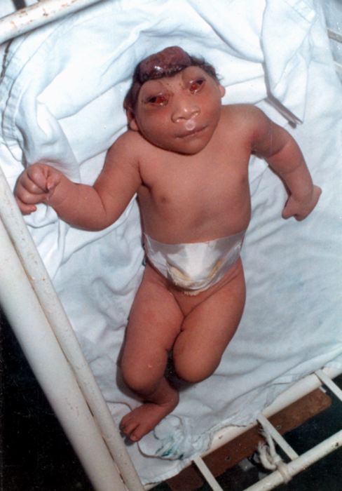
Anencephaly is the absence of a major portion of the brain, skull, and scalp that occurs during embryonic development. It is a cephalic disorder that results from a neural tube defect that occurs when the rostral (head) end of the neural tube fails to close, usually between the 23rd and 26th day following conception. Strictly speaking, the Greek term translates as "without a brain" (or totally lacking the inside part of the head), but it is accepted that children born with this disorder usually only lack a telencephalon, the largest part of the brain consisting mainly of the cerebral hemispheres, including the neocortex, which is responsible for cognition. The remaining structure is usually covered only by a thin layer of membrane—skin, bone, meninges, etc., are all lacking. With very few exceptions, infants with this disorder do not survive longer than a few hours or days after birth. Signs and symptoms The National Institute of Neurological Disorders and Stroke (NINDS) descr ... [...More Info...] [...Related Items...] OR: [Wikipedia] [Google] [Baidu] |
Agenesis Of The Corpus Callosum
Agenesis of the corpus callosum (ACC) is a rare birth defect in which there is a complete or partial absence of the corpus callosum. It occurs when the development of the corpus callosum, the band of white matter connecting the two hemispheres in the brain, in the embryo is disrupted. The result of this is that the fibers that would otherwise form the corpus callosum are instead longitudinally oriented along the ipsilateral ventricular wall and form structures called Probst bundles. In addition to agenesis, other degrees of callosal defects exist, including hypoplasia (underdevelopment or thinness), hypogenesis (partial agenesis) or dysgenesis (malformation). ACC is found in many syndromes and can often present alongside hypoplasia of the cerebellar vermis. When this is the case, there can also be an enlarged fourth ventricle or hydrocephalus; this is called Dandy–Walker malformation. Signs and symptoms Signs and symptoms of ACC and other callosal disorders vary greatly ... [...More Info...] [...Related Items...] OR: [Wikipedia] [Google] [Baidu] |
Genetic Disorder
A genetic disorder is a health problem caused by one or more abnormalities in the genome. It can be caused by a mutation in a single gene (monogenic) or multiple genes (polygenic) or by a chromosomal abnormality. Although polygenic disorders are the most common, the term is mostly used when discussing disorders with a single genetic cause, either in a gene or chromosome. The mutation responsible can occur spontaneously before embryonic development (a ''de novo'' mutation), or it can be Heredity, inherited from two parents who are carriers of a faulty gene (autosomal recessive inheritance) or from a parent with the disorder (autosomal dominant inheritance). When the genetic disorder is inherited from one or both parents, it is also classified as a hereditary disease. Some disorders are caused by a mutation on the X chromosome and have X-linked inheritance. Very few disorders are inherited on the Y linkage, Y chromosome or Mitochondrial disease#Causes, mitochondrial DNA (due to t ... [...More Info...] [...Related Items...] OR: [Wikipedia] [Google] [Baidu] |
Exencephaly
Exencephaly is a type of cephalic disorder wherein the brain is located outside of the skull. This condition is usually found in embryos as an early stage of anencephaly. As an exencephalic pregnancy progresses, the neural tissue gradually degenerates. The prognosis for infants born with exencephaly is extremely poor. It is rare to find an infant born with exencephaly, as most cases that are not early stages of anencephaly are usually stillborn. Those infants who are born with the condition usually die within hours or minutes. Caused by the failure of cranial neuropore to properly fuse, between the 3rd and 4th week post conception. The calvarium doesn't develop/fuse properly, therefore the brain extrudes from the cranium. Pathophysiology Until recently, the medical literature did not indicate a connection among many genetic disorders, both genetic syndromes and genetic diseases, that are now being found to be related. As a result of new genetic research, some of these are, in fact, ... [...More Info...] [...Related Items...] OR: [Wikipedia] [Google] [Baidu] |
Cerebellar Vermis
The cerebellar vermis (from Latin ''vermis,'' "worm") is located in the medial, cortico-nuclear zone of the cerebellum, which is in the posterior fossa of the cranium. The primary fissure in the vermis curves ventrolaterally to the superior surface of the cerebellum, dividing it into anterior and posterior lobes. Functionally, the vermis is associated with bodily posture and locomotion. The vermis is included within the spinocerebellum and receives somatic sensory input from the head and proximal body parts via ascending spinal pathways. The cerebellum develops in a rostro-caudal manner, with rostral regions in the midline giving rise to the vermis, and caudal regions developing into the cerebellar hemispheres. By 4 months of prenatal development, the vermis becomes fully foliated, while development of the hemispheres lags by 30–60 days. Postnatally, proliferation and organization of the cellular components of the cerebellum continues, with completion of the f ... [...More Info...] [...Related Items...] OR: [Wikipedia] [Google] [Baidu] |
Caroli Disease
Caroli disease (communicating cavernous ectasia, or congenital cystic dilatation of the intrahepatic biliary tree) is a rare inherited disorder characterized by cystic dilatation (or ectasia) of the bile ducts within the liver. There are two patterns of Caroli disease: focal or simple Caroli disease consists of abnormally widened bile ducts affecting an isolated portion of liver. The second form is more diffuse, and when associated with portal hypertension and congenital hepatic fibrosis, is often referred to as "Caroli syndrome". The underlying differences between the two types are not well understood. Caroli disease is also associated with liver failure and polycystic kidney disease. The disease affects about one in 1,000,000 people, with more reported cases of Caroli syndrome than of Caroli disease. Caroli disease is distinct from other diseases that cause ductal dilatation caused by obstruction, in that it is not one of the many choledochal cyst derivatives. Signs and symptom ... [...More Info...] [...Related Items...] OR: [Wikipedia] [Google] [Baidu] |
Cirrhosis
Cirrhosis, also known as liver cirrhosis or hepatic cirrhosis, and end-stage liver disease, is the impaired liver function caused by the formation of scar tissue known as fibrosis due to damage caused by liver disease. Damage causes tissue repair and subsequent formation of scar tissue, which over time can replace normal functioning tissue, leading to the impaired liver function of cirrhosis. The disease typically develops slowly over months or years. Early symptoms may include tiredness, weakness, loss of appetite, unexplained weight loss, nausea and vomiting, and discomfort in the right upper quadrant of the abdomen. As the disease worsens, symptoms may include itchiness, swelling in the lower legs, fluid build-up in the abdomen, jaundice, bruising easily, and the development of spider-like blood vessels in the skin. The fluid build-up in the abdomen may become spontaneously infected. More serious complications include hepatic encephalopathy, bleeding from dilated veins ... [...More Info...] [...Related Items...] OR: [Wikipedia] [Google] [Baidu] |
Hydrocephalus
Hydrocephalus is a condition in which an accumulation of cerebrospinal fluid (CSF) occurs within the brain. This typically causes increased intracranial pressure, pressure inside the skull. Older people may have headaches, double vision, poor balance, urinary incontinence, personality changes, or mental impairment. In babies, it may be seen as a rapid increase in head size. Other symptoms may include vomiting, sleepiness, seizures, and Parinaud's syndrome, downward pointing of the eyes. Hydrocephalus can occur due to birth defects or be acquired later in life. Associated birth defects include neural tube defects and those that result in aqueductal stenosis. Other causes include meningitis, brain tumors, traumatic brain injury, intraventricular hemorrhage, and subarachnoid hemorrhage. The four types of hydrocephalus are communicating, noncommunicating, ''ex vacuo'', and normal pressure hydrocephalus, normal pressure. Diagnosis is typically made by physical examination and medic ... [...More Info...] [...Related Items...] OR: [Wikipedia] [Google] [Baidu] |


