|
Choroid Diseases
The choroid, also known as the choroidea or choroid coat, is a part of the uvea, the vascular layer of the eye, and contains connective tissues, and lies between the retina and the sclera. The human choroid is thickest at the far extreme rear of the eye (at 0.2 mm), while in the outlying areas it narrows to 0.1 mm. The choroid provides oxygen and nourishment to the outer layers of the retina. Along with the ciliary body and iris, the choroid forms the uveal tract. The structure of the choroid is generally divided into four layers (classified in order of furthest away from the retina to closest): *Haller's layer - outermost layer of the choroid consisting of larger diameter blood vessels; * Sattler's layer - layer of medium diameter blood vessels; *Choriocapillaris - layer of capillaries; and * Bruch's membrane (synonyms: Lamina basalis, Complexus basalis, Lamina vitra) - innermost layer of the choroid. Blood supply There are two circulations of the eye: the ... [...More Info...] [...Related Items...] OR: [Wikipedia] [Google] [Baidu] |
Human Eye
The human eye is a sensory organ, part of the sensory nervous system, that reacts to visible light and allows humans to use visual information for various purposes including seeing things, keeping balance, and maintaining circadian rhythm. The eye can be considered as a living optical device. It is approximately spherical in shape, with its outer layers, such as the outermost, white part of the eye (the sclera) and one of its inner layers (the pigmented choroid) keeping the eye essentially light tight except on the eye's optic axis. In order, along the optic axis, the optical components consist of a first lens (the cornea—the clear part of the eye) that accomplishes most of the focussing of light from the outside world; then an aperture (the pupil) in a diaphragm (the iris—the coloured part of the eye) that controls the amount of light entering the interior of the eye; then another lens (the crystalline lens) that accomplishes the remaining focussing of light into ... [...More Info...] [...Related Items...] OR: [Wikipedia] [Google] [Baidu] |
Internal Carotid Artery
The internal carotid artery (Latin: arteria carotis interna) is an artery in the neck which supplies the anterior circulation of the brain. In human anatomy, the internal and external carotids arise from the common carotid arteries, where these bifurcate at cervical vertebrae C3 or C4. The internal carotid artery supplies the brain, including the eyes, while the external carotid nourishes other portions of the head, such as the face, scalp, skull, and meninges. Classification Terminologia Anatomica in 1998 subdivided the artery into four parts: "cervical", "petrous", "cavernous", and "cerebral". However, in clinical settings, the classification system of the internal carotid artery usually follows the 1996 recommendations by Bouthillier, describing seven anatomical segments of the internal carotid artery, each with a corresponding alphanumeric identifier—C1 cervical, C2 petrous, C3 lacerum, C4 cavernous, C5 clinoid, C6 ophthalmic, and C7 communicating. The Bouthillier nomenclat ... [...More Info...] [...Related Items...] OR: [Wikipedia] [Google] [Baidu] |
Night Vision
Night vision is the ability to see in low-light conditions, either naturally with scotopic vision or through a night-vision device. Night vision requires both sufficient spectral range and sufficient intensity range. Humans have poor night vision compared to many animals such as cats, foxes and rabbits, in part because the human eye lacks a tapetum lucidum, tissue behind the retina that reflects light back through the retina thus increasing the light available to the photoreceptors. Types of ranges Spectral range Night-useful spectral range techniques can sense radiation that is invisible to a human observer. Human vision is confined to a small portion of the electromagnetic spectrum called visible light. Enhanced spectral range allows the viewer to take advantage of non-visible sources of electromagnetic radiation (such as near-infrared or ultraviolet radiation). Some animals such as the mantis shrimp and trout can see using much more of the infrared and/or ultraviolet sp ... [...More Info...] [...Related Items...] OR: [Wikipedia] [Google] [Baidu] |
Low Vision
Visual impairment, also known as vision impairment, is a medical definition primarily measured based on an individual's better eye visual acuity; in the absence of treatment such as correctable eyewear, assistive devices, and medical treatment– visual impairment may cause the individual difficulties with normal daily tasks including reading and walking. Low vision is a functional definition of visual impairment that is chronic, uncorrectable with treatment or correctable lenses, and impacts daily living. As such low vision can be used as a disability metric and varies based on an individual's experience, environmental demands, accommodations, and access to services. The American Academy of Ophthalmology defines visual impairment as the best-corrected visual acuity of less than 20/40 in the better eye, and the World Health Organization defines it as a presenting acuity of less than 6/12 in the better eye. The term blindness is used for complete or nearly complete vision loss. In ... [...More Info...] [...Related Items...] OR: [Wikipedia] [Google] [Baidu] |
Albino
Albinism is the congenital absence of melanin in an animal or plant resulting in white hair, feathers, scales and skin and pink or blue eyes. Individuals with the condition are referred to as albino. Varied use and interpretation of the terms mean that written reports of albinistic animals can be difficult to verify. Albinism can reduce the survivability of an animal; for example, it has been suggested that albino alligators have an average survival span of only 24 hours due to the lack of protection from UV radiation and their lack of camouflage to avoid predators. It is a common misconception that all albino animals have characteristic pink or red eyes (resulting from the lack of pigment in the iris allowing the blood vessels of the retina to be visible), however this is not the case for some forms of albinism. Familiar albino animals include in-bred strains of laboratory animals (rats, mice and rabbits), but populations of naturally occurring albino animals exist in the wil ... [...More Info...] [...Related Items...] OR: [Wikipedia] [Google] [Baidu] |
Primates
Primates are a diverse order of mammals. They are divided into the strepsirrhines, which include the lemurs, galagos, and lorisids, and the haplorhines, which include the tarsiers and the simians (monkeys and apes, the latter including humans). Primates arose 85–55 million years ago first from small terrestrial mammals, which adapted to living in the trees of tropical forests: many primate characteristics represent adaptations to life in this challenging environment, including large brains, visual acuity, color vision, a shoulder girdle allowing a large degree of movement in the shoulder joint, and dextrous hands. Primates range in size from Madame Berthe's mouse lemur, which weighs , to the eastern gorilla, weighing over . There are 376–524 species of living primates, depending on which classification is used. New primate species continue to be discovered: over 25 species were described in the 2000s, 36 in the 2010s, and three in the 2020s. Primates have large bra ... [...More Info...] [...Related Items...] OR: [Wikipedia] [Google] [Baidu] |
Melanin
Melanin (; from el, μέλας, melas, black, dark) is a broad term for a group of natural pigments found in most organisms. Eumelanin is produced through a multistage chemical process known as melanogenesis, where the oxidation of the amino acid tyrosine is followed by polymerization. The melanin pigments are produced in a specialized group of cells known as melanocytes. Functionally, eumelanin serves as protection against Ultraviolet, UV radiation. There are five basic types of melanin: eumelanin, pheomelanin, neuromelanin, allomelanin and pyomelanin. The most common type is eumelanin, of which there are two types— brown eumelanin and black eumelanin. Pheomelanin, which is produced when melanocytes are malfunctioning due to derivation of the gene to its recessive format is a cysteine-derivative that contains polybenzothiazine portions that are largely responsible for the of red yellow tint given to some skin or hair colors. Neuromelanin is found in the brain. Research ha ... [...More Info...] [...Related Items...] OR: [Wikipedia] [Google] [Baidu] |
Teleosts
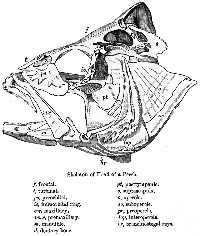
Teleostei (; Greek ''teleios'' "complete" + ''osteon'' "bone"), members of which are known as teleosts ), is, by far, the largest infraclass in the class Actinopterygii, the ray-finned fishes, containing 96% of all extant species of fish. Teleosts are arranged into about 40 orders and 448 families. Over 26,000 species have been described. Teleosts range from giant oarfish measuring or more, and ocean sunfish weighing over , to the minute male anglerfish ''Photocorynus spiniceps'', just long. Including not only torpedo-shaped fish built for speed, teleosts can be flattened vertically or horizontally, be elongated cylinders or take specialised shapes as in anglerfish and seahorses. The difference between teleosts and other bony fish lies mainly in their jaw bones; teleosts have a movable premaxilla and corresponding modifications in the jaw musculature which make it possible for them to protrude their jaws outwards from the mouth. This is of great advantage, enabling them to ... [...More Info...] [...Related Items...] OR: [Wikipedia] [Google] [Baidu] |
Laser Doppler Imaging
Laser Doppler imaging (LDI) is an imaging method that uses a laser beam to scan live tissue. When the laser light reaches the tissue, the moving blood cells generate doppler components in the reflected ( backscattered) light. The light that comes back is detected using a photodiode that converts it into an electrical signal. Then the signal is processed to calculate a signal that is proportional to the tissue perfusion in the scanned area. When the process is completed, the signal is processed to generate an image that shows the perfusion on a screen. The laser doppler effect was first used to measure microcirculation by Stern M.D. in 1975. And it is used widely in medicine, some representative research work about it are these: Use in Ophthalmology The eye offers a unique opportunity for the non-invasive exploration of cardiovascular diseases. LDI by digital holography can measure blood flow in the retina and choroid. In particular, the choroid is a highly vascularized tissue s ... [...More Info...] [...Related Items...] OR: [Wikipedia] [Google] [Baidu] |
Indocyanine Green Angiography
Indocyanine green angiography (ICGA) is a diagnostic procedure used to examine choroidal blood flow and associated pathology. Indocyanine green (ICG) is a water soluble cyanine dye which shows fluorescence in near-infrared (790–805 nm) range, with peak spectral absorption of 800-810 nm in blood. The near infrared light used in ICGA penetrates ocular pigments such as melanin and xanthophyll, as well as exudates and thin layers of sub-retinal vessels. Age-related macular degeneration is the third main cause of blindness worldwide, and it is the leading cause of blindness in industrialized countries. Indocyanine green angiography is widely used to study choroidal neovascularization in patients with exudative age-related macular degeneration. In nonexudative AMD, ICGA is used in classification of drusen and associated subretinal deposits. Indications Indications for indocyanine green angiography include: * Choroidal neovascularisation (CNV):Indocyanine green angiography is widely us ... [...More Info...] [...Related Items...] OR: [Wikipedia] [Google] [Baidu] |
Optical Coherence Tomography
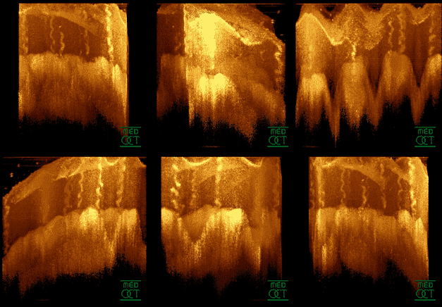
Optical coherence tomography (OCT) is an imaging technique that uses low-coherence light to capture micrometer-resolution, two- and three-dimensional images from within optical scattering media (e.g., biological tissue). It is used for medical imaging and industrial nondestructive testing (NDT). Optical coherence tomography is based on low-coherence interferometry, typically employing near-infrared light. The use of relatively long wavelength light allows it to penetrate into the scattering medium. Confocal microscopy, another optical technique, typically penetrates less deeply into the sample but with higher resolution. Depending on the properties of the light source ( superluminescent diodes, ultrashort pulsed lasers, and supercontinuum lasers have been employed), optical coherence tomography has achieved sub-micrometer resolution (with very wide-spectrum sources emitting over a ~100 nm wavelength range). Optical coherence tomography is one of a class of optical tom ... [...More Info...] [...Related Items...] OR: [Wikipedia] [Google] [Baidu] |


.jpg)


