|
Cervical Lymph Node
Cervical lymph nodes are lymph nodes found in the neck. Of the 800 lymph nodes in the human body, 300 are in the neck. Cervical lymph nodes are subject to a number of different pathological conditions including tumours, infection and inflammation. Classification There are approximately 300 lymph nodes in the neck, and they can be classified in a number of different ways. History The classification of the cervical lymph nodes is generally attributed to Henri Rouvière in his 1932 publication "Anatomie des Lymphatiques de l'Homme" Rouviere described the cervical lymph nodes as a collar which surrounded the upper aerodigestive tract, consisting of submental, facial, submandibular, parotid, mastoid, occipital and retropharyngeal nodes, together with two chains that run in the long axis of the neck, the anterior cervical and postero-lateral cervical groups. However, this system was based upon anatomical landmarks found in dissection, making it imperfectly suited to the need ... [...More Info...] [...Related Items...] OR: [Wikipedia] [Google] [Baidu] |
Cervix
The cervix or cervix uteri (Latin, 'neck of the uterus') is the lower part of the uterus (womb) in the human female reproductive system. The cervix is usually 2 to 3 cm long (~1 inch) and roughly cylindrical in shape, which changes during pregnancy. The narrow, central cervical canal runs along its entire length, connecting the uterine cavity and the lumen of the vagina. The opening into the uterus is called the internal os, and the opening into the vagina is called the external os. The lower part of the cervix, known as the vaginal portion of the cervix (or ectocervix), bulges into the top of the vagina. The cervix has been documented anatomically since at least the time of Hippocrates, over 2,000 years ago. The cervical canal is a passage through which sperm must travel to fertilize an egg cell after sexual intercourse. Several methods of contraception, including cervical caps and cervical diaphragms, aim to block or prevent the passage of sperm through the cervical ... [...More Info...] [...Related Items...] OR: [Wikipedia] [Google] [Baidu] |
Submandibular Triangle
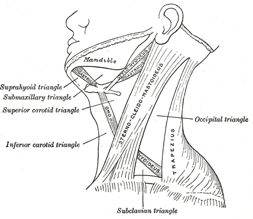
The submandibular triangle (or submaxillary or digastric triangle) corresponds to the region of the neck immediately beneath the body of the mandible. Boundaries and coverings It is bounded: * ''above'', by the lower border of the body of the mandible, and a line drawn from its angle to the mastoid process; * ''below'', by the posterior belly of the Digastricus; in front, by the anterior belly of the Digastricus. It is covered by the integument, superficial fascia, Platysma, and deep fascia, ramifying in which are branches of the facial nerve and ascending filaments of the cutaneous cervical nerve. Its floor is formed by the Mylohyoideus anteriorly, and by the hyoglossus posteriorly. Triangles * Beclard Triangle * Lesser Triangle * Pirogoff Triangle Divisions It is divided into an anterior and a posterior part by the stylomandibular ligament. Anterior part The anterior part contains the submandibular gland, superficial to which is the anterior facial vein, while imbedded ... [...More Info...] [...Related Items...] OR: [Wikipedia] [Google] [Baidu] |
Clavicle
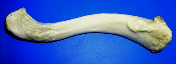
The clavicle, or collarbone, is a slender, S-shaped long bone approximately 6 inches (15 cm) long that serves as a strut between the shoulder blade and the sternum (breastbone). There are two clavicles, one on the left and one on the right. The clavicle is the only long bone in the body that lies horizontally. Together with the shoulder blade, it makes up the shoulder girdle. It is a palpable bone and, in people who have less fat in this region, the location of the bone is clearly visible. It receives its name from the Latin ''clavicula'' ("little key"), because the bone rotates along its axis like a key when the shoulder is abducted. The clavicle is the most commonly fractured bone. It can easily be fractured by impacts to the shoulder from the force of falling on outstretched arms or by a direct hit. Structure The collarbone is a thin doubly curved long bone that connects the arm to the trunk of the body. Located directly above the first rib, it acts as a strut to k ... [...More Info...] [...Related Items...] OR: [Wikipedia] [Google] [Baidu] |
Lower Jugular Nodes
The deep cervical lymph nodes are a group of cervical lymph nodes found near the internal jugular vein in the neck. Structure The deep cervical lymph nodes can be divided into upper and lower groups, or superior and inferior groups. Alternatively, they can be divided into deep anterior cervical lymph nodes and deep lateral cervical lymph nodes. They can also be divided into three groups: "superior deep jugular", "middle deep jugular", and "inferior deep jugular". Location The deep cervical lymph nodes are contained in the carotid sheath in the neck, close to the internal jugular vein. They connect to the meningeal lymphatic vessels superiorly. Function The deep cervical lymph nodes provide lymphatic drainage to many parts of the head, including the pharynx, mouth, and meninges In anatomy, the meninges (, ''singular:'' meninx ( or ), ) are the three membranes that envelop the brain and spinal cord. In mammals, the meninges are the dura mater, the arachnoid mater, and ... [...More Info...] [...Related Items...] OR: [Wikipedia] [Google] [Baidu] |
Sternohyoid Muscle
The sternohyoid muscle is a thin, narrow muscle attaching the hyoid bone to the sternum. It is one of the paired strap muscles of the infrahyoid muscles. It is supplied by the ansa cervicalis. It depresses the hyoid bone. Structure The sternohyoid muscle is one of the paired strap muscles of the infrahyoid muscles. It arises from the posterior border of the medial end of the clavicle, the posterior sternoclavicular ligament, and the upper and posterior part of the manubrium of the sternum. Passing upward and medially, it is inserted by short tendinous fibers into the lower border of the body of the hyoid bone. It runs lateral to the trachea. Nerve supply The sternohyoid muscle is supplied by a branch of the ansa cervicalis. Variations The sternohyoid muscle may be doubled, have accessory slips (Cleidohyoideus) or be completely absent in some people. It sometimes presents a transverse tendinous inscription immediately above its origin. Function The sternohyoid muscle p ... [...More Info...] [...Related Items...] OR: [Wikipedia] [Google] [Baidu] |
Cricoid Cartilage
The cricoid cartilage , or simply cricoid (from the Greek ''krikoeides'' meaning "ring-shaped") or cricoid ring, is the only complete ring of cartilage around the trachea. It forms the back part of the voice box and functions as an attachment site for muscles, cartilages, and ligaments involved in opening and closing the airway and in producing speech. Structure The cricoid cartilage sits just inferior to the thyroid cartilage in the neck, at the level of the C6 vertebra, and is joined to it medially by the median cricothyroid ligament and postero-laterally by the cricothyroid joints. Inferior to it are the rings of cartilage around the trachea (which are not continuous – rather they are C-shaped with a gap posteriorly). The cricoid is joined to the first tracheal ring by the cricotracheal ligament, and this can be felt as a more yielding area between the firm thyroid cartilage and firmer cricoid. It is also anatomically related to the thyroid gland; although the ... [...More Info...] [...Related Items...] OR: [Wikipedia] [Google] [Baidu] |
Middle Jugular Nodes
The deep cervical lymph nodes are a group of cervical lymph nodes found near the internal jugular vein in the neck. Structure The deep cervical lymph nodes can be divided into upper and lower groups, or superior and inferior groups. Alternatively, they can be divided into deep anterior cervical lymph nodes and deep lateral cervical lymph nodes. They can also be divided into three groups: "superior deep jugular", "middle deep jugular", and "inferior deep jugular". Location The deep cervical lymph nodes are contained in the carotid sheath in the neck, close to the internal jugular vein. They connect to the meningeal lymphatic vessels superiorly. Function The deep cervical lymph nodes provide lymphatic drainage to many parts of the head, including the pharynx, mouth, and meninges In anatomy, the meninges (, ''singular:'' meninx ( or ), ) are the three membranes that envelop the brain and spinal cord. In mammals, the meninges are the dura mater, the arachnoid mater, and ... [...More Info...] [...Related Items...] OR: [Wikipedia] [Google] [Baidu] |
Submandibular Gland
The paired submandibular glands (historically known as submaxillary glands) are major salivary glands located beneath the floor of the mouth. They each weigh about 15 grams and contribute some 60–67% of unstimulated saliva secretion; on stimulation their contribution decreases in proportion as the parotid secretion rises to 50%. The average length of the normal human submandibular salivary gland is approximately 27mm, while the average width is approximately 14.3mm. Structure Lying superior to the digastric muscles, each submandibular gland is divided into superficial and deep lobes, which are separated by the mylohyoid muscle: * The superficial lobe comprises most of the gland, with the mylohyoid muscle runs under it * The deep lobe is the smaller part Secretions are delivered into the submandibular duct on the deep portion after which they hook around the posterior edge of the mylohyoid muscle and proceed on the superior surface laterally. The excretory ducts are then cross ... [...More Info...] [...Related Items...] OR: [Wikipedia] [Google] [Baidu] |
Sternocleidomastoid Muscle
The sternocleidomastoid muscle is one of the largest and most superficial cervical muscles. The primary actions of the muscle are rotation of the head to the opposite side and flexion of the neck. The sternocleidomastoid is innervated by the accessory nerve. Etymology and location It is given the name ''sternocleidomastoid'' because it originates at the manubrium of the sternum (''sterno-'') and the clavicle (''cleido-'') and has an insertion at the mastoid process of the temporal bone of the skull. Structure The sternocleidomastoid muscle originates from two locations: the manubrium of the sternum and the clavicle. It travels obliquely across the side of the neck and inserts at the mastoid process of the temporal bone of the skull by a thin aponeurosis. The sternocleidomastoid is thick and narrow at its centre, and broader and thinner at either end. The sternal head is a round fasciculus, tendinous in front, fleshy behind, arising from the upper part of the front of the manubriu ... [...More Info...] [...Related Items...] OR: [Wikipedia] [Google] [Baidu] |
Base Of The Skull
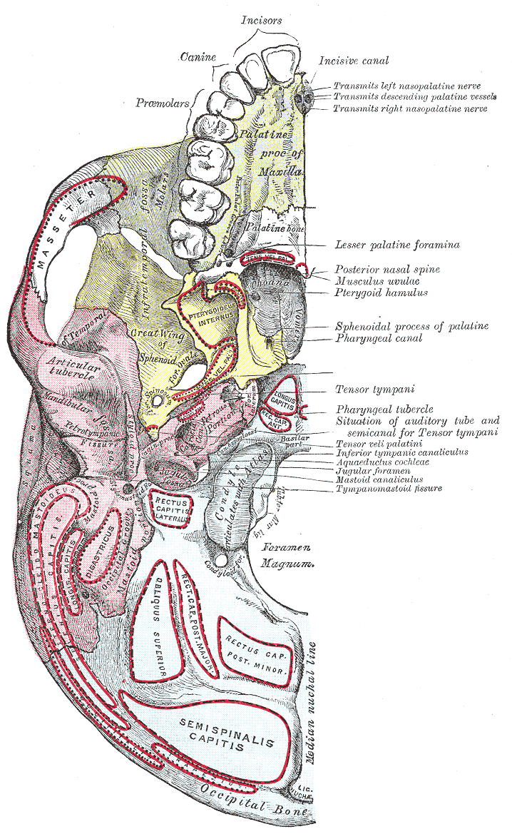
The base of skull, also known as the cranial base or the cranial floor, is the most inferior area of the skull. It is composed of the endocranium and the lower parts of the calvaria. Structure Structures found at the base of the skull are for example: Bones There are five bones that make up the base of the skull: *Ethmoid bone * Sphenoid bone * Occipital bone *Frontal bone *Temporal bone Sinuses *Occipital sinus * Superior sagittal sinus *Superior petrosal sinus Foramina of the skull * Foramen cecum *Optic foramen *Foramen lacerum *Foramen rotundum * Foramen magnum * Foramen ovale *Jugular foramen *Internal auditory meatus *Mastoid foramen *Sphenoidal emissary foramen *Foramen spinosum Sutures *Frontoethmoidal suture *Sphenofrontal suture *Sphenopetrosal suture *Sphenoethmoidal suture * Petrosquamous suture *Sphenosquamosal suture Other *Sphenoidal lingula *Subarcuate fossa *Dorsum sellae *Jugular process *Petro-occipital fissure *Condylar canal * Jugular tubercle * ... [...More Info...] [...Related Items...] OR: [Wikipedia] [Google] [Baidu] |
Accessory Nerve
The accessory nerve, also known as the eleventh cranial nerve, cranial nerve XI, or simply CN XI, is a cranial nerve that supplies the sternocleidomastoid and trapezius muscles. It is classified as the eleventh of twelve pairs of cranial nerves because part of it was formerly believed to originate in the brain. The sternocleidomastoid muscle tilts and rotates the head, whereas the trapezius muscle, connecting to the scapula, acts to shrug the shoulder. Traditional descriptions of the accessory nerve divide it into a spinal part and a cranial part. The cranial component rapidly joins the vagus nerve, and there is ongoing debate about whether the cranial part should be considered part of the accessory nerve proper. Consequently, the term "accessory nerve" usually refers only to nerve supplying the sternocleidomastoid and trapezius muscles, also called the spinal accessory nerve. Strength testing of these muscles can be measured during a neurological examination to assess funct ... [...More Info...] [...Related Items...] OR: [Wikipedia] [Google] [Baidu] |
Internal Jugular Vein
The internal jugular vein is a paired jugular vein that collects blood from the brain and the superficial parts of the face and neck. This vein runs in the carotid sheath with the common carotid artery and vagus nerve. It begins in the posterior compartment of the jugular foramen, at the base of the skull. It is somewhat dilated at its origin, which is called the ''superior bulb''. This vein also has a common trunk into which drains the anterior branch of the retromandibular vein, the facial vein, and the lingual vein. It runs down the side of the neck in a vertical direction, being at one end lateral to the internal carotid artery, and then lateral to the common carotid artery, and at the root of the neck, it unites with the subclavian vein to form the brachiocephalic vein (innominate vein); a little above its termination is a second dilation, the ''inferior bulb''. Above, it lies upon the rectus capitis lateralis, behind the internal carotid artery and the nerves passing ... [...More Info...] [...Related Items...] OR: [Wikipedia] [Google] [Baidu] |


