|
Cerebral Angiography
Cerebral angiography is a form of angiography which provides images of blood vessels in and around the brain, thereby allowing detection of abnormalities such as arteriovenous malformations and aneurysms. It was pioneered in 1927 by the Portuguese neurologist Egas Moniz at the University of Lisbon, who also helped develop thorotrast for use in the procedure. Typically a catheter is inserted into a large artery (such as the femoral artery) and threaded through the circulatory system to the carotid artery, where a contrast agent is injected. A series of radiographs are taken as the contrast agent spreads through the brain's arterial system, then a second series as it reaches the venous system. For some applications cerebral angiography may yield better images than less invasive methods such as computed tomography angiography and magnetic resonance angiography. In addition, cerebral angiography allows certain treatments to be performed immediately, based on its findings. In ... [...More Info...] [...Related Items...] OR: [Wikipedia] [Google] [Baidu] |
Transverse Plane
The transverse plane (also known as the horizontal plane, axial plane and transaxial plane) is an anatomical plane that divides the body into superior and inferior sections. It is perpendicular to the coronal and sagittal planes. List of clinically relevant anatomical planes * Transverse ''thoracic plane'' * '' Xiphosternal plane'' (or xiphosternal junction) * ''Transpyloric plane'' * ''Subcostal plane'' * '' Umbilical plane'' (or transumbilical plane) * '' Supracristal plane'' * ''Intertubercular plane'' (or transtubercular plane) * '' Interspinous plane'' Clinically relevant anatomical planes with associated structures * The transverse ''thoracic plane'' ** Plane through T4 & T5 vertebral junction and sternal angle of Louis. ** Marks the: *** Attachment of costal cartilage of rib 2 at the sternal angle; *** Aortic arch (beginning and end); *** Upper margin of SVC; *** Thoracic duct crossing; *** Tracheal bifurcation; *** Pulmonary trunk bifurcation; * The '' xiphost ... [...More Info...] [...Related Items...] OR: [Wikipedia] [Google] [Baidu] |
Connective Tissue
Connective tissue is one of the four primary types of animal tissue, along with epithelial tissue, muscle tissue, and nervous tissue. It develops from the mesenchyme derived from the mesoderm the middle embryonic germ layer. Connective tissue is found in between other tissues everywhere in the body, including the nervous system. The three meninges, membranes that envelop the brain and spinal cord are composed of connective tissue. Most types of connective tissue consists of three main components: elastic and collagen fibers, ground substance, and cells. Blood, and lymph are classed as specialized fluid connective tissues that do not contain fiber. All are immersed in the body water. The cells of connective tissue include fibroblasts, adipocytes, macrophages, mast cells and leucocytes. The term "connective tissue" (in German, ''Bindegewebe'') was introduced in 1830 by Johannes Peter Müller. The tissue was already recognized as a distinct class in the 18th century. ... [...More Info...] [...Related Items...] OR: [Wikipedia] [Google] [Baidu] |
Meningioma
Meningioma, also known as meningeal tumor, is typically a slow-growing tumor that forms from the meninges, the membranous layers surrounding the brain and spinal cord. Symptoms depend on the location and occur as a result of the tumor pressing on nearby tissue. Many cases never produce symptoms. Occasionally seizures, dementia, trouble talking, vision problems, one sided weakness, or loss of bladder control may occur. Risk factors include exposure to ionizing radiation such as during radiation therapy, a family history of the condition, and neurofibromatosis type 2. As of 2014 they do not appear to be related to cell phone use. They appear to be able to form from a number of different types of cells including arachnoid cells. Diagnosis is typically by medical imaging. If there are no symptoms, periodic observation may be all that is required. Most cases that result in symptoms can be cured by surgery. Following complete removal fewer than 20% recur. If surgery is not possib ... [...More Info...] [...Related Items...] OR: [Wikipedia] [Google] [Baidu] |
Embolisation
Embolization refers to the passage and lodging of an embolus within the circulatory system, bloodstream. It may be of natural origin (pathological), in which word sense, sense it is also called embolism, for example a pulmonary embolism; or it may be artificially induced (therapy, therapeutic), as a hemostasis, hemostatic treatment for bleeding or as a treatment for some types of cancer by deliberately blocking blood vessels to starve the neoplasm, tumor cells. In the management of cancer, cancer management application, the embolus, besides blocking the blood supply to the tumor, also often includes an ingredient to attack the tumor chemically or with irradiation. When it bears a chemotherapy drug, the process is called chemoembolization. Transcatheter arterial chemoembolization (TACE) is the usual form. When the embolus bears a medicinal radiocompounds, radiopharmaceutical for unsealed source radiotherapy, the process is called radioembolization or selective internal radiatio ... [...More Info...] [...Related Items...] OR: [Wikipedia] [Google] [Baidu] |
Dural Arteriovenous Fistula
A dural arteriovenous fistula (DAVF) or malformation is an abnormal direct connection (fistula) between a meningeal artery and a meningeal vein or dural venous sinus. Signs and symptoms The most common signs/symptoms of DAVFs are: # Pulsatile tinnitus # Occipital bruit # Headache # Visual impairment # Papilledema Pulsatile tinnitus is the most common symptom in patients, and it is associated with transverse-sigmoid sinus DAVFs. Carotid-cavernous DAVFs, on the other hand, are more closely associated with pulsatile exophthalmos. DAVFs may also be asymptomatic (e.g. cavernous sinus DAVFs). Location Most commonly found adjacent to dural sinuses in the following locations: # Transverse (lateral) sinus, left-sided slightly more common than right # Intratentorial # From the posterior cavernous sinus, usually draining to the transverse or sigmoid sinuses # Vertebral artery (posterior meningeal branch) Causes It is still unclear whether DAVFs are congenital or acquired. Current evi ... [...More Info...] [...Related Items...] OR: [Wikipedia] [Google] [Baidu] |
Cerebral Arteriovenous Malformation
A cerebral arteriovenous malformation (cerebral AVM, CAVM, cAVM) is an abnormal connection between the arteries and veins in the brain—specifically, an arteriovenous malformation in the cerebrum. Signs and symptoms The most frequently observed problems, related to an AVM, are headaches and seizures, cranial nerve deficits, backaches, neckaches and eventual nausea, as the coagulated blood makes its way down to be dissolved in the individual's spinal fluid. It is supposed that 15% of the population, at detection, have no symptoms at all. Other common symptoms are a pulsing noise in the head, progressive weakness and numbness and vision changes as well as debilitating, excruciating pain. In serious cases, the blood vessels rupture and there is bleeding within the brain (intracranial hemorrhage). Nevertheless, in more than half of patients with AVM, hemorrhage is the first symptom. Symptoms due to bleeding include loss of consciousness, sudden and severe headache, nausea, vomiting, ... [...More Info...] [...Related Items...] OR: [Wikipedia] [Google] [Baidu] |
Cerebral Vasospasm
Cerebral vasospasm is the prolonged, intense vasoconstriction of the larger conducting arteries in the subarachnoid space which is initially surrounded by a clot. Significant narrowing of the blood vessels in the brain develops gradually over the first few days after the aneurysm An aneurysm is an outward bulging, likened to a bubble or balloon, caused by a localized, abnormal, weak spot on a blood vessel wall. Aneurysms may be a result of a hereditary condition or an acquired disease. Aneurysms can also be a nidus ( ...al rupture. This kind of narrowing usually is maximal in about a week's time following intracerebral haemorrhage. Vasospasm is the one of the leading causes of death after the aneurysmal rupture along with the effect of the initial haemorrhage and later bleeding. References {{Reflist Brain disorders ... [...More Info...] [...Related Items...] OR: [Wikipedia] [Google] [Baidu] |
Stroke
A stroke is a disease, medical condition in which poor cerebral circulation, blood flow to the brain causes cell death. There are two main types of stroke: brain ischemia, ischemic, due to lack of blood flow, and intracranial hemorrhage, hemorrhagic, due to bleeding. Both cause parts of the brain to stop functioning properly. Signs and symptoms of a stroke may include an hemiplegia, inability to move or feel on one side of the body, receptive aphasia, problems understanding or expressive aphasia, speaking, dizziness, or Homonymous hemianopsia, loss of vision to one side. Signs and symptoms often appear soon after the stroke has occurred. If symptoms last less than one or two hours, the stroke is a transient ischemic attack (TIA), also called a mini-stroke. A subarachnoid hemorrhage, hemorrhagic stroke may also be associated with a thunderclap headache, severe headache. The symptoms of a stroke can be permanent. Long-term complications may include pneumonia and Urinary incontin ... [...More Info...] [...Related Items...] OR: [Wikipedia] [Google] [Baidu] |
Intracranial Aneurysm
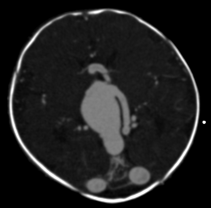
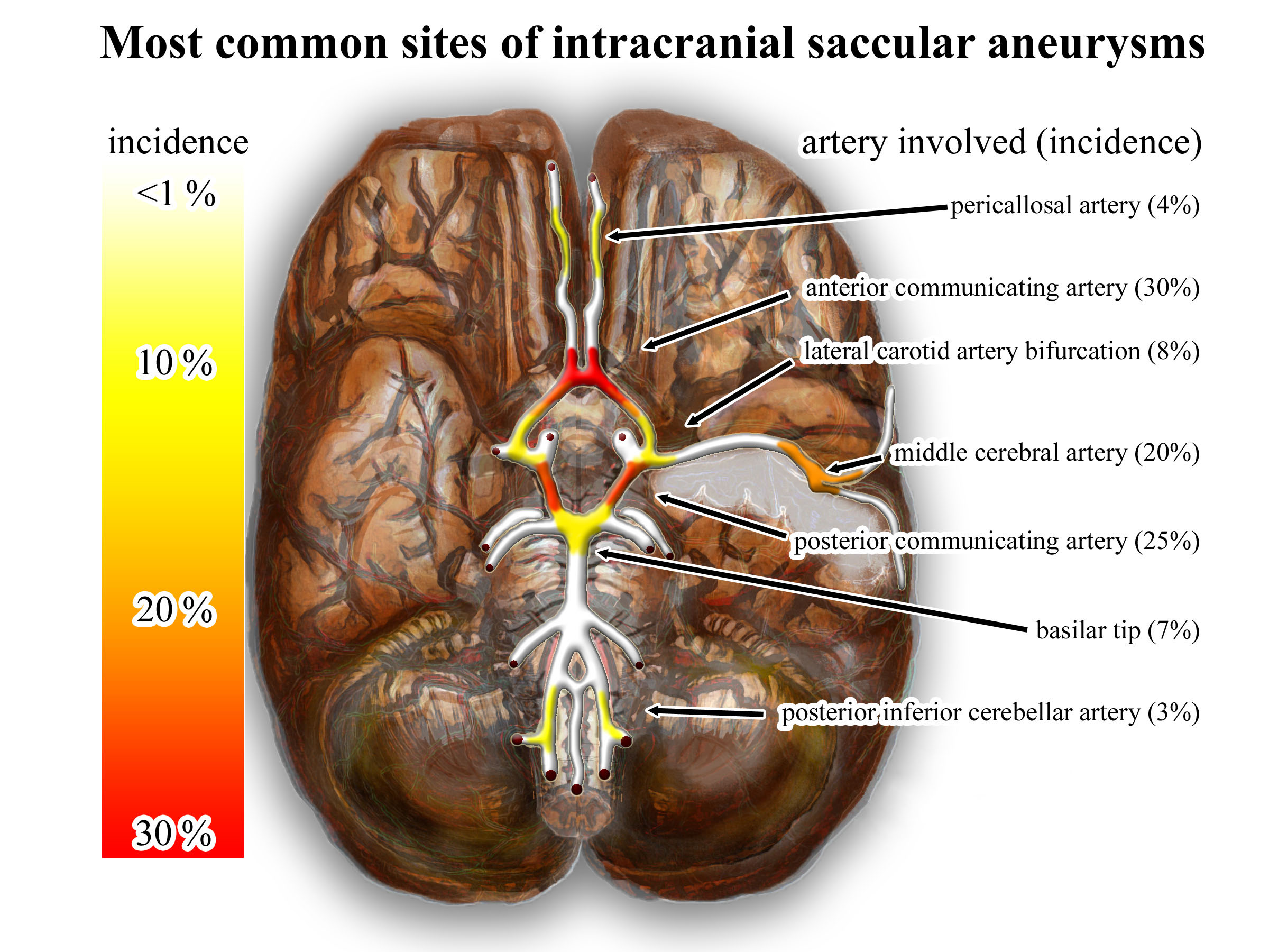
An intracranial aneurysm, also known as a brain aneurysm, is a cerebrovascular disorder in which weakness in the wall of a cerebral artery or vein causes a localized dilation or ballooning of the blood vessel. Aneurysms in the posterior circulation (basilar artery, vertebral arteries and posterior communicating artery) have a higher risk of rupture. Basilar artery aneurysms represent only 3–5% of all intracranial aneurysms but are the most common aneurysms in the posterior circulation. Classification Cerebral aneurysms are classified both by size and shape. Small aneurysms have a diameter of less than 15 mm. Larger aneurysms include those classified as large (15 to 25 mm), giant (25 to 50 mm), and super-giant (over 50 mm). Berry (saccular) aneurysms Saccular aneurysms, also known as berry aneurysms, appear as a round outpouching and are the most common form of cerebral aneurysm. Causes include connective tissue disorders, polycystic kidney disease, ar ... [...More Info...] [...Related Items...] OR: [Wikipedia] [Google] [Baidu] |
Intracerebral Haemorrhage
Intracerebral hemorrhage (ICH), also known as cerebral bleed, intraparenchymal bleed, and hemorrhagic stroke, or haemorrhagic stroke, is a sudden bleeding into the tissues of the brain, into its ventricles, or into both. It is one kind of bleeding within the skull and one kind of stroke. Symptoms can include headache, one-sided weakness, vomiting, seizures, decreased level of consciousness, and neck stiffness. Often, symptoms get worse over time. Fever is also common. Causes include brain trauma, aneurysms, arteriovenous malformations, and brain tumors. The biggest risk factors for spontaneous bleeding are high blood pressure and amyloidosis. Other risk factors include alcoholism, low cholesterol, blood thinners, and cocaine use. Diagnosis is typically by CT scan. Other conditions that may present similarly include ischemic stroke. Treatment should typically be carried out in an intensive care unit. Guidelines recommend decreasing the blood pressure to a systolic of 140& ... [...More Info...] [...Related Items...] OR: [Wikipedia] [Google] [Baidu] |
Subarachnoid Haemorrhage
Subarachnoid hemorrhage (SAH) is bleeding into the subarachnoid space—the area between the arachnoid membrane and the pia mater surrounding the brain. Symptoms may include a severe headache of rapid onset, vomiting, decreased level of consciousness, fever, and sometimes seizures. Neck stiffness or neck pain are also relatively common. In about a quarter of people a small bleed with resolving symptoms occurs within a month of a larger bleed. SAH may occur as a result of a head injury or spontaneously, usually from a ruptured cerebral aneurysm. Risk factors for spontaneous cases include high blood pressure, smoking, family history, alcoholism, and cocaine use. Generally, the diagnosis can be determined by a CT scan of the head if done within six hours of symptom onset. Occasionally, a lumbar puncture is also required. After confirmation further tests are usually performed to determine the underlying cause. Treatment is by prompt neurosurgery or endovascular coiling. M ... [...More Info...] [...Related Items...] OR: [Wikipedia] [Google] [Baidu] |
Mass Effect (medicine)
In medicine, a mass effect is the effect of a growing mass that results in secondary pathological effects by pushing on or displacing surrounding tissue. In oncology, the mass typically refers to a tumor. For example, cancer of the thyroid gland may cause symptoms due to compressions of certain structures of the head and neck; pressure on the laryngeal nerves may cause voice changes, narrowing of the windpipe may cause stridor, pressure on the gullet may cause dysphagia and so on. Surgical removal or debulking is sometimes used to palliate symptoms of the mass effect even if the underlying pathology is not curable. In neurology, a mass effect is the effect exerted by any mass, including, for example, hydrocephalus (cerebrospinal fluid buildup) or an evolving intracranial hemorrhage (bleeding within the skull) presenting with a clinically significant hematoma. The hematoma can exert a mass effect on the brain, increasing intracranial pressure and potentially causing midline ... [...More Info...] [...Related Items...] OR: [Wikipedia] [Google] [Baidu] |


