|
Central Pattern Generator
Central pattern generators (CPGs) are self-organizing biological neural circuits that produce rhythmic outputs in the absence of rhythmic input. They are the source of the tightly-coupled patterns of neural activity that drive rhythmic and stereotyped motor behaviors like walking, swimming, breathing, or chewing. The ability to function without input from higher brain areas still requires modulatory inputs, and their outputs are not fixed. Flexibility in response to sensory input is a fundamental quality of CPG-driven behavior. To be classified as a rhythmic generator, a CPG requires: # "two or more processes that interact such that each process sequentially increases and decreases, and # that, as a result of this interaction, the system repeatedly returns to its starting condition." CPGs have been found in invertebrates, and practically all vertebrate species investigated, including humans. General anatomy and physiology Intrinsic properties of CPG neurons CPG neurons can ha ... [...More Info...] [...Related Items...] OR: [Wikipedia] [Google] [Baidu] |
Self-organizing
Self-organization, also called spontaneous order in the social sciences, is a process where some form of overall order arises from local interactions between parts of an initially disordered system. The process can be spontaneous when sufficient energy is available, not needing control by any external agent. It is often triggered by seemingly random fluctuations, amplified by positive feedback. The resulting organization is wholly decentralized, distributed over all the components of the system. As such, the organization is typically robust and able to survive or self-repair substantial perturbation. Chaos theory discusses self-organization in terms of islands of predictability in a sea of chaotic unpredictability. Self-organization occurs in many physical, chemical, biological, robotic, and cognitive systems. Examples of self-organization include crystallization, thermal convection of fluids, chemical oscillation, animal swarming, neural circuits, and black market ... [...More Info...] [...Related Items...] OR: [Wikipedia] [Google] [Baidu] |
Brainstem
The brainstem (or brain stem) is the posterior stalk-like part of the brain that connects the cerebrum with the spinal cord. In the human brain the brainstem is composed of the midbrain, the pons, and the medulla oblongata. The midbrain is continuous with the thalamus of the diencephalon through the tentorial notch, and sometimes the diencephalon is included in the brainstem. The brainstem is very small, making up around only 2.6 percent of the brain's total weight. It has the critical roles of regulating cardiac, and respiratory function, helping to control heart rate and breathing rate. It also provides the main motor and sensory nerve supply to the face and neck via the cranial nerves. Ten pairs of cranial nerves come from the brainstem. Other roles include the regulation of the central nervous system and the body's sleep cycle. It is also of prime importance in the conveyance of motor and sensory pathways from the rest of the brain to the body, and from the body back to t ... [...More Info...] [...Related Items...] OR: [Wikipedia] [Google] [Baidu] |
Pylorus
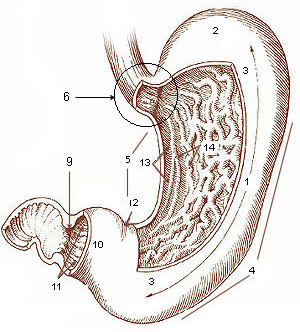
The pylorus ( or ), or pyloric part, connects the stomach to the duodenum. The pylorus is considered as having two parts, the ''pyloric antrum'' (opening to the body of the stomach) and the ''pyloric canal'' (opening to the duodenum). The ''pyloric canal'' ends as the ''pyloric orifice'', which marks the junction between the stomach and the duodenum. The orifice is surrounded by a sphincter, a band of muscle, called the ''pyloric sphincter''. The word ''pylorus'' comes from Greek πυλωρός, via Latin. The word ''pylorus'' in Greek means "gatekeeper", related to "gate" ( el, pyle) and is thus linguistically related to the word " pylon". Structure The pylorus is the furthest part of the stomach that connects to the duodenum. It is divided into two parts, the ''antrum'', which connects to the body of the stomach, and the ''pyloric canal'', which connects to the duodenum. Antrum The ''pyloric antrum'' is the initial portion of the pylorus. It is near the bottom of the stomach, ... [...More Info...] [...Related Items...] OR: [Wikipedia] [Google] [Baidu] |
Stomatogastric Ganglion
The stomatogastric ganglion (STG) is a much studied ganglion (collection of neurons) found in arthropods and studied extensively in decapod crustaceans. It is part of the stomatogastric nervous system. See also * Central pattern generator Central pattern generators (CPGs) are self-organizing biological neural circuits that produce rhythmic outputs in the absence of rhythmic input. They are the source of the tightly-coupled patterns of neural activity that drive rhythmic and stereot ... References Crustacean anatomy {{decapod-stub ... [...More Info...] [...Related Items...] OR: [Wikipedia] [Google] [Baidu] |
Whisking In Animals
Vibrissae (; singular: vibrissa; ), more generally called Whiskers, are a type of stiff, functional hair used by mammals to sense their environment. These hairs are finely specialised for this purpose, whereas other types of hair are coarser as tactile sensors. Although whiskers are specifically those found around the face, vibrissae are known to grow in clusters at various places around the body. Most mammals have them, including all non-human primates and especially nocturnal mammals. Whiskers are sensitive tactile hairs that aid navigation, locomotion, exploration, hunting, social touch and perform other functions. This article is primarily about the specialised sensing hairs of mammals, but some birds, fish, insects, crustaceans and other arthropods are known to have similar structures also used to sense the environment. Etymology Vibrissae (from Latin 'to vibrate') from the characteristic motion seen in a small rodent that is otherwise sitting still. In medicine, the ... [...More Info...] [...Related Items...] OR: [Wikipedia] [Google] [Baidu] |
Nucleus Tractus Solitarii
In the human brainstem, the solitary nucleus, also called nucleus of the solitary tract, nucleus solitarius, and nucleus tractus solitarii, (SN or NTS) is a series of purely sensory nuclei (clusters of nerve cell bodies) forming a vertical column of grey matter embedded in the medulla oblongata. Through the center of the SN runs the solitary tract, a white bundle of nerve fibers, including fibers from the facial, glossopharyngeal and vagus nerves, that innervate the SN. The SN projects to, among other regions, the reticular formation, parasympathetic preganglionic neurons, hypothalamus and thalamus, forming circuits that contribute to autonomic regulation. Cells along the length of the SN are arranged roughly in accordance with function; for instance, cells involved in taste are located in the rostral part, while those receiving information from cardio-respiratory and gastrointestinal processes are found in the caudal part. Inputs * Taste information from the facial nerve via the ... [...More Info...] [...Related Items...] OR: [Wikipedia] [Google] [Baidu] |
Respiratory Group
The respiratory center is located in the medulla oblongata and pons, in the brainstem. The respiratory center is made up of three major respiratory groups of neurons, two in the medulla and one in the pons. In the medulla they are the dorsal respiratory group, and the ventral respiratory group. In the pons, the pontine respiratory group includes two areas known as the pneumotaxic centre and the apneustic centre. The respiratory centre is responsible for generating and maintaining the rhythm of respiration, and also of adjusting this in homeostatic response to physiological changes. The respiratory center receives input from chemoreceptors, mechanoreceptors, the cerebral cortex, and the hypothalamus in order to regulate the rate and depth of breathing. Input is stimulated by altered levels of oxygen, carbon dioxide, and blood pH, by hormonal changes relating to stress and anxiety from the hypothalamus, and also by signals from the cerebral cortex to give a conscious control of r ... [...More Info...] [...Related Items...] OR: [Wikipedia] [Google] [Baidu] |
Retrotrapezoid Nucleus
The medulla oblongata or simply medulla is a long stem-like structure which makes up the lower part of the brainstem. It is anterior and partially inferior to the cerebellum. It is a cone-shaped neuronal mass responsible for autonomic (involuntary) functions, ranging from vomiting to sneezing. The medulla contains the cardiac, respiratory, vomiting and vasomotor centers, and therefore deals with the autonomic functions of breathing, heart rate and blood pressure as well as the sleep–wake cycle. During embryonic development, the medulla oblongata develops from the myelencephalon. The myelencephalon is a secondary vesicle which forms during the maturation of the rhombencephalon, also referred to as the hindbrain. The bulb is an archaic term for the medulla oblongata. In modern clinical usage, the word bulbar (as in bulbar palsy) is retained for terms that relate to the medulla oblongata, particularly in reference to medical conditions. The word bulbar can refer to the nerves ... [...More Info...] [...Related Items...] OR: [Wikipedia] [Google] [Baidu] |
Golgi Tendon Organ
The Golgi tendon organ (GTO) (also called Golgi organ, tendon organ, neurotendinous organ or neurotendinous spindle) is a proprioceptor – a type of sensory receptor that senses changes in muscle tension. It lies at the interface between a muscle and its tendon known as the musculotendinous junction also known as the myotendinous junction. It provides the sensory component of the Golgi tendon reflex. The Golgi tendon organ is one of several eponymous terms named after the Italian physician Camillo Golgi. Structure The body of the Golgi tendon organ is made up of braided strands of collagen (intrafusal fasciculi) that are less compact than elsewhere in the tendon and are encapsulated. The capsule is connected in series (along a single path) with a group of muscle fibers () at one end, and merge into the tendon proper at the other. Each capsule is about long, has a diameter of about , and is perforated by one or more afferent type Ib sensory nerve fibers ( Aɑ fiber), which ... [...More Info...] [...Related Items...] OR: [Wikipedia] [Google] [Baidu] |
Locomotor CPG Schematic
Locomotor may refer to: * ''Locomotor'', the Dutch equivalent of the German ''Kleinlokomotive'', a locomotive of small size and low power for light shunting duties * Locomotor activity * Locomotor ataxia * Locomotor effects of shoes * Locomotor stimulation * Locomotor system (other) See also * Locomotion (other) Locomotion means the act or ability of something to transport or move itself from place to place. Locomotion may refer to: Motion * Motion (physics) * Robot locomotion, of man-made devices By environment * Aquatic locomotion * Flight * Locomot ... * Locomotive (other) {{disambiguation ... [...More Info...] [...Related Items...] OR: [Wikipedia] [Google] [Baidu] |
Motor Pool (neuroscience)
A motor pool consists of all individual motor neurons that innervate a single muscle. Each individual muscle fiber is innervated by only one motor neuron, but one motor neuron may innervate several muscle fibers. This distinction is physiologically significant because the size of a given motor pool determines the activity of the muscle it innervates: for example, muscles responsible for finer movements are innervated by motor pools consisting of higher numbers of individual motor neurons. Motor pools are also distinguished by the different classes of motor neurons that they contain. The size, composition, and anatomical location of each motor pool is tightly controlled by complex developmental pathways.Carp, J. S. and Wolpaw, J. R. 2010. Motor Neurons and Spinal Control of Movement. eLDOI: 10.1002/9780470015902.a0000156.pub2/ref> Anatomy Distinct skeletal muscles are controlled by groups of individual motor units. Such motor units are made up of a single motor neuron and the muscle ... [...More Info...] [...Related Items...] OR: [Wikipedia] [Google] [Baidu] |
_self-organization2.jpg)


