|
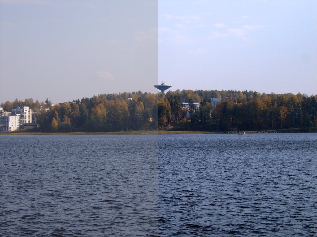
Center-surround Antagonism
Center-surround antagonism refers to antagonistic interactions between center and surround regions of the receptive fields of photoreceptor cells in the retina. Center surround antagonism enables edge detection and contrast enhancement within the visual cortex The visual cortex of the brain is the area of the cerebral cortex that processes visual information. It is located in the occipital lobe. Sensory input originating from the eyes travels through the lateral geniculate nucleus in the thalamus and .... References * Signal transduction {{eye-stub ... [...More Info...] [...Related Items...] OR: [Wikipedia] [Google] [Baidu] |
Photoreceptor Cell
A photoreceptor cell is a specialized type of neuroepithelial cell found in the retina that is capable of visual phototransduction. The great biological importance of photoreceptors is that they convert light (visible electromagnetic radiation) into signals that can stimulate biological processes. To be more specific, photoreceptor proteins in the cell absorb photons, triggering a change in the cell's membrane potential. There are currently three known types of photoreceptor cells in mammalian eyes: rods, cones, and intrinsically photosensitive retinal ganglion cells. The two classic photoreceptor cells are rods and cones, each contributing information used by the visual system to form an image of the environment, sight. Rods primarily mediate scotopic vision (dim conditions) whereas cones primarily mediate to photopic vision (bright conditions), but the processes in each that supports phototransduction is similar. A third class of mammalian photoreceptor cell was discovered ... [...More Info...] [...Related Items...] OR: [Wikipedia] [Google] [Baidu] |
Retina
The retina (from la, rete "net") is the innermost, light-sensitive layer of tissue of the eye of most vertebrates and some molluscs. The optics of the eye create a focused two-dimensional image of the visual world on the retina, which then processes that image within the retina and sends nerve impulses along the optic nerve to the visual cortex to create visual perception. The retina serves a function which is in many ways analogous to that of the film or image sensor in a camera. The neural retina consists of several layers of neurons interconnected by synapses and is supported by an outer layer of pigmented epithelial cells. The primary light-sensing cells in the retina are the photoreceptor cells, which are of two types: rods and cones. Rods function mainly in dim light and provide monochromatic vision. Cones function in well-lit conditions and are responsible for the perception of colour through the use of a range of opsins, as well as high-acuity vision used for task ... [...More Info...] [...Related Items...] OR: [Wikipedia] [Google] [Baidu] |
Edge Detection
Edge detection includes a variety of mathematical methods that aim at identifying edges, curves in a digital image at which the image brightness changes sharply or, more formally, has discontinuities. The same problem of finding discontinuities in one-dimensional signals is known as ''step detection'' and the problem of finding signal discontinuities over time is known as ''change detection''. Edge detection is a fundamental tool in image processing, machine vision and computer vision, particularly in the areas of feature detection and feature extraction. Motivations The purpose of detecting sharp changes in image brightness is to capture important events and changes in properties of the world. It can be shown that under rather general assumptions for an image formation model, discontinuities in image brightness are likely to correspond to: * discontinuities in depth, * discontinuities in surface orientation, * changes in material properties and * variations in scene illumi ... [...More Info...] [...Related Items...] OR: [Wikipedia] [Google] [Baidu] |
Contrast (vision)
Contrast is the contradiction in luminance or colour that makes an object (or its representation in an image or display) distinguishable. In visual perception of the real world, contrast is determined by the difference in the colour and brightness of the object and other objects within the same field of view. The human visual system is more sensitive to contrast than absolute luminance; we can perceive the world similarly regardless of the huge changes in illumination over the day or from place to place. The maximum ''contrast'' of an image is the contrast ratio or dynamic range. Images with a contrast ratio close to their medium's maximum possible contrast ratio experience a ''conservation of contrast'', wherein any increase in contrast in some parts of the image must necessarily result in a decrease in contrast elsewhere. Brightening an image will increase contrast in dark areas but decrease contrast in bright areas, while darkening the image will have the opposite effect. B ... [...More Info...] [...Related Items...] OR: [Wikipedia] [Google] [Baidu] |
Visual Cortex
The visual cortex of the brain is the area of the cerebral cortex that processes visual information. It is located in the occipital lobe. Sensory input originating from the eyes travels through the lateral geniculate nucleus in the thalamus and then reaches the visual cortex. The area of the visual cortex that receives the sensory input from the lateral geniculate nucleus is the primary visual cortex, also known as visual area 1 ( V1), Brodmann area 17, or the striate cortex. The extrastriate areas consist of visual areas 2, 3, 4, and 5 (also known as V2, V3, V4, and V5, or Brodmann area 18 and all Brodmann area 19). Both hemispheres of the brain include a visual cortex; the visual cortex in the left hemisphere receives signals from the right visual field, and the visual cortex in the right hemisphere receives signals from the left visual field. Introduction The primary visual cortex (V1) is located in and around the calcarine fissure in the occipital lobe. Each hemisphere's V1 ... [...More Info...] [...Related Items...] OR: [Wikipedia] [Google] [Baidu] |


