|
Cathepsin E
Cathepsin E is an enzyme () that in humans is encoded by the ''CTSE'' gene. The enzyme is also known as ''slow-moving proteinase'', ''erythrocyte membrane aspartic proteinase'', ''SMP'', ''EMAP'', ''non-pepsin proteinase'', ''cathepsin D-like acid proteinase'', ''cathepsin E-like acid proteinase'', ''cathepsin D-type proteinase'') is an enzyme. Cathepsin E is a protease found in animals, as well as various other organisms, that belongs to the aspartic protease group. In humans it is encoded by the CTSE gene located at 1q32 on chromosome 1. It is an intracellular non-lysosomal glycoprotein that is mainly found in the skin and in immune cells. The protein is an aspartyl protease that functions as a disulfide-linked homodimer, and has an oligosaccharide chain of the high-mannose type. It is a member of the peptidase A1 family, and therefore observes specificity similar to that of Pepsin A and Cathepsin D. Cathepsin E is an intracellular enzyme and does not appear to be involved in di ... [...More Info...] [...Related Items...] OR: [Wikipedia] [Google] [Baidu] |
Enzyme
Enzymes () are proteins that act as biological catalysts by accelerating chemical reactions. The molecules upon which enzymes may act are called substrates, and the enzyme converts the substrates into different molecules known as products. Almost all metabolic processes in the cell need enzyme catalysis in order to occur at rates fast enough to sustain life. Metabolic pathways depend upon enzymes to catalyze individual steps. The study of enzymes is called ''enzymology'' and the field of pseudoenzyme analysis recognizes that during evolution, some enzymes have lost the ability to carry out biological catalysis, which is often reflected in their amino acid sequences and unusual 'pseudocatalytic' properties. Enzymes are known to catalyze more than 5,000 biochemical reaction types. Other biocatalysts are catalytic RNA molecules, called ribozymes. Enzymes' specificity comes from their unique three-dimensional structures. Like all catalysts, enzymes increase the reaction ra ... [...More Info...] [...Related Items...] OR: [Wikipedia] [Google] [Baidu] |
Lymphatic System
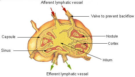
The lymphatic system, or lymphoid system, is an organ system in vertebrates that is part of the immune system, and complementary to the circulatory system. It consists of a large network of lymphatic vessels, lymph nodes, lymphatic or lymphoid organs, and lymphoid tissues. The vessels carry a clear fluid called lymph (the Latin word ''lympha'' refers to the deity of fresh water, "Lympha") back towards the heart, for re-circulation. Unlike the circulatory system that is a closed system, the lymphatic system is open. The human circulatory system processes an average of 20 litres of blood per day through capillary filtration, which removes plasma from the blood. Roughly 17 litres of the filtered blood is reabsorbed directly into the blood vessels, while the remaining three litres are left in the interstitial fluid. One of the main functions of the lymphatic system is to provide an accessory return route to the blood for the surplus three litres. The other main function is that of ... [...More Info...] [...Related Items...] OR: [Wikipedia] [Google] [Baidu] |
Metaplasia
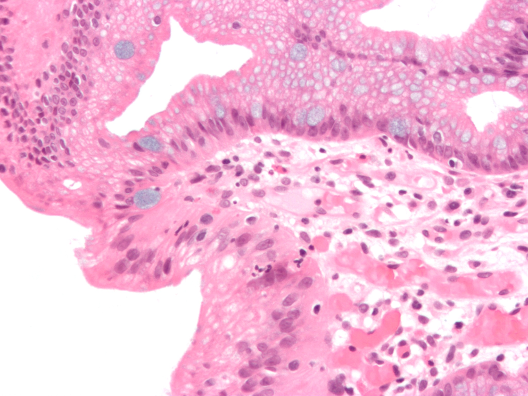
Metaplasia ( gr, "change in form") is the transformation of one differentiated cell type to another differentiated cell type. The change from one type of cell to another may be part of a normal maturation process, or caused by some sort of abnormal stimulus. In simplistic terms, it is as if the original cells are not robust enough to withstand their environment, so they transform into another cell type better suited to their environment. If the stimulus causing metaplasia is removed or ceases, tissues return to their normal pattern of differentiation. Metaplasia is not synonymous with dysplasia, and is not considered to be an actual cancer. It is also contrasted with heteroplasia, which is the spontaneous abnormal growth of cytologic and histologic elements. Today, metaplastic changes are usually considered to be an early phase of carcinogenesis, specifically for those with a history of cancers or who are known to be susceptible to carcinogenic changes. Metaplastic change is t ... [...More Info...] [...Related Items...] OR: [Wikipedia] [Google] [Baidu] |
Adenocarcinoma
Adenocarcinoma (; plural adenocarcinomas or adenocarcinomata ) (AC) is a type of cancerous tumor that can occur in several parts of the body. It is defined as neoplasia of epithelial tissue that has glandular origin, glandular characteristics, or both. Adenocarcinomas are part of the larger grouping of carcinomas, but are also sometimes called by more precise terms omitting the word, where these exist. Thus invasive ductal carcinoma, the most common form of breast cancer, is adenocarcinoma but does not use the term in its name—however, esophageal adenocarcinoma does to distinguish it from the other common type of esophageal cancer, esophageal squamous cell carcinoma. Several of the most common forms of cancer are adenocarcinomas, and the various sorts of adenocarcinoma vary greatly in all their aspects, so that few useful generalizations can be made about them. In the most specific usage (narrowest sense), the glandular origin or traits are exocrine; endocrine gland tumors, suc ... [...More Info...] [...Related Items...] OR: [Wikipedia] [Google] [Baidu] |
Cerebral Cortex
The cerebral cortex, also known as the cerebral mantle, is the outer layer of neural tissue of the cerebrum of the brain in humans and other mammals. The cerebral cortex mostly consists of the six-layered neocortex, with just 10% consisting of allocortex. It is separated into two cortices, by the longitudinal fissure that divides the cerebrum into the left and right cerebral hemispheres. The two hemispheres are joined beneath the cortex by the corpus callosum. The cerebral cortex is the largest site of neural integration in the central nervous system. It plays a key role in attention, perception, awareness, thought, memory, language, and consciousness. The cerebral cortex is part of the brain responsible for cognition. In most mammals, apart from small mammals that have small brains, the cerebral cortex is folded, providing a greater surface area in the confined volume of the cranium. Apart from minimising brain and cranial volume, cortical folding is crucial for the brain ... [...More Info...] [...Related Items...] OR: [Wikipedia] [Google] [Baidu] |
Striatum
The striatum, or corpus striatum (also called the striate nucleus), is a nucleus (a cluster of neurons) in the subcortical basal ganglia of the forebrain. The striatum is a critical component of the motor and reward systems; receives glutamatergic and dopaminergic inputs from different sources; and serves as the primary input to the rest of the basal ganglia. Functionally, the striatum coordinates multiple aspects of cognition, including both motor and action planning, decision-making, motivation, reinforcement, and reward perception. The striatum is made up of the caudate nucleus and the lentiform nucleus. The lentiform nucleus is made up of the larger putamen, and the smaller globus pallidus. Strictly speaking the globus pallidus is part of the striatum. It is common practice, however, to implicitly exclude the globus pallidus when referring to striatal structures. In primates, the striatum is divided into a ventral striatum, and a dorsal striatum, subdivisions that are ... [...More Info...] [...Related Items...] OR: [Wikipedia] [Google] [Baidu] |
Ischemia
Ischemia or ischaemia is a restriction in blood supply to any tissue, muscle group, or organ of the body, causing a shortage of oxygen that is needed for cellular metabolism (to keep tissue alive). Ischemia is generally caused by problems with blood vessels, with resultant damage to or dysfunction of tissue i.e. hypoxia and microvascular dysfunction. It also implies local hypoxia in a part of a body resulting from constriction (such as vasoconstriction, thrombosis, or embolism). Ischemia causes not only insufficiency of oxygen, but also reduced availability of nutrients and inadequate removal of metabolic wastes. Ischemia can be partial (poor perfusion) or total blockage. The inadequate delivery of oxygenated blood to the organs must be resolved either by treating the cause of the inadequate delivery or reducing the oxygen demand of the system that needs it. For example, patients with myocardial ischemia have a decreased blood flow to the heart and are prescribed with medi ... [...More Info...] [...Related Items...] OR: [Wikipedia] [Google] [Baidu] |
MHC Class II
MHC Class II molecules are a class of major histocompatibility complex (MHC) molecules normally found only on professional antigen-presenting cells such as dendritic cells, mononuclear phagocytes, some endothelial cells, thymic epithelial cells, and B cells. These cells are important in initiating immune responses. The antigens presented by class II peptides are derived from extracellular proteins (not cytosolic as in MHC class I). Loading of a MHC class II molecule occurs by phagocytosis; extracellular proteins are endocytosed, digested in lysosomes, and the resulting epitopic peptide fragments are loaded onto MHC class II molecules prior to their migration to the cell surface. In humans, the MHC class II protein complex is encoded by the human leukocyte antigen gene complex (HLA). HLAs corresponding to MHC class II are HLA-DP, HLA-DM, HLA-DOA, HLA-DOB, HLA-DQ, and HLA-DR. Mutations in the HLA gene complex can lead to bare lymphocyte syndrome (BLS), which is a type of MHC ... [...More Info...] [...Related Items...] OR: [Wikipedia] [Google] [Baidu] |
Endoplasmic Reticulum
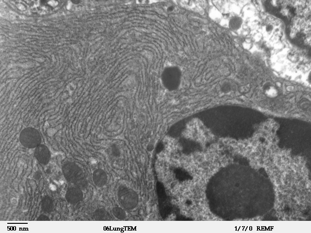
The endoplasmic reticulum (ER) is, in essence, the transportation system of the eukaryotic cell, and has many other important functions such as protein folding. It is a type of organelle made up of two subunits – rough endoplasmic reticulum (RER), and smooth endoplasmic reticulum (SER). The endoplasmic reticulum is found in most eukaryotic cells and forms an interconnected network of flattened, membrane-enclosed sacs known as cisternae (in the RER), and tubular structures in the SER. The membranes of the ER are continuous with the outer nuclear membrane. The endoplasmic reticulum is not found in red blood cells, or spermatozoa. The two types of ER share many of the same proteins and engage in certain common activities such as the synthesis of certain lipids and cholesterol. Different types of cells contain different ratios of the two types of ER depending on the activities of the cell. RER is found mainly toward the nucleus of cell and SER towards the cell membrane or plasma ... [...More Info...] [...Related Items...] OR: [Wikipedia] [Google] [Baidu] |
Cisterna
A cisterna (plural cisternae) is a flattened membrane vesicle found in the endoplasmic reticulum and Golgi apparatus. Cisternae are an integral part of the packaging and modification processes of proteins occurring in the Golgi. Function Proteins begin on the cis side of the Golgi (the side facing the ER) and exit on the trans side (the side facing the plasma membrane). Throughout their journey in the cisterna, the proteins are packaged and are modified for transport throughout the cell. The number of cisterna in the Golgi stack is dependent on the organism and cell type. The structure, composition, and function of each of the cisternae may be different inside the Golgi stack. These different variations of Golgi cisternae are categorized into 3 groups; cis Golgi network, medial, and trans Golgi network. The cis Golgi network is the first step in the cisternal structure of a protein being packaged, while the trans Golgi network is the last step in the cisternal structure when the ... [...More Info...] [...Related Items...] OR: [Wikipedia] [Google] [Baidu] |
Lymphoblast
__NOTOC__ A lymphoblast is a modified naive lymphocyte with altered cell morphology. It occurs when the lymphocyte is activated by an antigen (from antigen-presenting cells) and increased in volume by nucleus and cytoplasm growth as well as new mRNA and protein synthesis. The lymphoblast then starts dividing two to four times every 24 hours for three to five days, with a single lymphoblast making approximately 1000 clones of its original naive lymphocyte, with each clone sharing the originally unique antigen specificity. Finally the dividing cells differentiate into effector cells, known as plasma cells (for B cells), cytotoxic T cells, and helper T cells. Lymphoblasts can also refer to immature cells which typically differentiate to form mature lymphocytes. Normally lymphoblasts are found in the bone marrow, but in acute lymphoblastic leukemia (ALL), lymphoblasts proliferate uncontrollably and are found in large numbers in the peripheral blood. The size is between 10 and 20 μm. ... [...More Info...] [...Related Items...] OR: [Wikipedia] [Google] [Baidu] |
Osteoclast
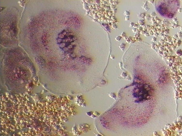
An osteoclast () is a type of bone cell that breaks down bone tissue. This function is critical in the maintenance, repair, and remodeling of bones of the vertebral skeleton. The osteoclast disassembles and digests the composite of hydrated protein and mineral at a molecular level by secreting acid and a collagenase, a process known as ''bone resorption''. This process also helps regulate the level of blood calcium. Osteoclasts are found on those surfaces of bone that are undergoing resorption. On such surfaces, the osteoclasts are seen to be located in shallow depressions called ''resorption bays (Howship's lacunae)''. The resorption bays are created by the erosive action of osteoclasts on the underlying bone. The border of the lower part of an osteoclast exhibits finger-like processes due to the presence of deep infoldings of the cell membrane; this border is called ''ruffled border''. The ruffled border lies in contact with the bone surface within a resorption bay. The periph ... [...More Info...] [...Related Items...] OR: [Wikipedia] [Google] [Baidu] |


