|
Cartilage Of The Septum
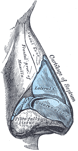
The septal nasal cartilage (cartilage of the septum or quadrangular cartilage) is composed of hyaline cartilage. It is somewhat quadrilateral in form, thicker at its margins than at its center, and completes the separation between the nasal cavities in front. Its anterior margin, thickest above, is connected with the nasal bones, and is continuous with the anterior margins of the lateral cartilages; below, it is connected to the medial crura of the major alar cartilages by fibrous tissue. Its posterior margin is connected with the perpendicular plate of the ethmoid The ethmoid bone (; from grc, ἡθμός, hēthmós, sieve) is an unpaired bone in the skull that separates the nasal cavity from the brain. It is located at the roof of the nose, between the two orbits. The cubical bone is lightweight due to a ...; its inferior margin with the vomer and the palatine processes of the maxillae. References External links * - "Nasal septum, lateral view" * - "Diagram of ske ... [...More Info...] [...Related Items...] OR: [Wikipedia] [Google] [Baidu] |
Hyaline Cartilage
Hyaline cartilage is the glass-like (hyaline) and translucent cartilage found on many joint surfaces. It is also most commonly found in the ribs, nose, larynx, and trachea. Hyaline cartilage is pearl-gray in color, with a firm consistency and has a considerable amount of collagen. It contains no nerves or blood vessels, and its structure is relatively simple. Structure Hyaline cartilage is covered externally by a fibrous membrane known as the perichondrium or, when it's along articulating surfaces, the synovial membrane. This membrane contains vessels that provide the cartilage with nutrition through diffusion. Hyaline cartilage matrix is primarily made of type II collagen and chondroitin sulphate, both of which are also found in elastic cartilage. Hyaline cartilage exists on the sternal ends of the ribs, in the larynx, trachea, and bronchi, and on the articulating surfaces of bones. It gives the structures a definite but pliable form. The presence of collagen fibres makes suc ... [...More Info...] [...Related Items...] OR: [Wikipedia] [Google] [Baidu] |
Nasal Cavities
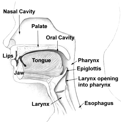
The nasal cavity is a large, air-filled space above and behind the nose in the middle of the face. The nasal septum divides the cavity into two cavities, also known as fossae. Each cavity is the continuation of one of the two nostrils. The nasal cavity is the uppermost part of the respiratory system and provides the nasal passage for inhaled air from the nostrils to the nasopharynx and rest of the respiratory tract. The paranasal sinuses surround and drain into the nasal cavity. Structure The term "nasal cavity" can refer to each of the two cavities of the nose, or to the two sides combined. The lateral wall of each nasal cavity mainly consists of the maxilla. However, there is a deficiency that is compensated for by the perpendicular plate of the palatine bone, the medial pterygoid plate, the labyrinth of ethmoid and the inferior concha. The paranasal sinuses are connected to the nasal cavity through small orifices called ostia. Most of these ostia communicate with the nos ... [...More Info...] [...Related Items...] OR: [Wikipedia] [Google] [Baidu] |
Nasal Bone
The nasal bones are two small oblong bones, varying in size and form in different individuals; they are placed side by side at the middle and upper part of the face and by their junction, form the bridge of the upper one third of the nose. Each has two surfaces and four borders. Structure The two nasal bones are joined at the midline internasal suture and make up the bridge of the nose. Surfaces The ''outer surface'' is concavo-convex from above downward, convex from side to side; it is covered by the procerus and nasalis muscles, and perforated about its center by a foramen, for the transmission of a small vein. The ''inner surface'' is concave from side to side, and is traversed from above downward, by a groove for the passage of a branch of the nasociliary nerve. Articulations The nasal articulates with four bones: two of the cranium, the frontal and ethmoid, and two of the face, the opposite nasal and the maxilla. Other animals In primitive bony fish and tetrapod ... [...More Info...] [...Related Items...] OR: [Wikipedia] [Google] [Baidu] |
Lateral Cartilage
The lateral cartilage (upper lateral cartilage, lateral process of septal nasal cartilage) is situated below the inferior margin of the nasal bone, and is flattened, and triangular in shape. Its anterior margin is thicker than the posterior, and is continuous above with the septal nasal cartilage, but separated from it below by a narrow fissure; its superior margin is attached to the nasal bone and the frontal process of the maxilla; its inferior margin is connected by fibrous tissue with the greater alar cartilage The major alar cartilage (greater alar cartilage) (lower lateral cartilage) is a thin, flexible plate, situated immediately below the lateral nasal cartilage, and bent upon itself in such a manner as to form the medial wall and lateral wall of th .... Where the lateral cartilage meets the greater alar cartilage, the lateral cartilage often curls up, to join with an inward curl of the greater alar cartilage. That curl of the inferior portion of the lateral cartilage is ... [...More Info...] [...Related Items...] OR: [Wikipedia] [Google] [Baidu] |
Major Alar Cartilage
The major alar cartilage (greater alar cartilage) (lower lateral cartilage) is a thin, flexible plate, situated immediately below the lateral nasal cartilage, and bent upon itself in such a manner as to form the medial wall and lateral wall of the nostril of its own side. The portion which forms the medial wall (crus mediale) is loosely connected with the corresponding portion of the opposite cartilage, the two forming, together with the thickened integument and subjacent tissue, the nasal septum. The part which forms the lateral wall (crus laterale) is curved to correspond with the ala of the nose; it is oval and flattened, narrow behind, where it is connected with the frontal process of the maxilla by a tough fibrous membrane, in which are found three or four small cartilaginous plates, the lesser alar cartilages (cartilagines alares minores; sesamoid cartilages). Above, it is connected by fibrous tissue to the lateral cartilage and front part of the cartilage of the septum; ... [...More Info...] [...Related Items...] OR: [Wikipedia] [Google] [Baidu] |
Ethmoid
The ethmoid bone (; from grc, ἡθμός, hēthmós, sieve) is an unpaired bone in the skull that separates the nasal cavity from the brain. It is located at the roof of the nose, between the two orbits. The cubical bone is lightweight due to a spongy construction. The ethmoid bone is one of the bones that make up the orbit of the eye. Structure The ethmoid bone is an anterior cranial bone located between the eyes. It contributes to the medial wall of the orbit, the nasal cavity, and the nasal septum. The ethmoid has three parts: cribriform plate, ethmoidal labyrinth, and perpendicular plate. The cribriform plate forms the roof of the nasal cavity and also contributes to formation of the anterior cranial fossa, the ethmoidal labyrinth consists of a large mass on either side of the perpendicular plate, and the perpendicular plate forms the superior two-thirds of the nasal septum. Between the orbital plate and the nasal conchae are the ethmoidal sinuses or ethmoidal air cells, w ... [...More Info...] [...Related Items...] OR: [Wikipedia] [Google] [Baidu] |
Palatine Process Of Maxilla
In human anatomy of the mouth, the palatine process of maxilla (palatal process), is a thick, horizontal process of the maxilla. It forms the anterior three quarters of the hard palate, the horizontal plate of the palatine bone making up the rest. Structure It is perforated by numerous foramina for the passage of the nutrient vessels; is channelled at the back part of its lateral border by a groove, sometimes a canal, for the transmission of the descending palatine vessels and the anterior palatine nerve from the spheno-palatine ganglion; and presents little depressions for the lodgement of the palatine glands. When the two maxillae are articulated, a funnel-shaped opening, the incisive foramen, is seen in the middle line, immediately behind the incisor teeth. In this opening the orifices of two lateral canals are visible; they are named the incisive canals or foramina of Stenson; through each of them passes the terminal branch of the descending palatine artery and the nasopa ... [...More Info...] [...Related Items...] OR: [Wikipedia] [Google] [Baidu] |


