|
Carotid Sinus Reflex
In human anatomy, the carotid sinus is a dilated area at the base of the internal carotid artery just superior to the bifurcation of the internal carotid and external carotid at the level of the superior border of thyroid cartilage. The carotid sinus extends from the bifurcation to the "true" internal carotid artery. The carotid sinus is sensitive to pressure changes in the arterial blood at this level. It is the major baroreception site in humans and most mammals. Structure The carotid sinus is the reflex area of the carotid artery, consisting of baroreceptors which monitor blood pressure. Function The carotid sinus contains numerous baroreceptors which function as a "sampling area" for many homeostatic mechanisms for maintaining blood pressure. The carotid sinus baroreceptors are innervated by the carotid sinus nerve, which is a branch of the glossopharyngeal nerve (CN IX). The neurons which innervate the carotid sinus centrally project to the solitary nucleus in the med ... [...More Info...] [...Related Items...] OR: [Wikipedia] [Google] [Baidu] |
Internal Carotid Artery
The internal carotid artery (Latin: arteria carotis interna) is an artery in the neck which supplies the anterior circulation of the brain. In human anatomy, the internal and external carotids arise from the common carotid arteries, where these bifurcate at cervical vertebrae C3 or C4. The internal carotid artery supplies the brain, including the eyes, while the external carotid nourishes other portions of the head, such as the face, scalp, skull, and meninges. Classification Terminologia Anatomica in 1998 subdivided the artery into four parts: "cervical", "petrous", "cavernous", and "cerebral". However, in clinical settings, the classification system of the internal carotid artery usually follows the 1996 recommendations by Bouthillier, describing seven anatomical segments of the internal carotid artery, each with a corresponding alphanumeric identifier—C1 cervical, C2 petrous, C3 lacerum, C4 cavernous, C5 clinoid, C6 ophthalmic, and C7 communicating. The Bouthillier nomenclat ... [...More Info...] [...Related Items...] OR: [Wikipedia] [Google] [Baidu] |
Innervate
A nerve is an enclosed, cable-like bundle of nerve fibers (called axons) in the peripheral nervous system. A nerve transmits electrical impulses. It is the basic unit of the peripheral nervous system. A nerve provides a common pathway for the electrochemical nerve impulses called action potentials that are transmitted along each of the axons to peripheral organs or, in the case of sensory nerves, from the periphery back to the central nervous system. Each axon, within the nerve, is an extension of an individual neuron, along with other supportive cells such as some Schwann cells that coat the axons in myelin. Within a nerve, each axon is surrounded by a layer of connective tissue called the endoneurium. The axons are bundled together into groups called fascicles, and each fascicle is wrapped in a layer of connective tissue called the perineurium. Finally, the entire nerve is wrapped in a layer of connective tissue called the epineurium. Nerve cells (often called neurons) are f ... [...More Info...] [...Related Items...] OR: [Wikipedia] [Google] [Baidu] |
Reflex
In biology, a reflex, or reflex action, is an involuntary, unplanned sequence or action and nearly instantaneous response to a stimulus. Reflexes are found with varying levels of complexity in organisms with a nervous system. A reflex occurs via neural pathways in the nervous system called reflex arcs. A stimulus initiates a neural signal, which is carried to a synapse. The signal is then transferred across the synapse to a motor neuron which evokes a target response. These neural signals do not always travel to the brain, so many reflexes are an automatic response to a stimulus that does not receive or need conscious thought. Many reflexes are fine-tuned to increase organism survival and self-defense. This is observed in reflexes such as the startle reflex, which provides an automatic response to an unexpected stimuli, and the feline righting reflex, which reorients a cat's body when falling to ensure safe landing. The simplest type of reflex, a short-latency reflex, has a s ... [...More Info...] [...Related Items...] OR: [Wikipedia] [Google] [Baidu] |
Vagus Nerve
The vagus nerve, also known as the tenth cranial nerve, cranial nerve X, or simply CN X, is a cranial nerve that interfaces with the parasympathetic control of the heart, lungs, and digestive tract. It comprises two nerves—the left and right vagus nerves—but they are typically referred to collectively as a single subsystem. The vagus is the longest nerve of the autonomic nervous system in the human body and comprises both sensory and motor fibers. The sensory fibers originate from neurons of the nodose ganglion, whereas the motor fibers come from neurons of the dorsal motor nucleus of the vagus and the nucleus ambiguus. The vagus was also historically called the pneumogastric nerve. Structure Upon leaving the medulla oblongata between the olive and the inferior cerebellar peduncle, the vagus nerve extends through the jugular foramen, then passes into the carotid sheath between the internal carotid artery and the internal jugular vein down to the neck, chest, and abdom ... [...More Info...] [...Related Items...] OR: [Wikipedia] [Google] [Baidu] |
Aortic Nerve
The aortic nerve, is a branch of the vagus nerve. It supplies autonomic afferent nerve fibers to the peripheral baroreceptors and chemoreceptors found in the aortic arch. Structure The aortic nerve is an autonomic afferent nerve fiber, and runs from the peripheral baroreceptors and chemoreceptors found in the aortic arch. It joins the vagus nerve. This allows for impulses to reach the solitary tract of the brainstem. Function The aortic nerve is part of the nerve pathway that allows for afferent impulses to be sent from the aortic arch to the medulla oblongata for control of the circulatory system. History The aortic nerve was allegedly discovered by Jewish Russian-French physiologist Elias von Cyon and German physician Carl Ludwig Carl Friedrich Wilhelm Ludwig (; 29 December 1816 – 23 April 1895) was a German physician and physiologist. His work as both a researcher and teacher had a major influence on the understanding, methods and apparatus used in almost all ... [...More Info...] [...Related Items...] OR: [Wikipedia] [Google] [Baidu] |
Aortic Arch
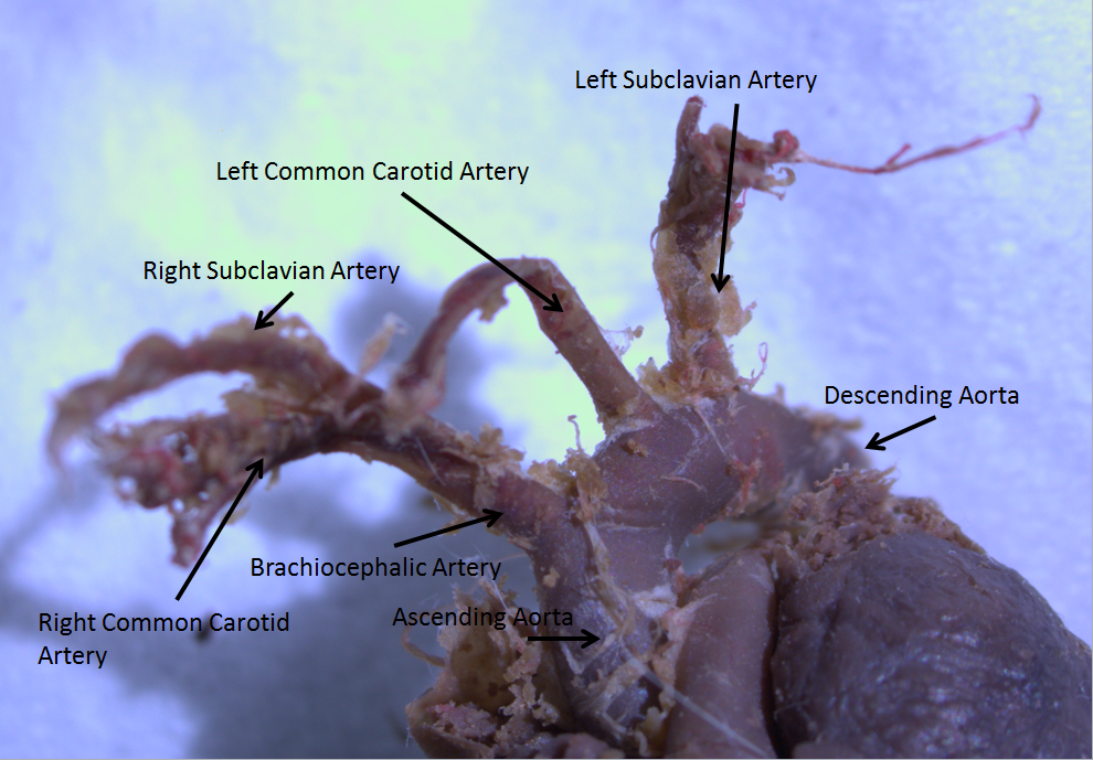
The aortic arch, arch of the aorta, or transverse aortic arch () is the part of the aorta between the ascending and descending aorta. The arch travels backward, so that it ultimately runs to the left of the trachea. Structure The aorta begins at the level of the upper border of the second/third sternocostal articulation of the right side, behind the ventricular outflow tract and pulmonary trunk. The right atrial appendage overlaps it. The first few centimeters of the ascending aorta and pulmonary trunk lies in the same pericardial sheath. and runs at first upward, arches over the pulmonary trunk, right pulmonary artery, and right main bronchus to lie behind the right second coastal cartilage. The right lung and sternum lies anterior to the aorta at this point. The aorta then passes posteriorly and to the left, anterior to the trachea, and arches over left main bronchus and left pulmonary artery, and reaches to the left side of the T4 vertebral body. Apart from T4 vertebral body ... [...More Info...] [...Related Items...] OR: [Wikipedia] [Google] [Baidu] |
Blood Vessel
The blood vessels are the components of the circulatory system that transport blood throughout the human body. These vessels transport blood cells, nutrients, and oxygen to the tissues of the body. They also take waste and carbon dioxide away from the tissues. Blood vessels are needed to sustain life, because all of the body's tissues rely on their functionality. There are five types of blood vessels: the arteries, which carry the blood away from the heart; the arterioles; the capillaries, where the exchange of water and chemicals between the blood and the tissues occurs; the venules; and the veins, which carry blood from the capillaries back towards the heart. The word ''vascular'', meaning relating to the blood vessels, is derived from the Latin ''vas'', meaning vessel. Some structures – such as cartilage, the epithelium, and the lens and cornea of the eye – do not contain blood vessels and are labeled ''avascular''. Etymology * artery: late Middle English; from Latin ... [...More Info...] [...Related Items...] OR: [Wikipedia] [Google] [Baidu] |
Heart
The heart is a muscular organ in most animals. This organ pumps blood through the blood vessels of the circulatory system. The pumped blood carries oxygen and nutrients to the body, while carrying metabolic waste such as carbon dioxide to the lungs. In humans, the heart is approximately the size of a closed fist and is located between the lungs, in the middle compartment of the chest. In humans, other mammals, and birds, the heart is divided into four chambers: upper left and right atria and lower left and right ventricles. Commonly the right atrium and ventricle are referred together as the right heart and their left counterparts as the left heart. Fish, in contrast, have two chambers, an atrium and a ventricle, while most reptiles have three chambers. In a healthy heart blood flows one way through the heart due to heart valves, which prevent backflow. The heart is enclosed in a protective sac, the pericardium, which also contains a small amount of fluid. The wall ... [...More Info...] [...Related Items...] OR: [Wikipedia] [Google] [Baidu] |
Autonomic Nervous System
The autonomic nervous system (ANS), formerly referred to as the vegetative nervous system, is a division of the peripheral nervous system that supplies viscera, internal organs, smooth muscle and glands. The autonomic nervous system is a control system that acts largely unconsciously and regulates bodily functions, such as the heart rate, its force of contraction, digestion, respiratory rate, pupillary dilation, pupillary response, Micturition, urination, and sexual arousal. This system is the primary mechanism in control of the fight-or-flight response. The autonomic nervous system is regulated by integrated reflexes through the brainstem to the spinal cord and organ (anatomy), organs. Autonomic functions include control of respiration, heart rate, cardiac regulation (the cardiac control center), vasomotor activity (the vasomotor center), and certain reflex, reflex actions such as coughing, sneezing, swallowing and vomiting. Those are then subdivided into other areas and are also ... [...More Info...] [...Related Items...] OR: [Wikipedia] [Google] [Baidu] |
Hypothalamus
The hypothalamus () is a part of the brain that contains a number of small nuclei with a variety of functions. One of the most important functions is to link the nervous system to the endocrine system via the pituitary gland. The hypothalamus is located below the thalamus and is part of the limbic system. In the terminology of neuroanatomy, it forms the ventral part of the diencephalon. All vertebrate brains contain a hypothalamus. In humans, it is the size of an almond. The hypothalamus is responsible for regulating certain metabolic processes and other activities of the autonomic nervous system. It synthesizes and secretes certain neurohormones, called releasing hormones or hypothalamic hormones, and these in turn stimulate or inhibit the secretion of hormones from the pituitary gland. The hypothalamus controls body temperature, hunger, important aspects of parenting and maternal attachment behaviours, thirst, fatigue, sleep, and circadian rhythms. Structure T ... [...More Info...] [...Related Items...] OR: [Wikipedia] [Google] [Baidu] |
Medulla Oblongata
The medulla oblongata or simply medulla is a long stem-like structure which makes up the lower part of the brainstem. It is anterior and partially inferior to the cerebellum. It is a cone-shaped neuronal mass responsible for autonomic (involuntary) functions, ranging from vomiting to sneezing. The medulla contains the cardiac, respiratory, vomiting and vasomotor centers, and therefore deals with the autonomic functions of breathing, heart rate and blood pressure as well as the sleep–wake cycle. During embryonic development, the medulla oblongata develops from the myelencephalon. The myelencephalon is a secondary vesicle which forms during the maturation of the rhombencephalon, also referred to as the hindbrain. The bulb is an archaic term for the medulla oblongata. In modern clinical usage, the word bulbar (as in bulbar palsy) is retained for terms that relate to the medulla oblongata, particularly in reference to medical conditions. The word bulbar can refer to the nerves ... [...More Info...] [...Related Items...] OR: [Wikipedia] [Google] [Baidu] |
Parasympathetic Nervous System
The parasympathetic nervous system (PSNS) is one of the three divisions of the autonomic nervous system, the others being the sympathetic nervous system and the enteric nervous system. The enteric nervous system is sometimes considered part of the autonomic nervous system, and sometimes considered an independent system. The autonomic nervous system is responsible for regulating the body's unconscious actions. The parasympathetic system is responsible for stimulation of "rest-and-digest" or "feed and breed" activities that occur when the body is at rest, especially after eating, including sexual arousal, salivation, lacrimation (tears), urination, digestion, and defecation. Its action is described as being complementary to that of the sympathetic nervous system, which is responsible for stimulating activities associated with the fight-or-flight response. Nerve fibres of the parasympathetic nervous system arise from the central nervous system. Specific nerves include several ... [...More Info...] [...Related Items...] OR: [Wikipedia] [Google] [Baidu] |


