|
Canals Of Hering
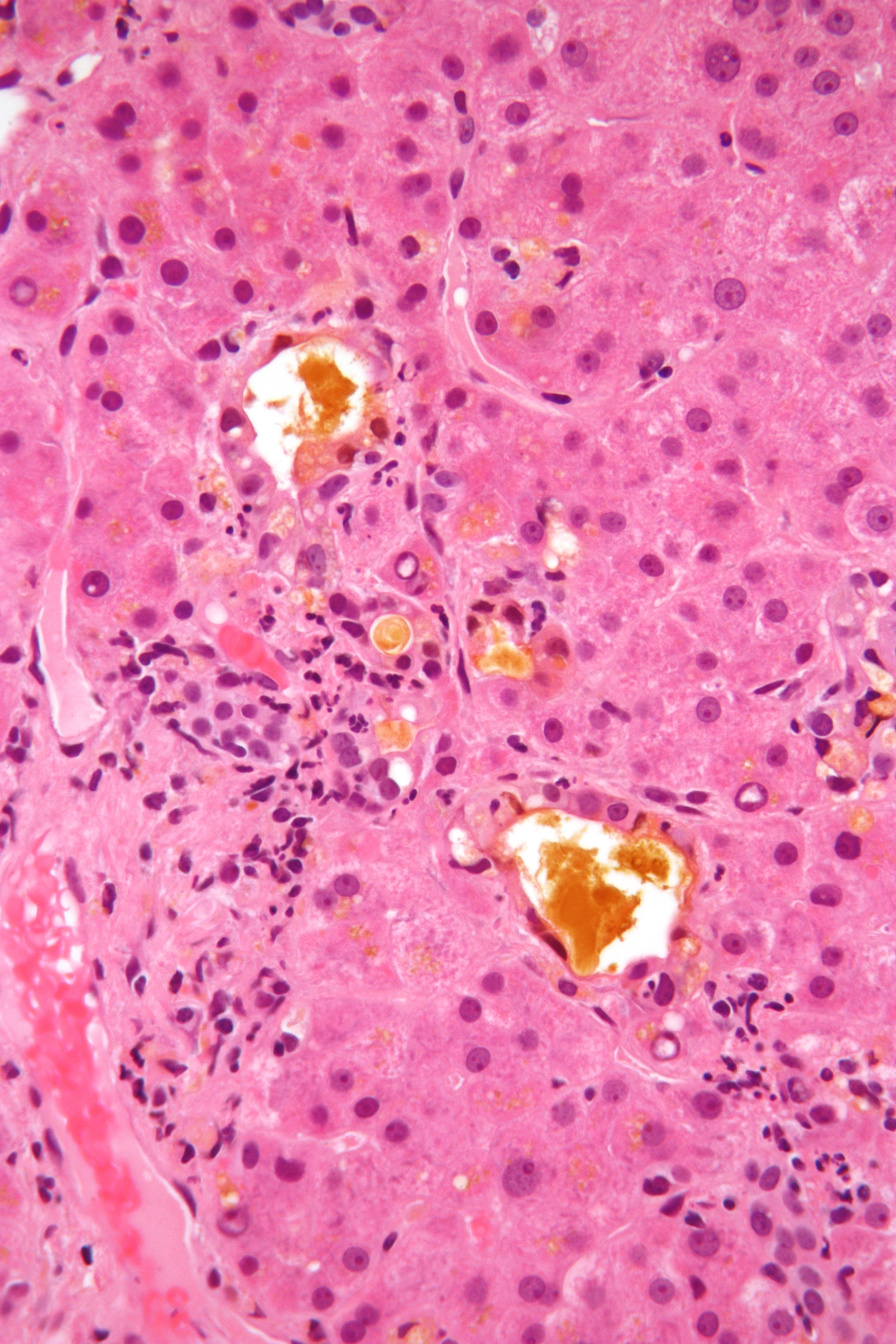
The canals of Hering, or intrahepatic bile ductules, are part of the outflow system of exocrine bile product from the liver. Liver stem cells are located in the canals of Hering. Structure They are found between the bile canaliculi and interlobular bile ducts near the outer edge of a classic liver lobule. Histology Histologically, the cells of the ductule are described as simple cuboidal epithelium, lined partially by cholangiocytes and hepatocytes. They may not be readily visible but can be differentially stained by cytokeratins CK19 and CK7. Clinical relevance The canals of Hering are destroyed early in primary biliary cholangitis and may be primary sites of scarring in methotrexate toxicity. Research has indicated the presence of intraorgan stem cells of the liver that can proliferate in disease states, so-called oval cells. History They are named for Ewald Hering Karl Ewald Konstantin Hering (5 August 1834 – 26 January 1918) was a German physiologist who did mu ... [...More Info...] [...Related Items...] OR: [Wikipedia] [Google] [Baidu] |
Bile Ductule
Bile (from Latin ''bilis''), or gall, is a dark-green-to-yellowish-brown fluid produced by the liver of most vertebrates that aids the digestion of lipids in the small intestine. In humans, bile is produced continuously by the liver (liver bile) and stored and concentrated in the gallbladder. After eating, this stored bile is discharged into the duodenum. The composition of hepatic bile is (97–98)% water, 0.7% bile salts, 0.2% bilirubin, 0.51% fats (cholesterol, fatty acids, and lecithin), and 200 meq/L inorganic salts. The two main pigments of bile are bilirubin, which is yellow, and its oxidised form biliverdin, which is green. When mixed, they are responsible for the brown color of feces. About 400 to 800 millilitres of bile is produced per day in adult human beings. Function Bile or gall acts to some extent as a surfactant, helping to emulsify the lipids in food. Bile salt anions are hydrophilic on one side and hydrophobic on the other side; consequently, they tend to ag ... [...More Info...] [...Related Items...] OR: [Wikipedia] [Google] [Baidu] |
Exocrine
Exocrine glands are glands that secrete substances on to an epithelial surface by way of a duct. Examples of exocrine glands include sweat, salivary, mammary, ceruminous, lacrimal, sebaceous, prostate and mucous. Exocrine glands are one of two types of glands in the human body, the other being endocrine glands, which secrete their products directly into the bloodstream. The liver and pancreas are both exocrine and endocrine glands; they are exocrine glands because they secrete products—bile and pancreatic juice—into the gastrointestinal tract through a series of ducts, and endocrine because they secrete other substances directly into the bloodstream. Exocrine sweat glands are part of the integumentary system; they have eccrine and apocrine types. Classification Structure Exocrine glands contain a glandular portion and a duct portion, the structures of which can be used to classify the gland. * The duct portion may be branched (called compound) or unbranched (called simple) ... [...More Info...] [...Related Items...] OR: [Wikipedia] [Google] [Baidu] |
Bile
Bile (from Latin ''bilis''), or gall, is a dark-green-to-yellowish-brown fluid produced by the liver of most vertebrates that aids the digestion of lipids in the small intestine. In humans, bile is produced continuously by the liver (liver bile) and stored and concentrated in the gallbladder. After eating, this stored bile is discharged into the duodenum. The composition of hepatic bile is (97–98)% water, 0.7% bile salts, 0.2% bilirubin, 0.51% fats (cholesterol, fatty acids, and lecithin), and 200 meq/L inorganic salts. The two main pigments of bile are bilirubin, which is yellow, and its oxidised form biliverdin, which is green. When mixed, they are responsible for the brown color of feces. About 400 to 800 millilitres of bile is produced per day in adult human beings. Function Bile or gall acts to some extent as a surfactant, helping to emulsify the lipids in food. Bile salt anions are hydrophilic on one side and hydrophobic on the other side; consequently, they tend ... [...More Info...] [...Related Items...] OR: [Wikipedia] [Google] [Baidu] |
Bile Canaliculi
Bile canaliculus (plural:bile canaliculi; also called bile capillaries) is a thin tube that collects bile secreted by hepatocytes. The bile canaliculi empty into a series of progressively larger bile ductules and ducts, which eventually become common hepatic duct. The bile canaliculi empty directly into the Canals of Hering. Hepatocytes are polyhedral in shape, therefore having no set shape or design, although they are made of cuboidal epithelial cells. They have surfaces facing the sinusoids, (called sinusoidal faces) and surfaces which contact other hepatocytes, (called lateral faces). Bile canaliculi are formed by grooves on some of the lateral faces of these hepatocytes A hepatocyte is a cell of the main parenchymal tissue of the liver. Hepatocytes make up 80% of the liver's mass. These cells are involved in: * Protein synthesis * Protein storage * Transformation of carbohydrates * Synthesis of cholesterol, .... Microvilli are present in the canaliculi. External link ... [...More Info...] [...Related Items...] OR: [Wikipedia] [Google] [Baidu] |
Interlobular Bile Ducts
The interlobular bile ducts (or interlobular ductules) carry bile in the liver between the Canals of Hering and the interlobar bile ducts. They are part of the interlobular portal triad and can be easily localized by looking for the much larger portal vein. The cells of the ducts are described as cuboidal epithelium with increasing amounts of connective tissue Connective tissue is one of the four primary types of animal tissue, along with epithelial tissue, muscle tissue, and nervous tissue. It develops from the mesenchyme derived from the mesoderm the middle embryonic germ layer. Connective tiss ... around it. References Hepatology {{digestive-stub ... [...More Info...] [...Related Items...] OR: [Wikipedia] [Google] [Baidu] |
Hepatic Lobule
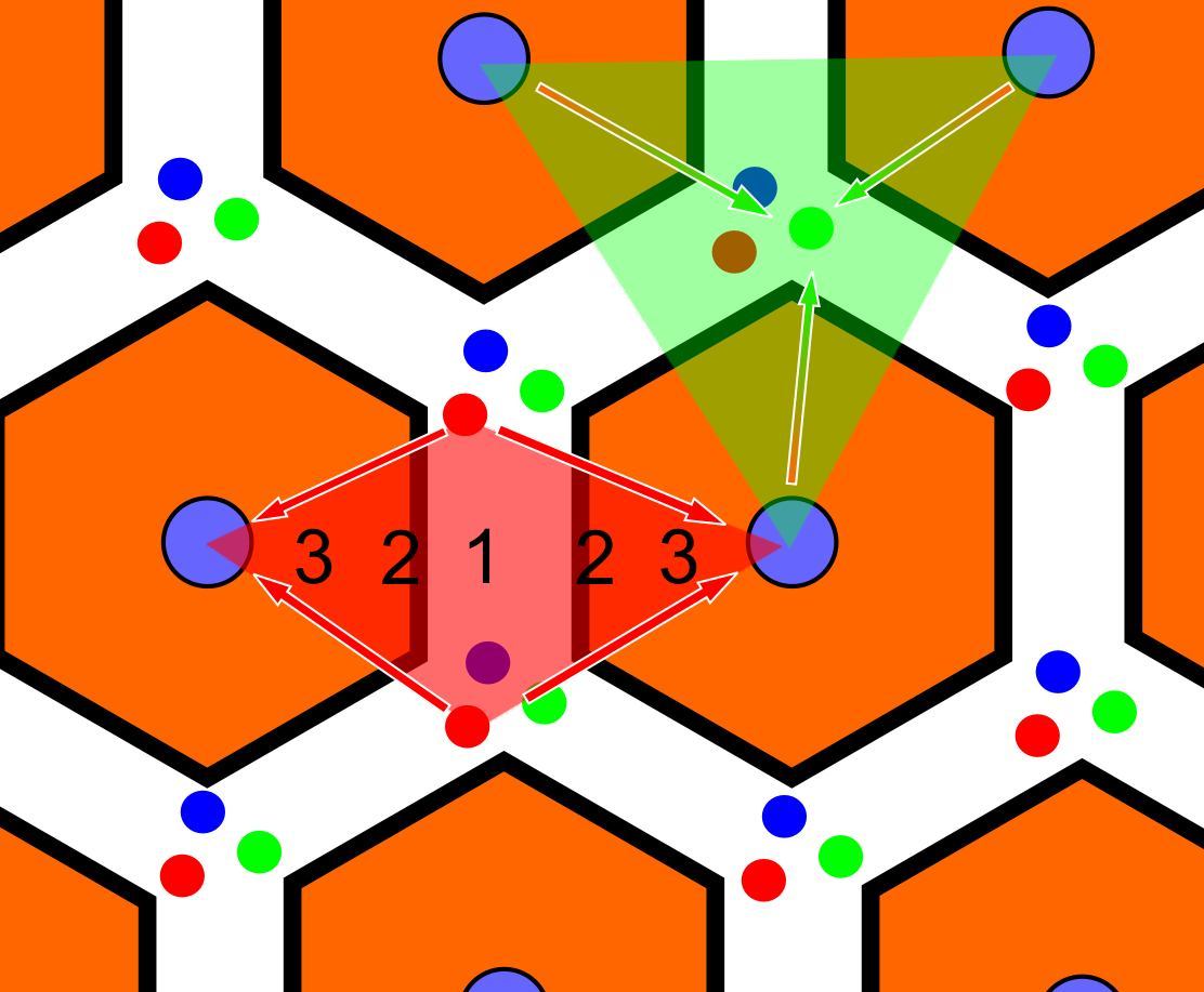
In histology (microscopic anatomy), the lobules of liver, or hepatic lobules, are small divisions of the liver defined at the microscopic scale. The hepatic lobule is a building block of the liver tissue, consisting of a portal triad, hepatocytes arranged in linear cords between a capillary network, and a central vein. Lobules are different from the lobes of liver: they are the smaller divisions of the lobes. The two-dimensional microarchitecture of the liver can be viewed from different perspectives: The term "hepatic lobule", without qualification, typically refers to the classical lobule. Structure The hepatic lobule can be described in terms of metabolic "zones", describing the hepatic acinus (terminal acinus). Each zone is centered on the line connecting two portal triads and extends outwards to the two adjacent central veins. The periportal zone I is nearest to the entering vascular supply and receives the most oxygenated blood, making it least sensitive to ischemic in ... [...More Info...] [...Related Items...] OR: [Wikipedia] [Google] [Baidu] |
Simple Cuboidal Epithelium
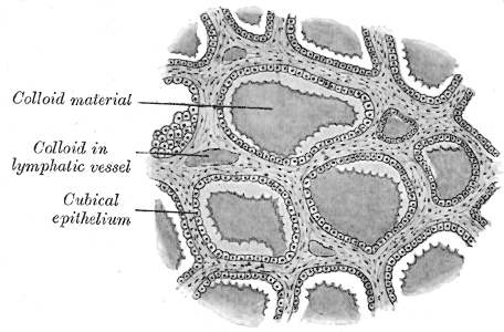
Simple cuboidal epithelium is a type of epithelium that consists of a single layer of cuboidal (cube-like) cells which have large, spherical and central nuclei. Simple cuboidal epithelium is found on the surface of ovaries, the lining of nephrons, the walls of the renal tubules, parts of the eye and thyroid, and in salivary glands. On these surfaces, the cells perform secretion and filtration. Location Simple cuboidal cells are also found in renal tubules of nephrons, glandular ducts, and thyroid follicles. Simple cuboidal cells are found in single rows with their spherical nuclei in the center of the cells and are directly attached to the basal surface. Simple ciliated cuboidal cells are also present in the respiratory bronchioles. Germinal cuboidal epithelial lines the ovaries and seminiferous tubules of testes. They undergo meiosis to form gametes A gamete (; , ultimately ) is a haploid cell that fuses with another haploid cell during fertilization in organisms th ... [...More Info...] [...Related Items...] OR: [Wikipedia] [Google] [Baidu] |
Cholangiocytes
Cholangiocytes are the epithelial cells of the bile duct. They are cuboidal epithelium in the small interlobular bile ducts, but become columnar and HCO3:-secreting in larger bile ducts approaching the porta hepatis and the extrahepatic ducts. They contribute to hepatocyte survival by transporting bile acids. Function In the healthy liver, cholangiocytes contribute to bile secretion via release of bicarbonate and water. Several hormones and locally acting mediators are known to contribute to cholangiocyte fluid/electrolyte secretion. These include secretin, acetylcholine, ATP, and bombesin. Cholangiocytes act through bile-acid independent bile flow, which is driven by the active transport of electrolytes. In contrast, hepatocytes secrete bile through bile-acid dependent bile flow, which is coupled to canalicular secretion of bile acids via ATP-driven transporters. This results in passive transcellular and paracellular secretion of fluid and electrolytes through an osmotic effect ... [...More Info...] [...Related Items...] OR: [Wikipedia] [Google] [Baidu] |
Hepatocytes
A hepatocyte is a cell of the main parenchymal tissue of the liver. Hepatocytes make up 80% of the liver's mass. These cells are involved in: * Protein synthesis * Protein storage * Transformation of carbohydrates * Synthesis of cholesterol, bile salts and phospholipids * Detoxification, modification, and excretion of exogenous and endogenous substances * Initiation of formation and secretion of bile Structure The typical hepatocyte is cubical with sides of 20-30 μm, (in comparison, a human hair has a diameter of 17 to 180 μm).The diameter of human hair ranges from 17 to 181 μm. The typical volume of a hepatocyte is 3.4 x 10⁻⁹ cm3. Smooth endoplasmic reticulum is abundant in hepatocytes, in contrast to most other cell types. Microanatomy Hepatocytes display an eosinophilic cytoplasm, reflecting numerous mitochondria, and basophilic stippling due to large amounts of smooth endoplasmic reticulum and free ribosomes. Brown lipofuscin granules are also observed ( ... [...More Info...] [...Related Items...] OR: [Wikipedia] [Google] [Baidu] |
Differential Staining
Differential staining is a staining process which uses more than one chemical staining, stain. Using multiple stains can better differentiate between different microorganisms or structures/cellular components of a single organism. Differential staining is used to detect abnormalities in the proportion of different white blood cells in the blood. The process or results are called a WBC differential. This test is useful because many diseases alter the proportion of certain white blood cells. By analyzing these differences in combination with a clinical exam and other lab tests, medical professionals can diagnose disease. One commonly recognizable use of differential staining is the Gram stain. Gram staining uses two dyes: Crystal violet and Fuchsin or Safranin (the counterstain) to differentiate between Gram-positive bacteria (large Peptidoglycan layer on outer surface of cell) and Gram-negative bacteria. Acid-fast stains are also differential stains. Further reading *http://ww ... [...More Info...] [...Related Items...] OR: [Wikipedia] [Google] [Baidu] |
Primary Biliary Cirrhosis
Primary biliary cholangitis (PBC), previously known as primary biliary cirrhosis, is an autoimmune disease of the liver. It results from a slow, progressive destruction of the small bile ducts of the liver, causing bile and other toxins to build up in the liver, a condition called cholestasis. Further slow damage to the liver tissue can lead to scarring, fibrosis, and eventually cirrhosis. Common symptoms are tiredness, itching, and in more advanced cases, jaundice. In early cases, the only changes may be those seen in blood tests. PBC is a relatively rare disease, affecting up to one in 3,000–4,000 people. It is much more common in women, with a sex ratio of at least 9:1 female to male. The condition has been recognised since at least 1851, and was named "primary biliary cirrhosis" in 1949. Because cirrhosis is a feature only of advanced disease, a change of its name to "primary biliary cholangitis" was proposed by patient advocacy groups in 2014. Signs and symptoms Pe ... [...More Info...] [...Related Items...] OR: [Wikipedia] [Google] [Baidu] |
Methotrexate
Methotrexate (MTX), formerly known as amethopterin, is a chemotherapy agent and immune-system suppressant. It is used to treat cancer, autoimmune diseases, and ectopic pregnancies. Types of cancers it is used for include breast cancer, leukemia, lung cancer, lymphoma, gestational trophoblastic disease, and osteosarcoma. Types of autoimmune diseases it is used for include psoriasis, rheumatoid arthritis, and Crohn's disease. It can be given by mouth or by injection. Common side effects include nausea, feeling tired, fever, increased risk of infection, low white blood cell counts, and breakdown of the skin inside the mouth. Other side effects may include liver disease, lung disease, lymphoma, and severe skin rashes. People on long-term treatment should be regularly checked for side effects. It is not safe during breastfeeding. In those with kidney problems, lower doses may be needed. It acts by blocking the body's use of folic acid. Methotrexate was first made in 1947 and ... [...More Info...] [...Related Items...] OR: [Wikipedia] [Google] [Baidu] |


