|
Calcifediol
Calcifediol, also known as calcidiol, 25-hydroxycholecalciferol, or 25-hydroxyvitamin D3 (abbreviated 25(OH)D3), is a form of vitamin D produced in the liver by hydroxylation of vitamin D3 (cholecalciferol) by the enzyme vitamin D 25-hydroxylase. Calcifediol can be further hydroxylated by the enzyme 25(OH)D-1α-hydroxylase, primarily in the kidney, to form calcitriol (1,25-(OH)2D3), which is the active hormonal form of vitamin D. Calcifediol is strongly bound in blood by the vitamin D-binding protein. Measurement of serum calcifediol is the usual test performed to determine a person's vitamin D status, to show vitamin D deficiency or sufficiency. Calcifediol is available as an oral medication in some countries to supplement vitamin D status. Biology Calcifediol is the precursor for calcitriol, the active form of vitamin D. It is synthesized in the liver, by hydroxylation of cholecalciferol (vitamin D3) at the 25-position. This enzymatic 25-hydroxylase reaction is mostly due t ... [...More Info...] [...Related Items...] OR: [Wikipedia] [Google] [Baidu] |
Vitamin D
Vitamin D is a group of fat-soluble secosteroids responsible for increasing intestinal absorption of calcium, magnesium, and phosphate, and many other biological effects. In humans, the most important compounds in this group are vitamin D3 (cholecalciferol) and vitamin D2 (ergocalciferol). The major natural source of the vitamin is synthesis of cholecalciferol in the lower layers of epidermis of the skin through a chemical reaction that is dependent on sun exposure (specifically UVB radiation). Cholecalciferol and ergocalciferol can be ingested from the diet and supplements. Only a few foods, such as the flesh of fatty fish, naturally contain significant amounts of vitamin D. In the U.S. and other countries, cow's milk and plant-derived milk substitutes are fortified with vitamin D, as are many breakfast cereals. Mushrooms exposed to ultraviolet light contribute useful amounts of vitamin D2. Dietary recommendations typically assume that all of a person's vitamin D is taken ... [...More Info...] [...Related Items...] OR: [Wikipedia] [Google] [Baidu] |
CYP2R1
CYP2R1 is cytochrome P450 2R1, an enzyme which is the principal vitamin D 25-hydroxylase. In humans it is encoded by the ''CYP2R1'' gene located on chromosome 11p15.2. It is expressed in the endoplasmic reticulum in liver, where it performs the first step in the activation of vitamin D by catalyzing the formation of 25-hydroxyvitamin D. Vitamin D 25-hydroxylase activity is also possessed by some other cytochrome P450 enzymes, in particular CYP27A1, which is found in mitochondria. Function CYP2R1 is a member of the cytochrome P450 superfamily of enzymes. The cytochrome P450 proteins are mono-oxygenases which catalyze many reactions involved in drug metabolism and synthesis of cholesterol, steroids and other lipids. CYP2R1 is present in the endoplasmic reticulum of the liver (the microsomal fraction). It has 25-hydroxylase activity, which converts cholecalciferol (vitamin D3) into calcifediol (25-hydroxyvitamin D3, also known as calcidiol), the major circulatory form of the vi ... [...More Info...] [...Related Items...] OR: [Wikipedia] [Google] [Baidu] |
Vitamin D 25-hydroxylase
CYP2R1 is cytochrome P450 2R1, an enzyme which is the principal vitamin D 25-hydroxylase. In humans it is encoded by the ''CYP2R1'' gene located on chromosome 11p15.2. It is expressed in the endoplasmic reticulum in liver, where it performs the first step in the activation of vitamin D by catalyzing the formation of 25-hydroxyvitamin D. Vitamin D 25-hydroxylase activity is also possessed by some other cytochrome P450 enzymes, in particular CYP27A1, which is found in mitochondria. Function CYP2R1 is a member of the cytochrome P450 superfamily of enzymes. The cytochrome P450 proteins are mono-oxygenases which catalyze many reactions involved in drug metabolism and synthesis of cholesterol, steroids and other lipids. CYP2R1 is present in the endoplasmic reticulum of the liver (the microsomal fraction). It has 25-hydroxylase activity, which converts cholecalciferol (vitamin D3) into calcifediol (25-hydroxyvitamin D3, also known as calcidiol), the major circulatory form of the vit ... [...More Info...] [...Related Items...] OR: [Wikipedia] [Google] [Baidu] |
Calcitriol
Calcitriol is the active form of vitamin D, normally made in the kidney. It is also known as 1,25-dihydroxycholecalciferol. It is a hormone which binds to and activates the vitamin D receptor in the nucleus of the cell, which then increases the expression of many genes. Calcitriol increases blood calcium (Ca2+) mainly by increasing the uptake of calcium from the intestines. It can be given as a medication for the treatment of low blood calcium and hyperparathyroidism due to kidney disease, low blood calcium due to hypoparathyroidism, osteoporosis, osteomalacia, and familial hypophosphatemia, and can be taken by mouth or by injection into a vein. Excessive amounts or intake can result in weakness, headache, nausea, constipation, urinary tract infections, and abdominal pain. Serious side effects may include high blood calcium and anaphylaxis. Regular blood tests are recommended after the medication is started and when the dose is changed. Calcitriol was identified as the active ... [...More Info...] [...Related Items...] OR: [Wikipedia] [Google] [Baidu] |
Vitamin D Deficiency
Vitamin D deficiency or hypovitaminosis D is a vitamin D level that is below normal. It most commonly occurs in people when they have inadequate exposure to sunlight, particularly sunlight with adequate ultraviolet B rays (UVB). Vitamin D deficiency can also be caused by inadequate nutritional intake of vitamin D; disorders that limit vitamin D absorption; and disorders that impair the conversion of vitamin D to active metabolites, including certain liver, kidney, and hereditary disorders. Deficiency impairs bone mineralization, leading to bone-softening diseases, such as rickets in children. It can also worsen osteomalacia and osteoporosis in adults, increasing the risk of bone fractures. Muscle weakness is also a common symptom of vitamin D deficiency, further increasing the risk of fall and bone fractures in adults. Vitamin D deficiency is associated with the development of schizophrenia. Vitamin D can be synthesized in the skin under the exposure of UVB from sunlight. Oily ... [...More Info...] [...Related Items...] OR: [Wikipedia] [Google] [Baidu] |
Monocytes
Monocytes are a type of leukocyte or white blood cell. They are the largest type of leukocyte in blood and can differentiate into macrophages and conventional dendritic cells. As a part of the vertebrate innate immune system monocytes also influence adaptive immune responses and exert tissue repair functions. There are at least three subclasses of monocytes in human blood based on their phenotypic receptors. Structure Monocytes are amoeboid in appearance, and have nongranulated cytoplasm. Thus they are classified as agranulocytes, although they might occasionally display some azurophil granules and/or vacuoles. With a diameter of 15–22 μm, monocytes are the largest cell type in peripheral blood. Monocytes are mononuclear cells and the ellipsoidal nucleus is often lobulated/indented, causing a bean-shaped or kidney-shaped appearance. Monocytes compose 2% to 10% of all leukocytes in the human body. Development Monocytes are produced by the bone marrow from precursors cal ... [...More Info...] [...Related Items...] OR: [Wikipedia] [Google] [Baidu] |
Keratinocytes
Keratinocytes are the primary type of cell found in the epidermis, the outermost layer of the skin. In humans, they constitute 90% of epidermal skin cells. Basal cells in the basal layer (''stratum basale'') of the skin are sometimes referred to as basal keratinocytes. Keratinocytes form a barrier against environmental damage by heat, UV radiation, water loss, pathogenic bacteria, fungi, parasites, and viruses. A number of structural proteins, enzymes, lipids, and antimicrobial peptides contribute to maintain the important barrier function of the skin. Keratinocytes differentiate from epidermal stem cells in the lower part of the epidermis and migrate towards the surface, finally becoming corneocytes and eventually be shed off, which happens every 40 to 56 days in humans. Function The primary function of keratinocytes is the formation of a barrier against environmental damage by heat, UV radiation, water loss, pathogenic bacteria, fungi, parasites, and viruses. Pathogen ... [...More Info...] [...Related Items...] OR: [Wikipedia] [Google] [Baidu] |
Placenta
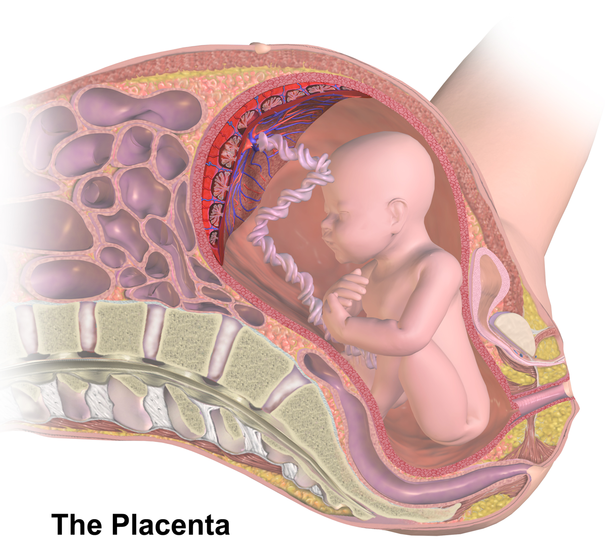
The placenta is a temporary embryonic and later fetal organ that begins developing from the blastocyst shortly after implantation. It plays critical roles in facilitating nutrient, gas and waste exchange between the physically separate maternal and fetal circulations, and is an important endocrine organ, producing hormones that regulate both maternal and fetal physiology during pregnancy. The placenta connects to the fetus via the umbilical cord, and on the opposite aspect to the maternal uterus in a species-dependent manner. In humans, a thin layer of maternal decidual (endometrial) tissue comes away with the placenta when it is expelled from the uterus following birth (sometimes incorrectly referred to as the 'maternal part' of the placenta). Placentas are a defining characteristic of placental mammals, but are also found in marsupials and some non-mammals with varying levels of development. Mammalian placentas probably first evolved about 150 million to 200 million years ... [...More Info...] [...Related Items...] OR: [Wikipedia] [Google] [Baidu] |
Parathyroid Gland
Parathyroid glands are small endocrine glands in the neck of humans and other tetrapods. Humans usually have four parathyroid glands, located on the back of the thyroid gland in variable locations. The parathyroid gland produces and secretes parathyroid hormone in response to a low blood calcium, which plays a key role in regulating the amount of calcium in the blood and within the bones. Parathyroid glands share a similar blood supply, venous drainage, and lymphatic drainage to the thyroid glands. Parathyroid glands are derived from the epithelial lining of the third and fourth pharyngeal pouches, with the superior glands arising from the fourth pouch and the inferior glands arising from the higher third pouch. The relative position of the inferior and superior glands, which are named according to their final location, changes because of the migration of embryological tissues. Hyperparathyroidism and hypoparathyroidism, characterized by alterations in the blood calcium levels ... [...More Info...] [...Related Items...] OR: [Wikipedia] [Google] [Baidu] |
CYP24A1
Cytochrome P450 family 24 subfamily A member 1 (abbreviated CYP24A1) is a member of the cytochrome P450 superfamily of enzymes encoded by the ''CYP24A1'' gene. It is a mitochondrial monooxygenase which catalyzes reactions including 24-hydroxylation of calcitriol (1,25-dihydroxyvitamin D3). It has also been identified as vitamin D3 24-hydroxylase.() Function CYP24A1 is an enzyme expressed in the mitochondrion of humans and other species. It catalyzes hydroxylation reactions which lead to the degradation of 1,25-dihydroxyvitamin D3, the physiologically active form of vitamin D. Hydroxylation of the side chain produces calcitroic acid and other metabolites which are excreted in bile. CYP24A1 was identified in the early 1970s and was first thought to be involved in vitamin D metabolism as the renal 25-hydroxyvitamin D3-24-hydroxylase, modifying calcifediol (25-hydroxyvitamin D) to produce 24,25-dihydroxycholecalciferol (24,25-dihydroxyvitamin D). Subsequent studies using r ... [...More Info...] [...Related Items...] OR: [Wikipedia] [Google] [Baidu] |
FGF23
Fibroblast growth factor 23 (FGF23) is a protein and member of the fibroblast growth factor (FGF) family which participates in the regulation of phosphate in plasma and vitamin D metabolism. In humans it is encoded by the gene. FGF23 decreases reabsorption of phosphate in the kidney. Mutations in ''FGF23'' can lead to its increased activity, resulting in autosomal dominant hypophosphatemic rickets. Description Fibroblast growth factor 23 (FGF23) is a protein which in humans is encoded by the gene. FGF23 is a member of the fibroblast growth factor (FGF) family which participates in phosphate and vitamin D metabolism and regulation. Function FGF23´s main function is to regulate the phosphate concentration in plasma. It does this by decreasing reabsorption of phosphate in the kidney, which means phosphate is excreted in urine. FGF23 is secreted by osteocytes in response to increased calcitriol. FGF23 acts on the kidneys by decreasing the expression of NPT2, a sodium-phosphate co ... [...More Info...] [...Related Items...] OR: [Wikipedia] [Google] [Baidu] |
.png)
.png)


