|
Cytopathology
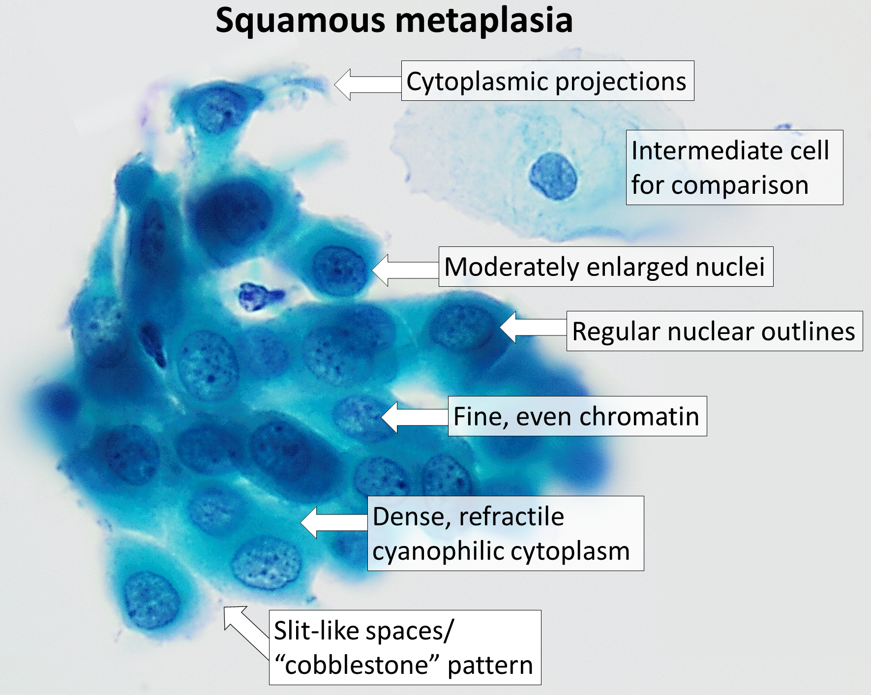
Cytopathology (from Greek , ''kytos'', "a hollow"; , ''pathos'', "fate, harm"; and , '' -logia'') is a branch of pathology that studies and diagnoses diseases on the cellular level. The discipline was founded by George Nicolas Papanicolaou in 1928. Cytopathology is generally used on samples of free cells or tissue fragments, in contrast to histopathology, which studies whole tissues. Cytopathology is frequently, less precisely, called "cytology", which means "the study of cells". Cytopathology is commonly used to investigate diseases involving a wide range of body sites, often to aid in the diagnosis of cancer but also in the diagnosis of some infectious diseases and other inflammatory conditions. For example, a common application of cytopathology is the Pap smear, a screening tool used to detect precancerous cervical lesions that may lead to cervical cancer. Cytopathologic tests are sometimes called smear tests because the samples may be smeared across a glass microscope slid ... [...More Info...] [...Related Items...] OR: [Wikipedia] [Google] [Baidu] |
Low-grade Squamous Intraepithelial Lesion
The Bethesda system (TBS), officially called The Bethesda System for Reporting Cervical Cytology, is a system for reporting cervical or vaginal cytologic diagnoses, used for reporting Pap smear results. It was introduced in 1988 and revised in 1991, 2001, and 2014. The name comes from the location (Bethesda, Maryland) of the conference, sponsored by the National Institutes of Health, that established the system. Since 2010, there is also a Bethesda system used for cytopathology of thyroid nodules, which is called The Bethesda System for Reporting Thyroid Cytopathology (TBSRTC or BSRTC). Like TBS, it was the result of a conference sponsored by the NIH and is published in book editions (currently by Springer). Mentions of "the Bethesda system" without further specification usually refer to the cervical system, unless the thyroid context of a discussion is implicit. Cervix Abnormal results include: * Atypical squamous cells ** Atypical squamous cells of undetermined significance (A ... [...More Info...] [...Related Items...] OR: [Wikipedia] [Google] [Baidu] |
Pap Smears
The Papanicolaou test (abbreviated as Pap test, also known as Pap smear (AE), cervical smear (BE), cervical screening (BE), or smear test (BE)) is a method of cervical screening used to detect potentially precancerous and cancerous processes in the cervix (opening of the uterus or womb) or colon (in both men and women). Abnormal findings are often followed up by more sensitive diagnostic procedures and, if warranted, interventions that aim to prevent progression to cervical cancer. The test was independently invented in the 1920s by Georgios Papanikolaou and Aurel Babeș and named after Papanikolaou. A simplified version of the test was introduced by Anna Marion Hilliard in 1957. A Pap smear is performed by opening the vagina with a speculum and collecting cells at the outer opening of the cervix at the transformation zone (where the outer squamous cervical cells meet the inner glandular endocervical cells), using an Ayre spatula or a cytobrush. A similar method is used to col ... [...More Info...] [...Related Items...] OR: [Wikipedia] [Google] [Baidu] |
George Nicolas Papanicolaou
Georgios Nikolaou Papanikolaou (or George Papanicolaou ; el, Γεώργιος Ν. Παπανικολάου ; 13 May 1883 – 19 February 1962) was a Greek physician who was a pioneer in cytopathology and early cancer detection, and inventor of the "Pap smear". After studying medicine in Greece and Germany, he emigrated in 1913 to the United States. He first reported that uterine cancer cells could be detected in vaginal smears in 1928, but his work was not widely recognized until the 1940s. An extensive trial of his techniques was carried out in the early 1950s. In 1961, he was invited to the University of Miami to lead and develop the Papanicolaou Cancer Research Institute there. Life Born in Kymi, Greece, Papanikolaou attended the University of Athens, where he studied literature, philosophy, languages and music. Urged by his father, he pursued a medical degree, which he received in 1904. Afterwards, he was conscripted into military service. When his obligation ended in 19 ... [...More Info...] [...Related Items...] OR: [Wikipedia] [Google] [Baidu] |
Pap Smear
The Papanicolaou test (abbreviated as Pap test, also known as Pap smear (AE), cervical smear (BE), cervical screening (BE), or smear test (BE)) is a method of cervical screening used to detect potentially precancerous and cancerous processes in the cervix (opening of the uterus or womb) or Colon (organ), colon (in both men and women). Abnormal findings are often followed up by more sensitive diagnostic procedures and, if warranted, interventions that aim to prevent progression to cervical cancer. The test was independently invented in the 1920s by Georgios Papanikolaou and Aurel Babeș and named after Papanikolaou. A simplified version of the test was introduced by Anna Marion Hilliard in 1957. A Pap smear is performed by opening the vagina with a speculum (medical), speculum and collecting cell (biology), cells at the outer opening of the cervix at the transformation zone (where the outer squamous cervical cells meet the inner glandular endocervical cells), using an Ayre spatula ... [...More Info...] [...Related Items...] OR: [Wikipedia] [Google] [Baidu] |
Papanicolaou Stain
Papanicolaou stain (also Papanicolaou's stain and Pap stain) is a multichromatic (multicolored) cytological staining technique developed by George Papanicolaou in 1942. The Papanicolaou stain is one of the most widely used stains in cytology, where it is used to aid pathologists in making a diagnosis. Although most notable for its use in the detection of cervical cancer in the Pap test or Pap smear, it is also used to stain non-gynecological specimen preparations from a variety of bodily secretions and from small needle biopsies of organs and tissues. Papanicolaou published three formulations of this stain in 1942, 1954, and 1960. Usage Pap staining is used to differentiate cells in smear preparations (in which samples are spread or smeared onto a glass microscope slide) from various bodily secretions and needle biopsies; the specimens may include gynecological smears (Pap smears), sputum, brushings, washings, urine, cerebrospinal fluid, abdominal fluid, pleural fluid, syno ... [...More Info...] [...Related Items...] OR: [Wikipedia] [Google] [Baidu] |
Pathology
Pathology is the study of the causes and effects of disease or injury. The word ''pathology'' also refers to the study of disease in general, incorporating a wide range of biology research fields and medical practices. However, when used in the context of modern medical treatment, the term is often used in a narrower fashion to refer to processes and tests that fall within the contemporary medical field of "general pathology", an area which includes a number of distinct but inter-related medical specialties that diagnose disease, mostly through analysis of tissue, cell, and body fluid samples. Idiomatically, "a pathology" may also refer to the predicted or actual progression of particular diseases (as in the statement "the many different forms of cancer have diverse pathologies", in which case a more proper choice of word would be " pathophysiologies"), and the affix ''pathy'' is sometimes used to indicate a state of disease in cases of both physical ailment (as in cardiomy ... [...More Info...] [...Related Items...] OR: [Wikipedia] [Google] [Baidu] |
Cell Biology
Cell biology (also cellular biology or cytology) is a branch of biology that studies the structure, function, and behavior of cells. All living organisms are made of cells. A cell is the basic unit of life that is responsible for the living and functioning of organisms. Cell biology is the study of structural and functional units of cells. Cell biology encompasses both prokaryotic and eukaryotic cells and has many subtopics which may include the study of cell metabolism, cell communication, cell cycle, biochemistry, and cell composition. The study of cells is performed using several microscopy techniques, cell culture, and cell fractionation. These have allowed for and are currently being used for discoveries and research pertaining to how cells function, ultimately giving insight into understanding larger organisms. Knowing the components of cells and how cells work is fundamental to all biological sciences while also being essential for research in biomedical fields such as ... [...More Info...] [...Related Items...] OR: [Wikipedia] [Google] [Baidu] |
Cytocentrifugation
A cytocentrifuge, sometimes referred to as a cytospin, is a specialized centrifuge used to concentrate cells in fluid specimens onto a microscope slide so that they can be stained and examined. Cytocentrifuges are used in various areas of the clinical laboratory, such as cytopathology, hematology and microbiology, as well as in biological research. The method can be used on many different types of specimens, including fine needle aspirates, cerebrospinal fluid, serous and synovial fluid, and urine. Procedure To prepare cytocentrifuge smears, a funnel assembly is attached to the front of a microscope slide. The surface of the funnel assembly that is in contact with the slide is lined with filter paper to absorb excess fluid. A few drops of fluid are placed in the funnel. The assembly is placed in the cytocentrifuge, which operates at a low force (600–800 x g) to preserve cellular structure. Centrifugal force pushes the fluid through the funnel's opening and concentrates the cells ... [...More Info...] [...Related Items...] OR: [Wikipedia] [Google] [Baidu] |
Cervical Intraepithelial Neoplasia
Cervical intraepithelial neoplasia (CIN), also known as cervical dysplasia, is the abnormal growth of cells on the surface of the cervix that could potentially lead to cervical cancer. More specifically, CIN refers to the potentially precancerous transformation of cells of the cervix. CIN most commonly occurs at the squamocolumnar junction of the cervix, a transitional area between the squamous epithelium of the vagina and the columnar epithelium of the endocervix. It can also occur in vaginal walls and vulvar epithelium. CIN is graded on a 1–3 scale, with 3 being the most abnormal (see classification section below). Human papillomavirus (HPV) infection is necessary for the development of CIN, but not all with this infection develop cervical cancer. Many women with HPV infection never develop CIN or cervical cancer. Typically, HPV resolves on its own. However, those with an HPV infection that lasts more than one or two years have a higher risk of developing a higher grade of CIN. ... [...More Info...] [...Related Items...] OR: [Wikipedia] [Google] [Baidu] |
Liquid-based Cytology
Liquid-based cytology is a method of preparing samples for examination in cytopathology. The sample is collected, normally by a small brush, in the same way as for a conventional smear test, but rather than the smear being transferred directly to a microscope slide, the sample is deposited into a small bottle of preservative liquid. At the laboratory, the liquid is treated to remove other elements such as mucus before a layer of cells is placed on a slide. History For many years, efforts have been made to develop methods that would enhance the sensitivity and specificity of the Papanicolaou smear (also called Pap smear). Emphasis has been placed on creating automated screening machines whose success depends on a representative sampling of cells on standardized slides containing a monolayer of well-stained, well-preserved cells. From this research and development, liquid-based gynecologic specimen collection has evolved. Its proponents argue that liquid-based preparations outperfo ... [...More Info...] [...Related Items...] OR: [Wikipedia] [Google] [Baidu] |
Serous Carcinoma 2c - Cytology
In physiology, serous fluid or serosal fluid (originating from the Medieval Latin word ''serosus'', from Latin ''serum'') is any of various body fluids resembling Serum (blood), serum, that are typically pale yellow or transparent and of a benign nature. The fluid fills the inside of body cavity, body cavities. Serous fluid originates from serous glands, with secretions enriched with proteins and water. Serous fluid may also originate from mixed glands, which contain both mucous cell, mucous and serous cells. A common trait of serous fluids is their role in assisting digestion, excretion, and respiratory system, respiration. In medical fields, especially cytopathology, serous fluid is a synonym for effusion fluids from various body cavities. Examples of effusion fluid are pleural effusion and pericardial effusion. There are many causes of effusions which include involvement of the cavity by cancer. Cancer in a serous cavity is called a serous carcinoma. Cytopathology evaluation is ... [...More Info...] [...Related Items...] OR: [Wikipedia] [Google] [Baidu] |
Screening (medicine)
Screening, in medicine, is a strategy used to look for as-yet-unrecognised conditions or risk markers. This testing can be applied to individuals or to a whole population. The people tested may not exhibit any signs or symptoms of a disease, or they might exhibit only one or two symptoms, which by themselves do not indicate a definitive diagnosis. Screening interventions are designed to identify conditions which could at some future point turn into disease, thus enabling earlier intervention and management in the hope to reduce mortality and suffering from a disease. Although screening may lead to an earlier diagnosis, not all screening tests have been shown to benefit the person being screened; overdiagnosis, misdiagnosis, and creating a false sense of security are some potential adverse effects of screening. Additionally, some screening tests can be inappropriately overused. For these reasons, a test used in a screening program, especially for a disease with low incidence, must ... [...More Info...] [...Related Items...] OR: [Wikipedia] [Google] [Baidu] |


.jpg)


.jpg)