|
Cuboid Bone
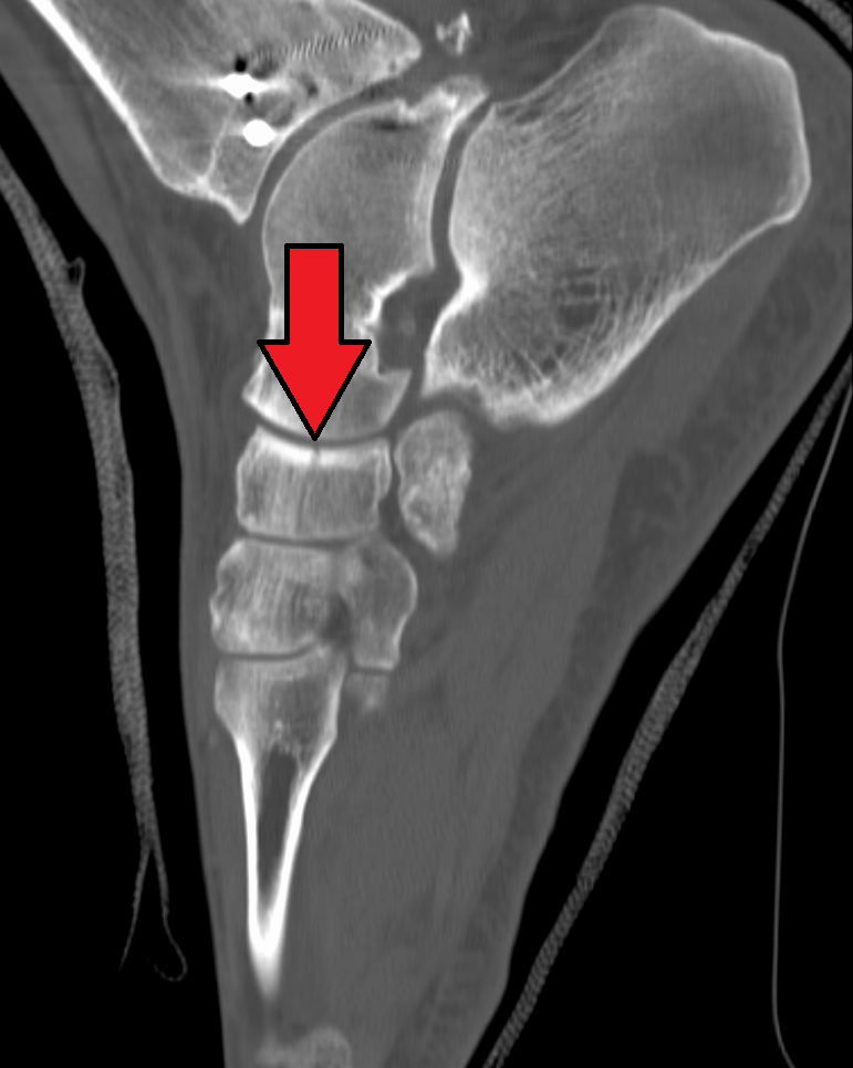
In the human body, the cuboid bone is one of the seven tarsal bones of the foot. Structure The cuboid bone is the most lateral of the bones in the distal row of the tarsus. It is roughly cubical in shape, and presents a prominence in its inferior (or plantar) surface, the tuberosity of the cuboid. The bone provides a groove where the tendon of the peroneus longus muscle passes to reach its insertion in the first metatarsal and medial cuneiform bones. Surfaces The dorsal surface, directed upward and lateralward, is rough, for the attachment of ligaments. The plantar surface presents in front a deep groove, the peroneal sulcus, which runs obliquely forward and medialward; it lodges the tendon of the peroneus longus, and is bounded behind by a prominent ridge, to which the long plantar ligament is attached. The ridge ends laterally in an eminence, the tuberosity, the surface of which presents an oval facet; on this facet glides the sesamoid bone or cartilage frequently found ... [...More Info...] [...Related Items...] OR: [Wikipedia] [Google] [Baidu] |
Calcaneocuboid Articulation
The calcaneocuboid joint is the joint between the calcaneus and the cuboid bone. Structure The calcaneocuboid joint is a type of saddle joint between the calcaneus and the cuboid bone. Ligaments There are five ligaments connecting the calcaneus and the cuboid bone, forming parts of the articular capsule: * the dorsal calcaneocuboid ligament. * part of the bifurcated ligament. * the long plantar ligament. * and the plantar calcaneocuboid ligament. Function The calcaneocuboid joint is conventionally described as among the least mobile joints in the human foot. The articular surfaces of the two bones are relatively flat with some irregular undulations, which seem to suggest movement limited to a single rotation and some translation. However, the cuboid rotates as much as 25° about an oblique axis during inversion-eversion in a movement that could be called involution. Clinical significance The calcaneocuboid joint may be affected by a calcaneal fracture. This may be a ... [...More Info...] [...Related Items...] OR: [Wikipedia] [Google] [Baidu] |
Tibialis Posterior
The tibialis posterior muscle is the most central of all the leg muscles, and is located in the deep posterior compartment of the leg. It is the key stabilizing muscle of the lower leg. Structure The tibialis posterior muscle originates on the inner posterior border of the fibula laterally. It is also attached to the interosseous membrane medially, which attaches to the tibia and fibula. The tendon of the tibialis posterior muscle (sometimes called the posterior tibial tendon) descends posterior to the medial malleolus. It terminates by dividing into plantar, main, and recurrent components. The main portion inserts into the tuberosity of the navicular bone. The smaller portion inserts into the plantar surface of the medial cuneiform. The plantar portion inserts into the bases of the second, third and fourth metatarsals, the intermediate and lateral cuneiforms and the cuboid. The recurrent portion inserts into the sustentaculum tali of the calcaneus. Blood is supplied ... [...More Info...] [...Related Items...] OR: [Wikipedia] [Google] [Baidu] |
Bones Of The Lower Limb
A bone is a rigid organ that constitutes part of the skeleton in most vertebrate animals. Bones protect the various other organs of the body, produce red and white blood cells, store minerals, provide structure and support for the body, and enable mobility. Bones come in a variety of shapes and sizes and have complex internal and external structures. They are lightweight yet strong and hard and serve multiple functions. Bone tissue (osseous tissue), which is also called bone in the uncountable sense of that word, is hard tissue, a type of specialized connective tissue. It has a honeycomb-like matrix internally, which helps to give the bone rigidity. Bone tissue is made up of different types of bone cells. Osteoblasts and osteocytes are involved in the formation and mineralization of bone; osteoclasts are involved in the resorption of bone tissue. Modified (flattened) osteoblasts become the lining cells that form a protective layer on the bone surface. The mineralized m ... [...More Info...] [...Related Items...] OR: [Wikipedia] [Google] [Baidu] |
Knucklebones
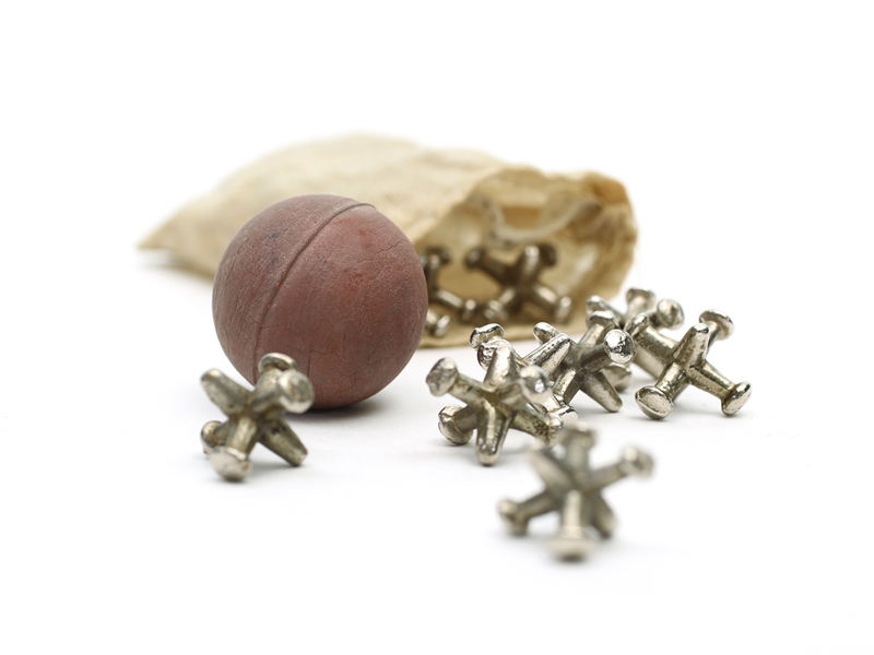
Knucklebones, also known as scatter jacks, snobs, astragalus, tali, dibs, fivestones, jacks, or jackstones, among many other names, is a game of dexterity played with a number of small objects that are thrown up, caught, and manipulated in various manners. It is ancient in origin and is found in various cultures worldwide. The name "knucklebones" is derived from the Ancient Greek version of the game, which uses the astragalus (a bone in the ankle, or hock) of a sheep. However, different variants of the game from various cultures use other objects, including stones, seashells, seeds, and cubes. Modern knucklebones consist of six points, or knobs, projecting from a common base and are usually made of metal or plastic. The winner is the first player to successfully complete a prescribed series of throws, which, though similar, differ widely in detail. The simplest throw consists in either tossing up one stone, the jack, or bouncing a ball and picking up one or more stones or k ... [...More Info...] [...Related Items...] OR: [Wikipedia] [Google] [Baidu] |
Terms For Anatomical Location
Standard anatomical terms of location are used to unambiguously describe the anatomy of animals, including humans. The terms, typically derived from Latin or Greek roots, describe something in its standard anatomical position. This position provides a definition of what is at the front ("anterior"), behind ("posterior") and so on. As part of defining and describing terms, the body is described through the use of anatomical planes and anatomical axes. The meaning of terms that are used can change depending on whether an organism is bipedal or quadrupedal. Additionally, for some animals such as invertebrates, some terms may not have any meaning at all; for example, an animal that is radially symmetrical will have no anterior surface, but can still have a description that a part is close to the middle ("proximal") or further from the middle ("distal"). International organisations have determined vocabularies that are often used as standard vocabularies for subdisciplines of ana ... [...More Info...] [...Related Items...] OR: [Wikipedia] [Google] [Baidu] |
Bone
A bone is a rigid organ that constitutes part of the skeleton in most vertebrate animals. Bones protect the various other organs of the body, produce red and white blood cells, store minerals, provide structure and support for the body, and enable mobility. Bones come in a variety of shapes and sizes and have complex internal and external structures. They are lightweight yet strong and hard and serve multiple functions. Bone tissue (osseous tissue), which is also called bone in the uncountable sense of that word, is hard tissue, a type of specialized connective tissue. It has a honeycomb-like matrix internally, which helps to give the bone rigidity. Bone tissue is made up of different types of bone cells. Osteoblasts and osteocytes are involved in the formation and mineralization of bone; osteoclasts are involved in the resorption of bone tissue. Modified (flattened) osteoblasts become the lining cells that form a protective layer on the bone surface. The mineralize ... [...More Info...] [...Related Items...] OR: [Wikipedia] [Google] [Baidu] |
Cuboid Syndrome
Cuboid syndrome or cuboid subluxation describes a condition that results from subtle injury to the calcaneocuboid joint and ligaments in the vicinity of the cuboid bone, one of seven tarsal bones of the human foot. This condition often manifests in the form of lateral (little toe side) foot pain and sometimes general foot weakness. Cuboid syndrome, which is relatively common but not well defined or recognized, is known by many other names, including lateral plantar neuritis, cuboid fault syndrome, peroneal cuboid syndrome, dropped cuboid, locked cuboid and subluxed cuboid. Signs and symptoms A patient with cuboid syndrome usually seeks medical advice and attention complaining of pain, discomfort, or weakness along the lateral aspect of the foot between the fourth and fifth metatarsals and the calcaneocuboid joint. The pain may radiate throughout the foot. Tenderness may be elicited over the tendon of the peroneus longus muscle and an antalgic gait may be observed. The pain may be ... [...More Info...] [...Related Items...] OR: [Wikipedia] [Google] [Baidu] |
Tibialis Posterior Muscle
The tibialis posterior muscle is the most central of all the leg muscles, and is located in the deep posterior compartment of the leg. It is the key stabilizing muscle of the lower leg. Structure The tibialis posterior muscle originates on the inner posterior border of the fibula laterally. It is also attached to the interosseous membrane medially, which attaches to the tibia and fibula. The tendon of the tibialis posterior muscle (sometimes called the posterior tibial tendon) descends posterior to the medial malleolus. It terminates by dividing into plantar, main, and recurrent components. The main portion inserts into the tuberosity of the navicular bone. The smaller portion inserts into the plantar surface of the medial cuneiform. The plantar portion inserts into the bases of the second, third and fourth metatarsals, the intermediate and lateral cuneiforms and the cuboid. The recurrent portion inserts into the sustentaculum tali of the calcaneus. Blood is supplied to t ... [...More Info...] [...Related Items...] OR: [Wikipedia] [Google] [Baidu] |
Navicular Bone
The navicular bone is a small bone found in the feet of most mammals. Human anatomy The navicular bone in humans is one of the tarsal bones, found in the foot. Its name derives from the human bone's resemblance to a small boat, caused by the strongly concave proximal articular surface. The term ''navicular bone'' or ''hand navicular bone'' was formerly used for the scaphoid bone, one of the carpal bones of the wrist. The navicular bone in humans is located on the medial side of the foot, and articulates proximally with the talus, distally with the three cuneiform bones, and laterally with the cuboid. It is the last of the foot bones to start ossification and does not tend to do so until the end of the third year in girls and the beginning of the fourth year in boys, although a large range of variation has been reported. The tibialis posterior is the only muscle that attaches to the navicular bone. The main portion of the muscle inserts into the tuberosity of the ... [...More Info...] [...Related Items...] OR: [Wikipedia] [Google] [Baidu] |
Lateral Cuneiform Bone
There are three cuneiform ("wedge-shaped") bones in the human foot: * the first or medial cuneiform * the second or intermediate cuneiform, also known as the middle cuneiform * the third or lateral cuneiform They are located between the navicular bone and the first, second and third metatarsal bones and are medial to the cuboid bone. Structure There are three cuneiform bones: # The medial cuneiform (also known as first cuneiform) is the largest of the cuneiforms. It is situated at the medial side of the foot, anterior to the navicular bone and posterior to the base of the first metatarsal. Lateral to it is the intermediate cuneiform. It articulates with four bones: the navicular, second cuneiform, and first and second metatarsals. The tibialis anterior and fibularis longus muscle inserts at the medial cuneiform bone. # The intermediate cuneiform (second cuneiform or middle cuneiform) is shaped like a wedge, the thin end pointing downwards. The intermediate cuneiform is situ ... [...More Info...] [...Related Items...] OR: [Wikipedia] [Google] [Baidu] |
Tarsometatarsal Joints
The tarsometatarsal joints (Lisfranc joints) are arthrodial joints in the foot. The tarsometatarsal joints involve the first, second and third cuneiform bones, the cuboid bone and the metatarsal bones. The eponym of Lisfranc joint is 18th-19th century surgeon and gynecologist, Jacques Lisfranc de St. Martin. Structure Bones The bones entering into their formation are the first, second, and third cuneiforms, and the cuboid bone, which articulate with the bases of the metatarsal bones. The first metatarsal bone articulates with the first cuneiform; the second is deeply wedged in between the first and third cuneiforms articulating by its base with the second cuneiform; the third articulates with the third cuneiform; the fourth, with the cuboid and third cuneiform; and the fifth, with the cuboid. The bones are connected by dorsal, plantar, and interosseous ligaments. Dorsal ligaments The dorsal ligaments are strong, flat bands. The first metatarsal is joined to the first cunei ... [...More Info...] [...Related Items...] OR: [Wikipedia] [Google] [Baidu] |
Calcaneocuboid Joint
The calcaneocuboid joint is the joint between the calcaneus and the cuboid bone. Structure The calcaneocuboid joint is a type of saddle joint between the calcaneus and the cuboid bone. Ligaments There are five ligaments connecting the calcaneus and the cuboid bone, forming parts of the articular capsule: * the dorsal calcaneocuboid ligament. * part of the bifurcated ligament. * the long plantar ligament. * and the plantar calcaneocuboid ligament. Function The calcaneocuboid joint is conventionally described as among the least mobile joints in the human foot. The articular surfaces of the two bones are relatively flat with some irregular undulations, which seem to suggest movement limited to a single rotation and some translation. However, the cuboid rotates as much as 25° about an oblique axis during inversion- eversion in a movement that could be called involution. Clinical significance The calcaneocuboid joint may be affected by a calcaneal fracture. This may be a s ... [...More Info...] [...Related Items...] OR: [Wikipedia] [Google] [Baidu] |


