|
Creatinine Kinase
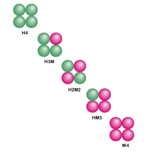
Creatine kinase (CK), also known as creatine phosphokinase (CPK) or phosphocreatine kinase, is an enzyme () expressed by various tissues and cell types. CK catalyses the conversion of creatine and uses adenosine triphosphate (ATP) to create phosphocreatine (PCr) and adenosine diphosphate (ADP). This CK enzyme reaction is reversible and thus ATP can be generated from PCr and ADP. In tissues and cells that consume ATP rapidly, especially skeletal muscle, but also brain, photoreceptor cells of the retina, hair cells of the inner ear, spermatozoa and smooth muscle, PCr serves as an energy reservoir for the rapid buffering and regeneration of ATP ''in situ'', as well as for intracellular energy transport by the PCr shuttle or circuit. Thus creatine kinase is an important enzyme in such tissues. Clinically, creatine kinase is assayed in blood tests as a marker of damage of CK-rich tissue such as in myocardial infarction (heart attack), rhabdomyolysis (severe muscle breakdown), muscular ... [...More Info...] [...Related Items...] OR: [Wikipedia] [Google] [Baidu] |
X-ray Crystallography
X-ray crystallography is the experimental science determining the atomic and molecular structure of a crystal, in which the crystalline structure causes a beam of incident X-rays to diffract into many specific directions. By measuring the angles and intensities of these diffracted beams, a crystallographer can produce a three-dimensional picture of the density of electrons within the crystal. From this electron density, the mean positions of the atoms in the crystal can be determined, as well as their chemical bonds, their crystallographic disorder, and various other information. Since many materials can form crystals—such as salts, metals, minerals, semiconductors, as well as various inorganic, organic, and biological molecules—X-ray crystallography has been fundamental in the development of many scientific fields. In its first decades of use, this method determined the size of atoms, the lengths and types of chemical bonds, and the atomic-scale differences among various mat ... [...More Info...] [...Related Items...] OR: [Wikipedia] [Google] [Baidu] |
Myositis
Myositis is a rare disease that involves inflammation of the muscles. This can present with a variety of symptoms such as skin involvement (i.e., rashes), muscle weakness, and other organ involvement. Systemic symptoms such as weight loss, fatigue, and low fever can also present. Causes Injury, medicines, infection, or an autoimmune disorder can lead to myositis. It can also be idiopathic. * Injury - A mild form of myositis can occur with hard exercise. A more severe form of muscle injury, called rhabdomyolysis, is also associated with myositis. This is a condition where injury to your muscles causes them to quickly break down. * Medicines - A variety of different medicines can cause myositis. One of the most common drug types that can cause myositis is statins. Statins are drugs that are used to help lower high cholesterol. One of the most common side effects of statin therapy is muscle pain. Rarely, statin therapy can lead to myositis. * Infection - The most common infectious c ... [...More Info...] [...Related Items...] OR: [Wikipedia] [Google] [Baidu] |
Myocardium
Cardiac muscle (also called heart muscle, myocardium, cardiomyocytes and cardiac myocytes) is one of three types of vertebrate muscle tissues, with the other two being skeletal muscle and smooth muscle. It is an involuntary, striated muscle that constitutes the main tissue of the wall of the heart. The cardiac muscle (myocardium) forms a thick middle layer between the outer layer of the heart wall (the pericardium) and the inner layer (the endocardium), with blood supplied via the coronary circulation. It is composed of individual cardiac muscle cells joined by intercalated discs, and encased by collagen fibers and other substances that form the extracellular matrix. Cardiac muscle contracts in a similar manner to skeletal muscle, although with some important differences. Electrical stimulation in the form of a cardiac action potential triggers the release of calcium from the cell's internal calcium store, the sarcoplasmic reticulum. The rise in calcium causes the cell's m ... [...More Info...] [...Related Items...] OR: [Wikipedia] [Google] [Baidu] |
CKMT2
Creatine kinase S-type, mitochondrial is an enzyme that in humans is encoded by the ''CKMT2'' gene. Mitochondrial creatine kinase (MtCK) is responsible for the transfer of high energy phosphate from mitochondria to the cytosolic carrier, creatine. The "energy-rich" gamma-phosphate group of ATP that is generated by oxidative phosphorylation inside mitochondria is trans-phosphorylated to creatine (Cr) to give phospho-creatine (PCr), which then is exported from the mitochondria into the cytosol, where it is made available to cytosolic creatine kinases (CK) for ''in situ'' regeneration of the ATP that has been used for cellular work. Cr then is returning to the mitochondria where it stimulates mitochondrial respiration and again is charged-up by mitochondrial ATP via MtCK. This process is termed the PCr/Cr-shuttle or circuit. MtCK belongs to the creatine kinase (CK) isoenzyme family. It exists as two isoenzymes, sarcomeric MtCK and ubiquitous MtCK, encoded by separate genes. Mitocho ... [...More Info...] [...Related Items...] OR: [Wikipedia] [Google] [Baidu] |
CKMT1B
Creatine kinase, mitochondrial 1B also known as CKMT1B is one of two genes which encode the ubiquitous mitochondrial creatine kinase (ubiquitous mtCK or CKMT1). Function Mitochondrial creatine (MtCK) kinase is responsible for the transfer of high energy phosphate from mitochondria to the cytosolic carrier, creatine. It belongs to the creatine kinase isoenzyme family. It exists as two isoenzymes, sarcomeric MtCK (CKMT2) and ubiquitous MtCK, encoded by separate genes. Mitochondrial creatine kinase occurs in two different oligomeric forms: dimers and octamers, in contrast to the exclusively dimeric cytosolic creatine kinase isoenzymes. Ubiquitous mitochondrial creatine kinase has 80% homology with the coding exons of sarcomeric mitochondrial creatine kinase. Two genes located near each other on chromosome 15 (CKMT1A Creatine kinase U-type, mitochondrial, also called ubiquitous mitochondrial creatine kinase (uMtCK), is in humans encoded by ''CKMT1A'' gene. CKMT1A catalyzes the rev ... [...More Info...] [...Related Items...] OR: [Wikipedia] [Google] [Baidu] |
CKMT1A
Creatine kinase U-type, mitochondrial, also called ubiquitous mitochondrial creatine kinase (uMtCK), is in humans encoded by ''CKMT1A'' gene. CKMT1A catalyzes the reversible transfer of the γ-phosphate group of ATP to the guanidino group of Cr to yield ADP and PCr. The impairment of CKMT1A has been reported in ischaemia, cardiomyopathy, and neurodegenerative disorders. Overexpression of CKMT1A has been reported related with several tumors. Structure Gene The ''CKMT1A'' gene lies on the chromosome location of 15q15.3 and consists of 11 exons. Protein CKMT1A consists of 417 amino acids and weighs 47037Da. CKMT1A is rich in amino acids with hydroxyl-containing and basic side chains. Function There are four distinct types of CK subunits in the tissue of mammals, which are expressed species specifically, developmental stage specifically, and tissue specifically. Ubiquitously expressed, CKMT1A is located in the mitochondrial intermembrane space and form both homodimeric ... [...More Info...] [...Related Items...] OR: [Wikipedia] [Google] [Baidu] |
CKM (gene)
Creatine kinase, muscle also known as MCK is a creatine kinase that in humans is encoded by the ''MCK'' gene. Structure In the figure to the right, the crystal structure of the muscle-type M-CK monomer is shown. In vivo, two such monomers arrange symmetrically to form the active MM-CK enzyme. Function The protein encoded by this gene is a cytoplasmic enzyme involved in cellular energy homeostasis. The encoded protein reversibly catalyzes the transfer of "energy-rich" phosphate between ATP and creatine and between phospho-creatine and ADP. Its functional entity is a MM-CK homodimer in striated (sarcomeric) skeletal and cardiac muscle. Clinical significance In heart, in addition to the MM-CK homodimer, also the heterodimer MB-CK consisting of one muscle (M-CK) and one brain-type ( B-CK) subunit is expressed. The latter may be an important serum marker for myocardial infarction A myocardial infarction (MI), commonly known as a heart attack, occurs when blood flow de ... [...More Info...] [...Related Items...] OR: [Wikipedia] [Google] [Baidu] |
CKB (gene)
Brain-type creatine kinase also known as CK-BB is a creatine kinase that in humans is encoded by the ''CKB'' gene. Function The protein encoded by this gene, CK-BB, consists of a homodimer of two identical brain-type CK-B subunits. BB-CK is a cytoplasmic enzyme involved in cellular energy homeostasis, with certain fractions of the enzyme being bound to cell membranes, ATPases, and a variety of ATP-requiring enzymes in the cell. There, CK-BB forms tightly coupled microcompartments for in situ regeneration of ATP that has been used up. The encoded protein reversibly catalyzes the transfer of "energy-rich" phosphate between ATP and creatine or between phospho-creatine (PCr) and ADP. Its functional entity is a homodimer (CK-BB) in brain and smooth muscle as well as in other tissues and cells such as neuronal cells, retina, kidney, bone, etc. In heart, a heterodimer (CK-MB) shahil consisting of one CK-B brain-type CK subunit and one CK-M muscle-type CK subunit is prominently expr ... [...More Info...] [...Related Items...] OR: [Wikipedia] [Google] [Baidu] |
Monomer
In chemistry, a monomer ( ; ''mono-'', "one" + '' -mer'', "part") is a molecule that can react together with other monomer molecules to form a larger polymer chain or three-dimensional network in a process called polymerization. Classification Monomers can be classified in many ways. They can be subdivided into two broad classes, depending on the kind of the polymer that they form. Monomers that participate in condensation polymerization have a different stoichiometry than monomers that participate in addition polymerization: : Other classifications include: *natural vs synthetic monomers, e.g. glycine vs caprolactam, respectively *polar vs nonpolar monomers, e.g. vinyl acetate vs ethylene, respectively *cyclic vs linear, e.g. ethylene oxide vs ethylene glycol, respectively The polymerization of one kind of monomer gives a homopolymer. Many polymers are copolymers, meaning that they are derived from two different monomers. In the case of condensation polymerizations, the r ... [...More Info...] [...Related Items...] OR: [Wikipedia] [Google] [Baidu] |
Mitochondrion
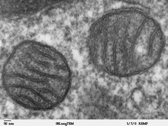
A mitochondrion (; ) is an organelle found in the cells of most Eukaryotes, such as animals, plants and fungi. Mitochondria have a double membrane structure and use aerobic respiration to generate adenosine triphosphate (ATP), which is used throughout the cell as a source of chemical energy. They were discovered by Albert von Kölliker in 1857 in the voluntary muscles of insects. The term ''mitochondrion'' was coined by Carl Benda in 1898. The mitochondrion is popularly nicknamed the "powerhouse of the cell", a phrase coined by Philip Siekevitz in a 1957 article of the same name. Some cells in some multicellular organisms lack mitochondria (for example, mature mammalian red blood cells). A large number of unicellular organisms, such as microsporidia, parabasalids and diplomonads, have reduced or transformed their mitochondria into other structures. One eukaryote, ''Monocercomonoides'', is known to have completely lost its mitochondria, and one multicellular organism, '' ... [...More Info...] [...Related Items...] OR: [Wikipedia] [Google] [Baidu] |
Chromosome
A chromosome is a long DNA molecule with part or all of the genetic material of an organism. In most chromosomes the very long thin DNA fibers are coated with packaging proteins; in eukaryotic cells the most important of these proteins are the histones. These proteins, aided by chaperone proteins, bind to and condense the DNA molecule to maintain its integrity. These chromosomes display a complex three-dimensional structure, which plays a significant role in transcriptional regulation. Chromosomes are normally visible under a light microscope only during the metaphase of cell division (where all chromosomes are aligned in the center of the cell in their condensed form). Before this happens, each chromosome is duplicated ( S phase), and both copies are joined by a centromere, resulting either in an X-shaped structure (pictured above), if the centromere is located equatorially, or a two-arm structure, if the centromere is located distally. The joined copies are now called si ... [...More Info...] [...Related Items...] OR: [Wikipedia] [Google] [Baidu] |
Isoenzyme
In biochemistry, isozymes (also known as isoenzymes or more generally as multiple forms of enzymes) are enzymes that differ in amino acid sequence but catalyze the same chemical reaction. Isozymes usually have different kinetic parameters (e.g. different ''K''M values), or are regulated differently. They permit the fine-tuning of metabolism to meet the particular needs of a given tissue or developmental stage. In many cases, isozymes are encoded by homologous genes that have diverged over time. Strictly speaking, enzymes with different amino acid sequences that catalyse the same reaction are isozymes if encoded by different genes, or allozymes if encoded by different alleles of the same gene; the two terms are often used interchangeably. Introduction Isozymes were first described by R. L. Hunter and Clement Markert (1957) who defined them as ''different variants of the same enzyme having identical functions and present in the same individual''. This definition encompasses (1) ... [...More Info...] [...Related Items...] OR: [Wikipedia] [Google] [Baidu] |


