|
Cranium
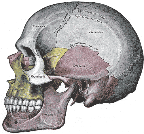
The skull is a bone protective cavity for the brain. The skull is composed of four types of bone i.e., cranial bones, facial bones, ear ossicles and hyoid bone. However two parts are more prominent: the cranium and the mandible. In humans, these two parts are the neurocranium and the viscerocranium (facial skeleton) that includes the mandible as its largest bone. The skull forms the anterior-most portion of the skeleton and is a product of cephalisation—housing the brain, and several sensory structures such as the eyes, ears, nose, and mouth. In humans these sensory structures are part of the facial skeleton. Functions of the skull include protection of the brain, fixing the distance between the eyes to allow stereoscopic vision, and fixing the position of the ears to enable sound localisation of the direction and distance of sounds. In some animals, such as horned ungulates (mammals with hooves), the skull also has a defensive function by providing the mount (on the ... [...More Info...] [...Related Items...] OR: [Wikipedia] [Google] [Baidu] |
Neurocranium
In human anatomy, the neurocranium, also known as the braincase, brainpan, or brain-pan is the upper and back part of the skull, which forms a protective case around the brain. In the human skull, the neurocranium includes the calvaria or skullcap. The remainder of the skull is the facial skeleton. In comparative anatomy, neurocranium is sometimes used synonymously with endocranium or chondrocranium. Structure The neurocranium is divided into two portions: * the membranous part, consisting of flat bones, which surround the brain; and * the cartilaginous part, or chondrocranium, which forms bones of the base of the skull. In humans, the neurocranium is usually considered to include the following eight bones: * 1 ethmoid bone * 1 frontal bone * 1 occipital bone * 2 parietal bones * 1 sphenoid bone * 2 temporal bones The ossicles (three on each side) are usually not included as bones of the neurocranium. There may variably also be extra sutural bones present. Below the ... [...More Info...] [...Related Items...] OR: [Wikipedia] [Google] [Baidu] |
Facial Skeleton
The facial skeleton comprises the ''facial bones'' that may attach to build a portion of the skull. The remainder of the skull is the braincase. In human anatomy and development, the facial skeleton is sometimes called the ''membranous viscerocranium'', which comprises the mandible and dermatocranial elements that are not part of the braincase. Structure In the human skull, the facial skeleton consists of fourteen bones in the face: * Inferior turbinal (2) * Lacrimal bones (2) * Mandible * Maxilla (2) * Nasal bones (2) * Palatine bones (2) * Vomer * Zygomatic bones (2) Variations Elements of the ''cartilaginous viscerocranium'' (i.e., splanchnocranial elements), such as the hyoid bone, are sometimes considered part of the facial skeleton. The ethmoid bone (or a part of it) and also the sphenoid bone are sometimes included, but otherwise considered part of the neurocranium. Because the maxillary bones are fused, they are often collectively listed as only one bone. T ... [...More Info...] [...Related Items...] OR: [Wikipedia] [Google] [Baidu] |
Cranial Cavity
The cranial cavity, also known as intracranial space, is the space within the skull that accommodates the brain. The skull minus the mandible is called the ''cranium''. The cavity is formed by eight cranial bones known as the neurocranium that in humans includes the skull cap and forms the protective case around the brain. The remainder of the skull is called the facial skeleton. Meninges are protective membranes that surround the brain to minimize damage of the brain when there is head trauma. Meningitis is the inflammation of meninges caused by bacterial or viral infections. Structure The capacity of an adult human cranial cavity is 1,200–1,700 cm3. The spaces between meninges and the brain are filled with a clear cerebrospinal fluid, increasing the protection of the brain. Facial bones of the skull are not included in the cranial cavity. There are only eight cranial bones: The occipital, sphenoid, frontal, ethmoid, two parietal, and two temporal bones are fused to ... [...More Info...] [...Related Items...] OR: [Wikipedia] [Google] [Baidu] |
Suture (joint)
In anatomy, fibrous joints are joints connected by fibrous tissue, consisting mainly of collagen. These are fixed joints where bones are united by a layer of white fibrous tissue of varying thickness. In the skull the joints between the bones are called sutures. Such immovable joints are also referred to as synarthroses. Types Most fibrous joints are also called "fixed" or "immovable". These joints have no joint cavity and are connected via fibrous connective tissue. The skull bones are connected by fibrous joints called '' sutures''. In fetal skulls the sutures are wide to allow slight movement during birth. They later become rigid ( synarthrodial). Some of the long bones in the body such as the radius and ulna in the forearm are joined by a '' syndesmosis'' (along the interosseous membrane). Syndemoses are slightly moveable ( amphiarthrodial). The distal tibiofibular joint is another example. A '' gomphosis'' is a joint between the root of a tooth and the socket in the ... [...More Info...] [...Related Items...] OR: [Wikipedia] [Google] [Baidu] |
Flat Bone
Flat bones are bones whose principal function is either extensive protection or the provision of broad surfaces for muscular attachment. These bones are expanded into broad, flat plates,'' Gray's Anatomy'' (1918). (See infobox) as in the cranium (skull), the ilium ( pelvis), sternum and the rib cage. The flat bones are: the occipital, parietal, frontal, nasal, lacrimal, vomer, hip bone (coxal bone), sternum, ribs, and scapulae. These bones are composed of two thin layers of compact bone enclosing between them a variable quantity of cancellous bone, which is the location of red bone marrow. In an adult, most red blood cells are formed in flat bones. In the cranial bones, the layers of compact tissue are familiarly known as the tables of the skull; the outer one is thick and tough; the inner is thin, dense, and brittle, and hence is termed the vitreous (glass-like) table. The intervening cancellous tissue is called the diploë, and this, in the nasal region of the skull ... [...More Info...] [...Related Items...] OR: [Wikipedia] [Google] [Baidu] |
Skeleton
A skeleton is the structural frame that supports the body of an animal. There are several types of skeletons, including the exoskeleton, which is the stable outer shell of an organism, the endoskeleton, which forms the support structure inside the body, and the hydroskeleton, a flexible internal skeleton supported by fluid pressure. Vertebrates are animals with a vertebral column, and their skeletons are typically composed of bone and cartilage. Invertebrates are animals that lack a vertebral column. The skeletons of invertebrates vary, including hard exoskeleton shells, plated endoskeletons, or spicules. Cartilage is a rigid connective tissue that is found in the skeletal systems of vertebrates and invertebrates. Etymology The term ''skeleton'' comes . ''Sceleton'' is an archaic form of the word. Classification Skeletons can be defined by several attributes. Solid skeletons consist of hard substances, such as bone, cartilage, or cuticle. These can be further divided by locat ... [...More Info...] [...Related Items...] OR: [Wikipedia] [Google] [Baidu] |
Skeletal System
A skeleton is the structural frame that supports the body of an animal. There are several types of skeletons, including the exoskeleton, which is the stable outer shell of an organism, the endoskeleton, which forms the support structure inside the body, and the hydroskeleton, a flexible internal skeleton supported by fluid pressure. Vertebrates are animals with a vertebral column, and their skeletons are typically composed of bone and cartilage. Invertebrates are animals that lack a vertebral column. The skeletons of invertebrates vary, including hard exoskeleton shells, plated endoskeletons, or spicules. Cartilage is a rigid connective tissue that is found in the skeletal systems of vertebrates and invertebrates. Etymology The term ''skeleton'' comes . ''Sceleton'' is an archaic form of the word. Classification Skeletons can be defined by several attributes. Solid skeletons consist of hard substances, such as bone, cartilage, or cuticle. These can be further divided by ... [...More Info...] [...Related Items...] OR: [Wikipedia] [Google] [Baidu] |
Process (anatomy)
In anatomy, a process ( la, processus) is a projection or outgrowth of tissue from a larger body. For instance, in a vertebra, a process may serve for muscle attachment and leverage (as in the case of the transverse and spinous processes), or to fit (forming a synovial joint), with another vertebra (as in the case of the articular processes).Moore, Keith L. et al. (2010) ''Clinically Oriented Anatomy'', 6th Ed, p.442 fig. 4.2 The word is used even at the microanatomic level, where cells can have processes such as cilia or pedicels. Depending on the tissue, processes may also be called by other terms, such as ''apophysis'', '' tubercle'', or ''protuberance''. Examples Examples of processes include: *The many processes of the human skull: ** The mastoid and styloid processes of the temporal bone ** The zygomatic process of the temporal bone ** The zygomatic process of the frontal bone ** The orbital, temporal, lateral, frontal, and maxillary processes of the zygomati ... [...More Info...] [...Related Items...] OR: [Wikipedia] [Google] [Baidu] |
List Of English Words Of Old Norse Origin
Words of Old Norse origin have entered the English language, primarily from the contact between Old Norse and Old English during colonisation of eastern and northern England between the mid 9th to the 11th centuries (see also Danelaw). Many of these words are part of English core vocabulary, such as ''egg'' or ''knife''. There are hundreds of such words, and the list below does not aim at completeness. To be distinguished from loan words which date back to the Old English period are modern Old Norse loans originating in the context of Old Norse philology, such as ''kenning'' (1871), and loans from modern Icelandic (such as ''geyser'', 1781). Yet another class comprises loans from Old Norse into Old French, which via Anglo-Norman were then indirectly loaned into Middle English; an example is ''flâneur'', via French from the Old Norse verb ''flana'' "to wander aimlessly". A ; ado: influenced by Norse "at" ("to", infinitive marker) which was used with English "do" in certain ... [...More Info...] [...Related Items...] OR: [Wikipedia] [Google] [Baidu] |
Sinus (anatomy)
A sinus is a sac or cavity in any organ or tissue, or an abnormal cavity or passage caused by the destruction of tissue. In common usage, "sinus" usually refers to the paranasal sinuses, which are air cavities in the cranial bones, especially those near the nose and connecting to it. Most individuals have four paired cavities located in the cranial bone or skull. Etymology ''Sinus'' is Latin for "bay", "pocket", "curve", or "bosom". In anatomy, the term is used in various contexts. The word "sinusitis" is used to indicate that one or more of the membrane linings found in the sinus cavities has become inflamed or infected. It is however distinct from a fistula, which is a tract connecting two epithelial surfaces. If left untreated, infections occurring in the sinus cavities can affect the chest and lungs. Sinuses in the body * Paranasal sinuses ** Maxillary ** Ethmoid ** Sphenoid ** Frontal * Dural venous sinuses ** Anterior midline *** Cavernous *** Superior petrosal ... [...More Info...] [...Related Items...] OR: [Wikipedia] [Google] [Baidu] |
Zoology
Zoology ()The pronunciation of zoology as is usually regarded as nonstandard, though it is not uncommon. is the branch of biology that studies the animal kingdom, including the structure, embryology, evolution, classification, habits, and distribution of all animals, both living and extinct, and how they interact with their ecosystems. The term is derived from Ancient Greek , ('animal'), and , ('knowledge', 'study'). Although humans have always been interested in the natural history of the animals they saw around them, and made use of this knowledge to domesticate certain species, the formal study of zoology can be said to have originated with Aristotle. He viewed animals as living organisms, studied their structure and development, and considered their adaptations to their surroundings and the function of their parts. The Greek physician Galen studied human anatomy and was one of the greatest surgeons of the ancient world, but after the fall of the Western Roman Empire ... [...More Info...] [...Related Items...] OR: [Wikipedia] [Google] [Baidu] |
Fossa (anatomy)
In anatomy, a fossa (; plural ''fossae'' ( or ); from Latin ''fossa'', "ditch" or "trench") is a depression or hollow, usually in a bone, such as the hypophyseal fossa (the depression in the sphenoid bone).Venieratos D, Anagnostopoulou S, Garidou A., A new morphometric method for the sella turcica and the hypophyseal fossa and its clinical relevance.;Folia Morphol (Warsz). 2005 Nov;64(4):240-7. Some examples include: In the Skull: * Cranial fossa ** Anterior cranial fossa ** Middle cranial fossa *** Interpeduncular fossa ** Posterior cranial fossa * Hypophyseal fossa * Temporal bone fossa ** Mandibular fossa ** Jugular fossa * Infratemporal fossa * Pterygopalatine fossa * Pterygoid fossa * Lacrimal fossa ** Fossa for lacrimal gland ** Fossa for lacrimal sac * Mandibular fossa * Scaphoid fossa * Jugular fossa * Condyloid fossa * Rhomboid fossa In the Mandible: * Retromolar fossa In the Torso: * Fossa ovalis (heart) * Infraclavicular fossa * Pyriform foss ... [...More Info...] [...Related Items...] OR: [Wikipedia] [Google] [Baidu] |


