|
Cranial Ultrasound
Cranial ultrasound is a technique for scanning the brain using high-frequency sound waves. It is used almost exclusively in babies because their fontanelle (the soft spot on the skull) provides an "acoustic window". A different form of ultrasound-based brain scanning, transcranial Doppler, can be used in any age group. This uses Doppler ultrasound to assess blood flow through the major arteries in the brain, and can scan through bone. It is not usual for this technique to be referred to simply as "cranial ultrasound". Additionally, cranial ultrasound can be used for intra-operative imaging in adults undergoing neurosurgery once the skull has been opened, for example to help identify the margins of a tumour. Uses Premature babies are especially vulnerable to certain conditions involving the brain. These include intraventricular haemorrhage (IVH), which often occurs during the first few days, and periventricular leukomalacia (PVL), which tends to occur later on. One of the ... [...More Info...] [...Related Items...] OR: [Wikipedia] [Google] [Baidu] |
Fontanelle
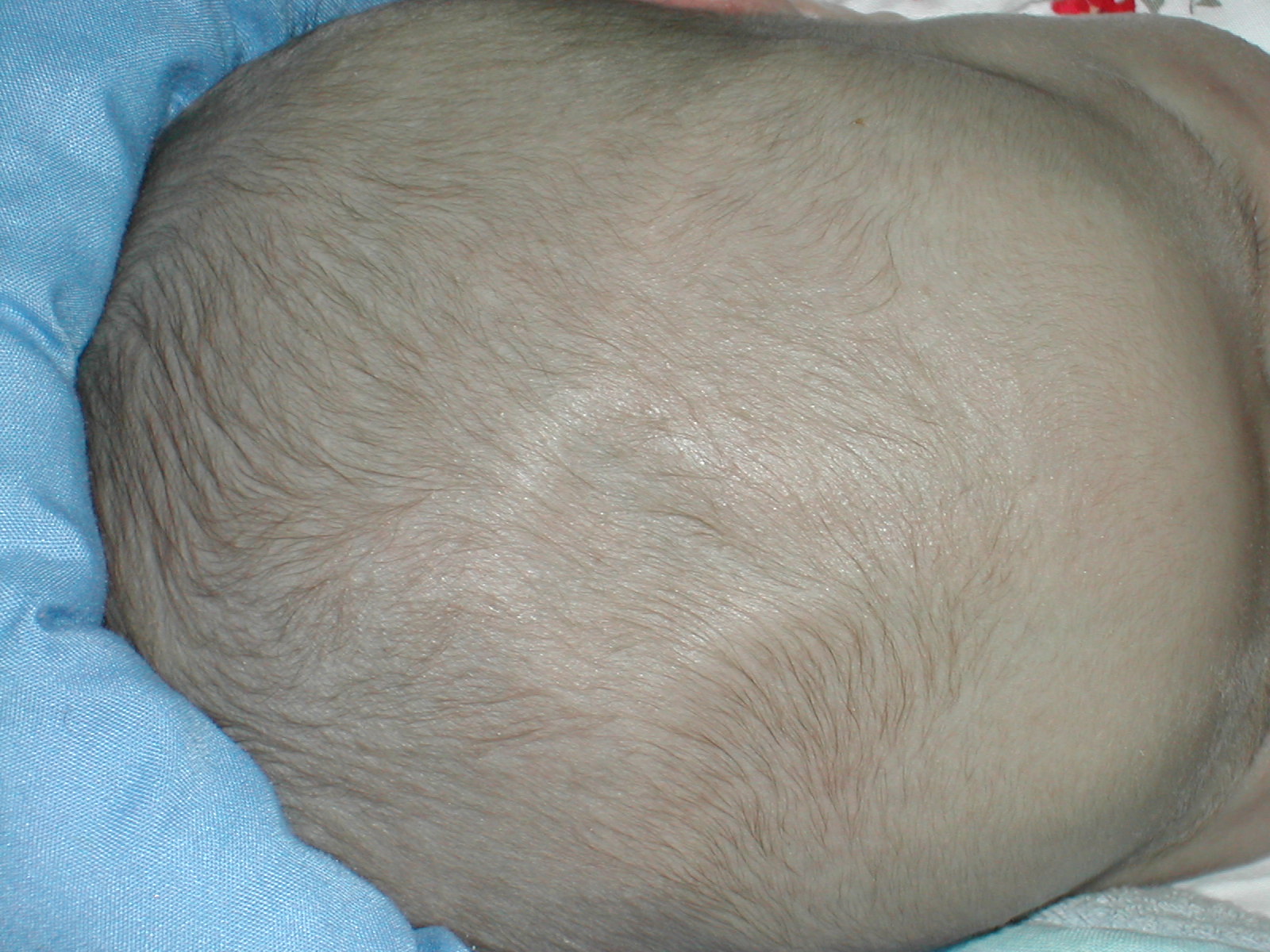
A fontanelle (or fontanel) (colloquially, soft spot) is an anatomical feature of the infant human skull comprising soft membranous gaps ( sutures) between the cranial bones that make up the calvaria of a fetus or an infant. Fontanelles allow for stretching and deformation of the neurocranium both during birth and later as the brain expands faster than the surrounding bone can grow. Premature complete ossification of the sutures is called craniosynostosis. After infancy, the anterior fontanelle is known as the bregma. Structure An infant's skull consists of five main bones: two frontal bones, two parietal bones, and one occipital bone. These are joined by fibrous sutures, which allow movement that facilitates childbirth and brain growth. * Posterior fontanelle is triangle-shaped. It lies at the junction between the sagittal suture and lambdoid suture. At birth, the skull features a small posterior fontanelle with an open area covered by a tough membrane, where the two pariet ... [...More Info...] [...Related Items...] OR: [Wikipedia] [Google] [Baidu] |
Coronal Plane
The coronal plane (also known as the frontal plane) is an anatomical plane that divides the body into dorsal and ventral sections. It is perpendicular to the sagittal and transverse planes. Details The coronal plane is an example of a longitudinal plane. For a human, the mid-coronal plane would transect a standing body into two halves (front and back, or anterior and posterior) in an imaginary line that cuts through both shoulders. The description of the coronal plane applies to most animals as well as humans even though humans walk upright and the various planes are usually shown in the vertical orientation. The sternal plane (''planum sternale'') is a coronal plane which transects the front of the sternum. Etymology The term is derived from Latin ''corona'' ('garland, crown'), from Ancient Greek κορώνη (''korōnē'', 'garland, wreath'). The coronal plane is so-called because it lies in the direction of Coronal suture. Additional images File:Coronal plane CT scan of t ... [...More Info...] [...Related Items...] OR: [Wikipedia] [Google] [Baidu] |
Parenchyma
Parenchyma () is the bulk of functional substance in an animal organ or structure such as a tumour. In zoology it is the name for the tissue that fills the interior of flatworms. Etymology The term ''parenchyma'' is New Latin from the word παρέγχυμα ''parenchyma'' meaning 'visceral flesh', and from παρεγχεῖν ''parenchyma'' meaning 'to pour in' from παρα- ''para-'' 'beside' + ἐν ''en-'' 'in' + χεῖν ''chyma'' 'to pour'. Originally, Erasistratus and other anatomists used it to refer to certain human tissues. Later, it was also applied to plant tissues by Nehemiah Grew. Structure The parenchyma is the ''functional'' parts of an organ (anatomy), organ, or of a structure such as a tumour in the body. This is in contrast to the Stroma (animal tissue), stroma, which refers to the ''structural'' tissue of organs or of structures, namely, the connective tissues. Brain The brain parenchyma refers to the functional tissue in the brain that is made up of t ... [...More Info...] [...Related Items...] OR: [Wikipedia] [Google] [Baidu] |
Cerebellum
The cerebellum (Latin for "little brain") is a major feature of the hindbrain of all vertebrates. Although usually smaller than the cerebrum, in some animals such as the mormyrid fishes it may be as large as or even larger. In humans, the cerebellum plays an important role in motor control. It may also be involved in some cognition, cognitive functions such as attention and language as well as emotion, emotional control such as regulating fear and pleasure responses, but its movement-related functions are the most solidly established. The human cerebellum does not initiate movement, but contributes to Motor coordination, coordination, precision, and accurate timing: it receives input from sensory systems of the spinal cord and from other parts of the brain, and integrates these inputs to fine-tune motor activity. Cerebellar damage produces disorders in Fine motor skill, fine movement, Equilibrioception, equilibrium, Human positions, posture, and motor learning in humans. Anatomica ... [...More Info...] [...Related Items...] OR: [Wikipedia] [Google] [Baidu] |
Ionising Radiation
Ionizing radiation (or ionising radiation), including nuclear radiation, consists of subatomic particles or electromagnetic waves that have sufficient energy to ionize atoms or molecules by detaching electrons from them. Some particles can travel up to 99% of the speed of light, and the electromagnetic waves are on the high-energy portion of the electromagnetic spectrum. Gamma rays, X-rays, and the higher energy ultraviolet part of the electromagnetic spectrum are ionizing radiation, whereas the lower energy ultraviolet, visible light, nearly all types of laser light, infrared, microwaves, and radio waves are non-ionizing radiation. The boundary between ionizing and non-ionizing radiation in the ultraviolet area is not sharply defined, as different molecules and atoms ionize at different energies. The energy of ionizing radiation starts between 10 electronvolts (eV) and 33 eV. Typical ionizing subatomic particles include alpha particles, beta particles, and neutrons. Th ... [...More Info...] [...Related Items...] OR: [Wikipedia] [Google] [Baidu] |
Ventriculomegaly
Ventriculomegaly is a brain condition that mainly occurs in the fetus when the lateral ventricles become dilated. The most common definition uses a width of the atrium of the lateral ventricle of greater than 10 mm. This occurs in around 1% of pregnancies. When this measurement is between 10 and 15 mm, the ventriculomegaly may be described as mild to moderate. When the measurement is greater than 15mm, the ventriculomegaly may be classified as more severe. Enlargement of the ventricles may occur for a number of reasons, such as loss of brain volume (perhaps due to infection or infarction), or impaired outflow or absorption of cerebrospinal fluid from the ventricles, called hydrocephalus or normal pressure hydrocephalus associated with conspicuous brain sulcus. Often, however, there is no identifiable cause. The interventricular foramen may be congenitally malformed, or may have become obstructed by infection, hemorrhage, or rarely tumor, which may impair the drainage ... [...More Info...] [...Related Items...] OR: [Wikipedia] [Google] [Baidu] |
Temporal Bone
The temporal bones are situated at the sides and base of the skull, and lateral to the temporal lobes of the cerebral cortex. The temporal bones are overlaid by the sides of the head known as the temples, and house the structures of the ears. The lower seven cranial nerves and the major vessels to and from the brain traverse the temporal bone. Structure The temporal bone consists of four parts— the squamous, mastoid, petrous and tympanic parts. The squamous part is the largest and most superiorly positioned relative to the rest of the bone. The zygomatic process is a long, arched process projecting from the lower region of the squamous part and it articulates with the zygomatic bone. Posteroinferior to the squamous is the mastoid part. Fused with the squamous and mastoid parts and between the sphenoid and occipital bones lies the petrous part, which is shaped like a pyramid. The tympanic part is relatively small and lies inferior to the squamous part, anterior to the mast ... [...More Info...] [...Related Items...] OR: [Wikipedia] [Google] [Baidu] |
Posterior Fontanelle
The posterior fontanelle (lambdoid fontanelle, occipital fontanelle) is a gap between bones in the human skull (known as fontanelle), triangular in form and situated at the junction of the sagittal suture and lambdoidal suture. It generally closes in 6–8 weeks from birth. The cranial point in adults corresponding the fontanelle is called 'lambda' A delay in closure is associated with congenital hypothyroidism. See also * Lambda (anatomy) The lambda is the meeting point of the sagittal suture and the lambdoid suture. This is also the point of the occipital angle. It is named after the Greek letter lambda. Structure The lambda is the meeting point of the sagittal suture and t ... References Skull {{musculoskeletal-stub ... [...More Info...] [...Related Items...] OR: [Wikipedia] [Google] [Baidu] |
Mastoid Fontanelle
The asterion is a meeting point between three sutures between bones of the skull. It is an important surgical landmark. Structure In human anatomy, the asterion is a visible (craniometric) point on the exposed skull. It is just posterior to the ear. It is the point where three cranial sutures meet: * the lambdoid suture. * parietomastoid suture. * occipitomastoid suture. It is also the point where three cranial bones meet: * the parietal bone. * the occipital bone. * the mastoid portion of the temporal bone. In the adult, it lies 4 cm behind and 12 mm above the center of the entrance to the ear canal. Its relation to other anatomical structures is fairly variable. Clinical significance Neurosurgeons may use the asterion to orient themselves, in order to plan safe entry into the skull for some operations, such as when using a retro-sigmoid approach. Etymology The asterion receives its name from the Greek ἀστέριον (''astērion''), meaning "star" or "starry". T ... [...More Info...] [...Related Items...] OR: [Wikipedia] [Google] [Baidu] |
Posterior Cranial Fossa
The posterior cranial fossa is part of the cranial cavity, located between the foramen magnum and tentorium cerebelli. It contains the brainstem and cerebellum. This is the most inferior of the fossae. It houses the cerebellum, medulla and pons. Anteriorly it extends to the apex of the petrous temporal. Posteriorly it is enclosed by the occipital bone. Laterally portions of the squamous temporal and mastoid part of the temporal bone form its walls. Features Foramen magnum The most conspicuous, large opening in the floor of the fossa. It transmits the medulla, the ascending portions of the spinal accessory nerve (XI), and the vertebral arteries. Internal acoustic meatus Lies in the anterior wall of the posterior cranial fossa. It transmits the facial (VII) and vestibulocochlear (VIII) cranial nerves into a canal in the petrous temporal bone. Jugular foramen Lies between the inferior edge of the petrous temporal bone and the adjacent occipital bone and transmits the internal ... [...More Info...] [...Related Items...] OR: [Wikipedia] [Google] [Baidu] |
Sonographers
A sonographer is an allied healthcare professional who specializes in the use of ultrasonic imaging devices to produce diagnostic images, scans, videos or three-dimensional volumes of anatomy and diagnostic data. The requirements for clinical practice vary greatly by country. Sonography requires specialized education and skills to acquire, analyze and optimize information in the image. Due to the high levels of decisional latitude and diagnostic input, sonographers have a high degree of responsibility in the diagnostic process. Many countries require medical sonographers to have professional certification. Sonographers have core knowledge in ultrasound physics, cross-sectional anatomy, physiology, and pathology. A sonologist is a medical doctor who has undergone formal medical ultrasound training to diagnose and treat diseases. Sonologist is licensed to perform and write ultrasound imaging reports independently or verifies a sonographer's report, prescribe medications and medica ... [...More Info...] [...Related Items...] OR: [Wikipedia] [Google] [Baidu] |
Radiologists
Radiology ( ) is the medical discipline that uses medical imaging to diagnose diseases and guide their treatment, within the bodies of humans and other animals. It began with radiography (which is why its name has a root referring to radiation), but today it includes all imaging modalities, including those that use no electromagnetic radiation (such as ultrasonography and magnetic resonance imaging), as well as others that do, such as computed tomography (CT), fluoroscopy, and nuclear medicine including positron emission tomography (PET). Interventional radiology is the performance of usually minimally invasive medical procedures with the guidance of imaging technologies such as those mentioned above. The modern practice of radiology involves several different healthcare professions working as a team. The radiologist is a medical doctor who has completed the appropriate post-graduate training and interprets medical images, communicates these findings to other physicians by me ... [...More Info...] [...Related Items...] OR: [Wikipedia] [Google] [Baidu] |


