|
Corona Glandis
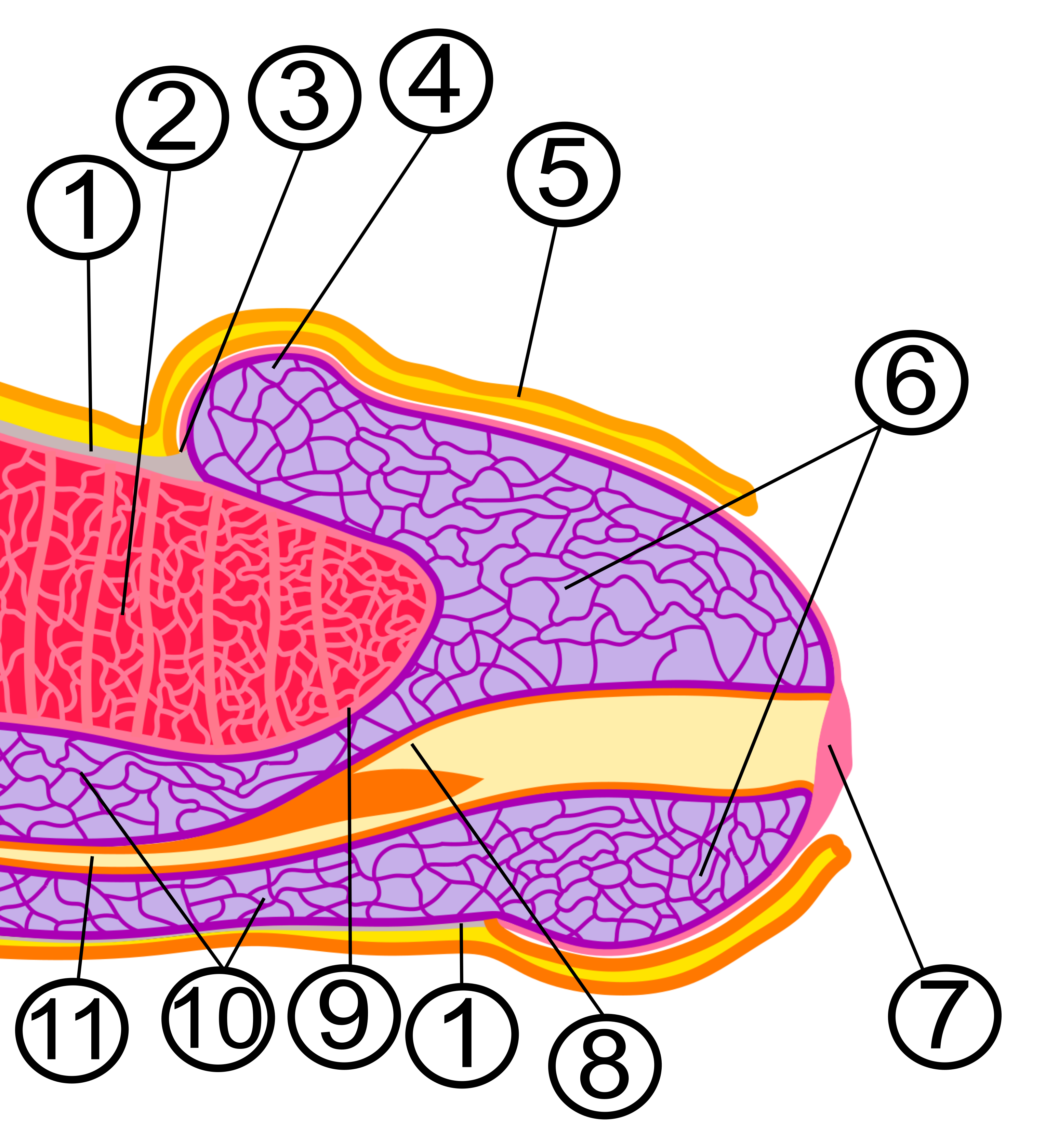
The corona of glans penis or penis crown refers to the rounded projecting border that forms at the base of the glans in human males. The corona overhangs a mucosal surface, known as the neck of the penis, which separates the shaft and the glans. The deep retro-glandular coronal sulcus forms between the corona and the neck of the penis. The corona and the neck are highly vascularized areas of the penis. The axial and dorsal penile arteries merge together at the neck before entering the glans. Small venous tributaries deriving from the corona drain the glans forming a venous retro-coronal plexus before merging with the dorsal veins. The circumference and the underside of the corona are densely innervated by several types of nerve terminals and are considered by males an erogenous area of the glans. In some males, small skin-colored bumps, known as pearly penile papules Pearly penile papules (PPP) are benign small bumps on the human penis. They vary in size from 1–4&nb ... [...More Info...] [...Related Items...] OR: [Wikipedia] [Google] [Baidu] |
Dorsal Artery Of The Penis
The dorsal artery of the penis is an artery on the top surface of the penis. It is a branch of the internal pudendal artery. It runs forward on the dorsum of the penis to the glans, where it divides into two branches to the glans penis and the foreskin (prepuce). The dorsal artery of the penis supplies the integument and fibrous sheath of the corpus cavernosum penis, the glans penis, the foreskin, and the skin of the distal shaft. It also branches with circumflex arteries that supply the corpus spongiosum. Its role in erectile function is unknown. The dorsal artery of the penis may be damaged in traumatic amputation of the penis and repairing the dorsal artery surgically prevents skin loss, but it is not essential for sexual and urinary function. Its hemodynamics and blood pressure can be assessed to test for sexual impairment. Structure The dorsal artery of the penis is a branch of the internal pudendal artery. It ascends between the crus penis and the pubic symphysis ... [...More Info...] [...Related Items...] OR: [Wikipedia] [Google] [Baidu] |
Dorsal Veins Of The Penis
In human anatomy, the dorsal veins of the penis comprise the superficial dorsal vein of the penis and the deep dorsal vein of the penis. Superficial dorsal vein The ''superficial dorsal vein of the penis'' drains the prepuce and skin of the penis, and, running backward in the subcutaneous tissue, inclines to the right or left, and opens into the corresponding superficial external pudendal vein, a tributary of the great saphenous vein. In contrast to the deep dorsal vein, it lies ''outside'' Buck's fascia. It is possible for the vein to rupture, which presents in a manner similar to penile fracture. Deep dorsal vein The ''deep dorsal vein of the penis'' lies beneath the deep fascia of the penis; it receives the blood from the glans penis and corpora cavernosa penis and courses backward in the middle line between the dorsal arteries; near the root of the penis it passes between the two parts of the suspensory ligament and then through an aperture between the arcuate pub ... [...More Info...] [...Related Items...] OR: [Wikipedia] [Google] [Baidu] |
Dorsal Nerve Of The Penis
The dorsal nerve of the penis is the deepest division of the pudendal nerve; it accompanies the internal pudendal artery along the ramus of the ischium; it then runs forward along the margin of the inferior ramus of the pubis, between the superior and inferior layers of the fascia of the urogenital diaphragm. Piercing the inferior layer it gives a branch to the corpus cavernosum penis, and passes forward, in company with the dorsal artery of the penis, between the layers of the suspensory ligament, on to the dorsum of the penis, and ends on the glans penis. It innervates the skin of the penis. Gallery File:Gray824.png, Deep and superficial dissection of the lumbar plexus File:Gray837.png , Sacral plexus of the right side (dorsal nerve of penis visible at bottom left) See also * Cavernous nerves of penis The cavernous nerves are post-ganglionic parasympathetic nerves that facilitate penile erection and clitoral erection. They arise from cell bodies in the inferior hyp ... [...More Info...] [...Related Items...] OR: [Wikipedia] [Google] [Baidu] |
Glans Penis
In male human anatomy, the glans penis, commonly referred to as the glans, is the bulbous structure at the distal end of the human penis that is the human male's most sensitive erogenous zone and their primary anatomical source of sexual pleasure. It is anatomically homologous to the clitoral glans. The glans penis is part of the male reproductive organs in humans and other mammals where it may appear smooth, spiny, elongated or divided. It is externally lined with mucosal tissue, which creates a smooth texture and glossy appearance. In humans, the glans is a continuation of the corpus spongiosum of the penis. At the summit appears the urinary meatus and at the base forms the corona glandis. An elastic band of tissue, known as the frenulum, runs on its ventral surface. In men who are not circumcised, it is completely or partially covered by the foreskin. In adults, the foreskin can generally be retracted over and past the glans manually or sometimes automatically during an ... [...More Info...] [...Related Items...] OR: [Wikipedia] [Google] [Baidu] |
Male Reproductive System
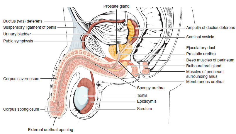
The male reproductive system consists of a number of sex organs that play a role in the process of human reproduction. These organs are located on the outside of the body and within the pelvis. The main male sex organs are the penis and the testicles which produce semen and sperm, which, as part of sexual intercourse, fertilize an ovum in the female's body; the fertilized ovum (zygote) develops into a fetus, which is later born as an infant. The corresponding system in females is the female reproductive system. External genital organs Penis The penis is the male intromittent organ. It has a long shaft and an enlarged bulbous-shaped tip called the glans penis, which supports and is protected by the foreskin. When the male becomes sexually aroused, the penis becomes erect and ready for sexual activity. Erection occurs because sinuses within the erectile tissue of the penis become filled with blood. The arteries of the penis are dilated while the veins are compressed so ... [...More Info...] [...Related Items...] OR: [Wikipedia] [Google] [Baidu] |
Mucous Membrane
A mucous membrane or mucosa is a membrane that lines various cavities in the body of an organism and covers the surface of internal organs. It consists of one or more layers of epithelial cells overlying a layer of loose connective tissue. It is mostly of endodermal origin and is continuous with the skin at body openings such as the eyes, eyelids, ears, inside the nose, inside the mouth, lips, the genital areas, the urethral opening and the anus. Some mucous membranes secrete mucus, a thick protective fluid. The function of the membrane is to stop pathogens and dirt from entering the body and to prevent bodily tissues from becoming dehydrated. Structure The mucosa is composed of one or more layers of epithelial cells that secrete mucus, and an underlying lamina propria of loose connective tissue. The type of cells and type of mucus secreted vary from organ to organ and each can differ along a given tract. Mucous membranes line the digestive, respiratory and reproductive trac ... [...More Info...] [...Related Items...] OR: [Wikipedia] [Google] [Baidu] |
Human Penis
The human penis is an external male intromittent organ that additionally serves as the urinary duct. The main parts are the root (radix); the body (corpus); and the epithelium of the penis including the shaft skin and the foreskin (prepuce) covering the glans penis. The body of the penis is made up of three columns of tissue: two corpora cavernosa on the dorsal side and corpus spongiosum between them on the ventral side. The human male urethra passes through the prostate gland, where it is joined by the ejaculatory duct, and then through the penis. The urethra traverses the corpus spongiosum, and its opening, the meatus (), lies on the tip of the glans penis. It is a passage both for urination and ejaculation of semen (''see'' male reproductive system.) Most of the penis develops from the same embryonic tissue as the clitoris in females. The skin around the penis and the urethra share the same embryonic origin as the labia minora in females. An erection is the stiffening e ... [...More Info...] [...Related Items...] OR: [Wikipedia] [Google] [Baidu] |
Sulcus (anatomy)
In biological morphology and anatomy, a sulcus (pl. ''sulci'') is a furrow or fissure (Latin ''fissura'', plural ''fissurae''). It may be a groove, natural division, deep furrow, elongated cleft, or tear in the surface of a limb or an organ, most notably on the surface of the brain, but also in the lungs, certain muscles (including the heart), as well as in bones, and elsewhere. Many sulci are the product of a surface fold or junction, such as in the gums, where they fold around the neck of the tooth. In invertebrate zoology, a sulcus is a fold, groove, or boundary, especially at the edges of sclerites or between segments. In pollen a grain that is grooved by a sulcus is termed sulcate. Examples in anatomy Liver *Ligamentum teres hepatis fissure *Ligamentum venosum fissure *Portal fissure, found in the under-surface of the liver *Transverse fissure of liver, found in the lower surface of the liver *Umbilical fissure, found in front of the liver Lung *Azygos fissure, of rig ... [...More Info...] [...Related Items...] OR: [Wikipedia] [Google] [Baidu] |
Vascularized
Angiogenesis is the physiological process through which new blood vessels form from pre-existing vessels, formed in the earlier stage of vasculogenesis. Angiogenesis continues the growth of the vasculature by processes of sprouting and splitting. Vasculogenesis is the embryonic formation of endothelial cells from mesoderm cell precursors, and from neovascularization, although discussions are not always precise (especially in older texts). The first vessels in the developing embryo form through vasculogenesis, after which angiogenesis is responsible for most, if not all, blood vessel growth during development and in disease. Angiogenesis is a normal and vital process in growth and development, as well as in wound healing and in the formation of granulation tissue. However, it is also a fundamental step in the transition of tumors from a benign state to a malignant one, leading to the use of angiogenesis inhibitors in the treatment of cancer. The essential role of angiogenesis in t ... [...More Info...] [...Related Items...] OR: [Wikipedia] [Google] [Baidu] |
Venous Plexus
In vertebrates, a venous plexus is a normal congregation anywhere in the body of multiple veins. A list of venous plexuses: * Basilar plexus * Batson venous plexus * Internal vertebral venous plexuses * Pterygoid plexus * Submucosal venous plexus of the nose * Uterine venous plexus * Vaginal venous plexus * Venous plexus of hypoglossal canal * Vesical venous plexus * Soleal venous plexus * Pampiniform venous plexus * Prostatic venous plexus * Rectal venous plexus The rectal venous plexus (or hemorrhoidal plexus) surrounds the rectum, and communicates in front with the vesical venous plexus in the male, and the vaginal venous plexus in the female. A free communication between the portal and systemic venous ... References Veins {{Circulatory-stub ... [...More Info...] [...Related Items...] OR: [Wikipedia] [Google] [Baidu] |
Nerve
A nerve is an enclosed, cable-like bundle of nerve fibers (called axons) in the peripheral nervous system. A nerve transmits electrical impulses. It is the basic unit of the peripheral nervous system. A nerve provides a common pathway for the electrochemical nerve impulses called action potentials that are transmitted along each of the axons to peripheral organs or, in the case of sensory nerves, from the periphery back to the central nervous system. Each axon, within the nerve, is an extension of an individual neuron, along with other supportive cells such as some Schwann cells that coat the axons in myelin. Within a nerve, each axon is surrounded by a layer of connective tissue called the endoneurium. The axons are bundled together into groups called fascicles, and each fascicle is wrapped in a layer of connective tissue called the perineurium. Finally, the entire nerve is wrapped in a layer of connective tissue called the epineurium. Nerve cells (often called neurons) are f ... [...More Info...] [...Related Items...] OR: [Wikipedia] [Google] [Baidu] |
Erogenous Zone
An erogenous zone (from Greek , ''érōs'' "love"; and English ''-genous'' "producing", from Greek , ''-genḗs'' "born") is an area of the human body that has heightened sensitivity, the stimulation of which may generate a sexual response, such as relaxation, sexual fantasies, sexual arousal and orgasm. Erogenous zones are located all over the human body, but the sensitivity of each varies, and depends on concentrations of nerve endings that can provide pleasurable sensations when stimulated. The touching of another person's erogenous zone is regarded as an act of physical intimacy. Whether a person finds stimulation in these areas to be pleasurable or objectionable depends on a range of factors, including their level of arousal, the circumstances in which it takes place, the cultural context, the nature of the relationship between the partners, and the partners' personal histories. Erogenous zones may be classified by the type of sexual response that they generate. Many peo ... [...More Info...] [...Related Items...] OR: [Wikipedia] [Google] [Baidu] |


