|
Constrictor Pharyngis Superior
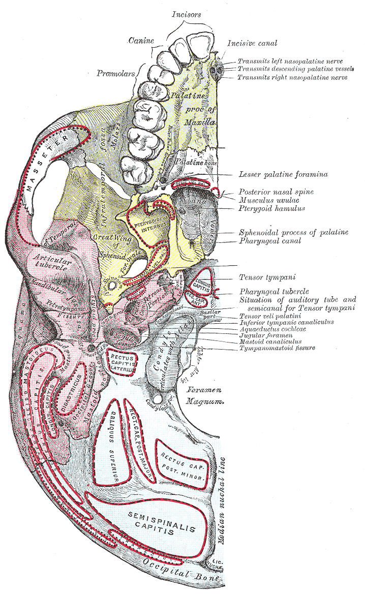
The superior pharyngeal constrictor muscle is a muscle in the pharynx. It is the highest located muscle of the three pharyngeal constrictors. The muscle is a quadrilateral muscle, thinner and paler than the inferior pharyngeal constrictor muscle and middle pharyngeal constrictor muscle. The muscle is divided into four parts: A pterygopharyngeal, buccopharyngeal, mylopharyngeal and a glossopharyngeal part. Origin and insertion The four parts of this muscle arise from: - the lower third of the posterior margin of the medial pterygoid plate and its hamulus (Pterygopharyngeal part) - from the pterygomandibular raphe (Buccopharyngeal part) - from the alveolar process of the mandible above the posterior end of the mylohyoid line (Mylopharyngeal part) - and by a few fibers from the side of the tongue (Glossopharyngeal part) The fibers curve backward to be inserted into the median raphe, being also prolonged by means of an aponeurosis to the pharyngeal spine on the basilar p ... [...More Info...] [...Related Items...] OR: [Wikipedia] [Google] [Baidu] |
Human Pharynx
The pharynx (plural: pharynges) is the part of the throat behind the mouth and nasal cavity, and above the oesophagus and trachea (the tubes going down to the stomach and the lungs). It is found in vertebrates and invertebrates, though its structure varies across species. The pharynx carries food and air to the esophagus and larynx respectively. The flap of cartilage called the epiglottis stops food from entering the larynx. In humans, the pharynx is part of the digestive system and the conducting zone of the respiratory system. (The conducting zone—which also includes the nostrils of the nose, the larynx, trachea, bronchi, and bronchioles—filters, warms and moistens air and conducts it into the lungs). The human pharynx is conventionally divided into three sections: the nasopharynx, oropharynx, and laryngopharynx. It is also important in vocalization. In humans, two sets of pharyngeal muscles form the pharynx and determine the shape of its lumen. They are arranged ... [...More Info...] [...Related Items...] OR: [Wikipedia] [Google] [Baidu] |
Human Mandible
In anatomy, the mandible, lower jaw or jawbone is the largest, strongest and lowest bone in the human facial skeleton. It forms the lower jaw and holds the lower teeth in place. The mandible sits beneath the maxilla. It is the only movable bone of the skull (discounting the ossicles of the middle ear). It is connected to the temporal bones by the temporomandibular joints. The bone is formed in the fetus from a fusion of the left and right mandibular prominences, and the point where these sides join, the mandibular symphysis, is still visible as a faint ridge in the midline. Like other symphyses in the body, this is a midline articulation where the bones are joined by fibrocartilage, but this articulation fuses together in early childhood.Illustrated Anatomy of the Head and Neck, Fehrenbach and Herring, Elsevier, 2012, p. 59 The word "mandible" derives from the Latin word ''mandibula'', "jawbone" (literally "one used for chewing"), from '' mandere'' "to chew" and ''-bula' ... [...More Info...] [...Related Items...] OR: [Wikipedia] [Google] [Baidu] |
Pharyngeal Branch Of Vagus Nerve
The pharyngeal branch of the vagus nerve, the principal motor nerve of the pharynx, arises from the upper part of the ganglion nodosum, and consists principally of filaments from the cranial portion of the accessory nerve. It passes across the internal carotid artery to the upper border of the Constrictor pharyngis medius, where it divides into numerous filaments, which join with branches from the glossopharyngeal, sympathetic, and external laryngeal to form the pharyngeal plexus. From the plexus, branches are distributed to the muscles and mucous membrane of the pharynx (except the stylopharyngeus The stylopharyngeus is a muscle in the head that stretches between the temporal styloid process and the pharynx. Structure The stylopharyngeus is a long, slender muscle, cylindrical above, flattened below. It arises from the medial side of the ..., which is innervated by the glossopharyngeal nerve (CN IX)) and the muscles of the soft palate, except the Tensor veli palati ... [...More Info...] [...Related Items...] OR: [Wikipedia] [Google] [Baidu] |
Esophagus
The esophagus (American English) or oesophagus (British English; both ), non-technically known also as the food pipe or gullet, is an organ in vertebrates through which food passes, aided by peristaltic contractions, from the pharynx to the stomach. The esophagus is a fibromuscular tube, about long in adults, that travels behind the trachea and heart, passes through the diaphragm, and empties into the uppermost region of the stomach. During swallowing, the epiglottis tilts backwards to prevent food from going down the larynx and lungs. The word ''oesophagus'' is from Ancient Greek οἰσοφάγος (oisophágos), from οἴσω (oísō), future form of φέρω (phérō, “I carry”) + ἔφαγον (éphagon, “I ate”). The wall of the esophagus from the lumen outwards consists of mucosa, submucosa (connective tissue), layers of muscle fibers between layers of fibrous tissue, and an outer layer of connective tissue. The mucosa is a stratified squamous epith ... [...More Info...] [...Related Items...] OR: [Wikipedia] [Google] [Baidu] |
Bolus (digestion)
In digestion, a bolus (from Latin ''bolus'', "ball") is a ball-like mixture of food and saliva that forms in the mouth during the process of chewing (which is largely an adaptation for plant-eating mammals). It has the same color as the food being eaten, and the saliva gives it an alkaline pH. Under normal circumstances, the bolus is swallowed, and travels down the esophagus to the stomach The stomach is a muscular, hollow organ in the gastrointestinal tract of humans and many other animals, including several invertebrates. The stomach has a dilated structure and functions as a vital organ in the digestive system. The stomach i ... for digestion. See also * Chyme * Chyle References Digestive system {{Digestive-stub ... [...More Info...] [...Related Items...] OR: [Wikipedia] [Google] [Baidu] |
Sinus Of Morgagni (pharynx)
In the pharynx, the sinus of Morgagni is the enclosed space between the upper border of the superior pharyngeal constrictor muscle, the base of the skull and the pharyngeal aponeurosis.Gray's Anatomy 1918, ChapterThe Pharynx Contents Structures passing through this sinus are: # Cartilaginous part of auditory tube # Levator veli palatini muscle # Ascending palatine artery # Palatine branch of Ascending pharyngeal artery # Tensor veli palatini muscle Clinical significance In nasopharyngeal carcinoma, the tumor A neoplasm () is a type of abnormal and excessive growth of tissue. The process that occurs to form or produce a neoplasm is called neoplasia. The growth of a neoplasm is uncoordinated with that of the normal surrounding tissue, and persists ... may extend laterally and involve this sinus involving the mandibular nerve. This produces a triad of symptoms known as Trotter's Triad. These symptoms are: # Conductive deafness (due to Eustachian tube obstruction) ... [...More Info...] [...Related Items...] OR: [Wikipedia] [Google] [Baidu] |
Pharyngeal Aponeurosis
As it descends it diminishes in thickness, and is gradually lost. It is strengthened posteriorly by a strong fibrous band, which is attached above to the pharyngeal spine on the under surface of the basilar portion of the occipital bone, and passes downward, forming a median raphé, which gives attachment to the Constrictores pharyngis. Additional images File:Slide1kuku.JPG, Larynx, pharynx and tongue.Deep dissection, posterior view. File:Slide2kuku.JPG, Larynx, pharynx and tongue.Deep dissection, posterior view. File:Slide3kuku.JPG, Larynx, pharynx and tongue.Deep dissection, Posterior view. References External links * * http://ect.downstate.edu/courseware/haonline/labs/l31/100101.htm * http://www.instantanatomy.net/headneck/areas/phpharyngobasilarfascia.html Fascial spaces of the head and neck {{Anatomy-stub ... [...More Info...] [...Related Items...] OR: [Wikipedia] [Google] [Baidu] |
Base Of The Skull
The base of skull, also known as the cranial base or the cranial floor, is the most inferior area of the skull. It is composed of the endocranium and the lower parts of the calvaria. Structure Structures found at the base of the skull are for example: Bones There are five bones that make up the base of the skull: * Ethmoid bone *Sphenoid bone *Occipital bone * Frontal bone * Temporal bone Sinuses * Occipital sinus *Superior sagittal sinus * Superior petrosal sinus Foramina of the skull * Foramen cecum * Optic foramen * Foramen lacerum * Foramen rotundum * Foramen magnum *Foramen ovale * Jugular foramen * Internal auditory meatus * Mastoid foramen * Sphenoidal emissary foramen * Foramen spinosum Sutures * Frontoethmoidal suture * Sphenofrontal suture *Sphenopetrosal suture * Sphenoethmoidal suture *Petrosquamous suture The petrosquamous suture is a cranial suture between the petrous portion and the squama of the temporal bone. It forms the Koerner's septum. The petr ... [...More Info...] [...Related Items...] OR: [Wikipedia] [Google] [Baidu] |
Eustachian Tube
In anatomy, the Eustachian tube, also known as the auditory tube or pharyngotympanic tube, is a tube that links the nasopharynx to the middle ear, of which it is also a part. In adult humans, the Eustachian tube is approximately long and in diameter. It is named after the sixteenth-century Italian anatomist Bartolomeo Eustachi. In humans and other tetrapods, both the middle ear and the ear canal are normally filled with air. Unlike the air of the ear canal, however, the air of the middle ear is not in direct contact with the atmosphere outside the body; thus, a pressure difference can develop between the atmospheric pressure of the ear canal and the middle ear. Normally, the Eustachian tube is collapsed, but it gapes open with swallowing and with positive pressure, allowing the middle ear's pressure to adjust to the atmospheric pressure. When taking off in an aircraft, the ambient air pressure goes from higher (on the ground) to lower (in the sky). The air in the middle ea ... [...More Info...] [...Related Items...] OR: [Wikipedia] [Google] [Baidu] |
Levator Veli Palatini
The levator veli palatini () is the elevator muscle of the soft palate in the human body. It is supplied via the pharyngeal plexus. During swallowing, it contracts, elevating the soft palate to help prevent food from entering the nasopharynx. Structure The levator veli palatini muscle is found in the soft palate of the mouth. It arises from the under surface of the apex of the petrous part of the temporal bone, and from the surface inferolateral to the medial lamina of the cartilage of the Eustachian tube. It does not connect with the medial lamina. It passes above the upper concave margin of the superior pharyngeal constrictor muscle. It spreads out in the palatine velum, its fibers extending obliquely downward and medially to the middle line, where they blend with those of the opposite side. It lies lateral to the choana. Nerve supply The levator veli palatini muscle is supplied by the pharyngeal plexus, which is supplied by the vagus nerve (CN X). Function The ... [...More Info...] [...Related Items...] OR: [Wikipedia] [Google] [Baidu] |
Occipital Bone
The occipital bone () is a cranial dermal bone and the main bone of the occiput (back and lower part of the skull). It is trapezoidal in shape and curved on itself like a shallow dish. The occipital bone overlies the occipital lobes of the cerebrum. At the base of skull in the occipital bone, there is a large oval opening called the foramen magnum, which allows the passage of the spinal cord. Like the other cranial bones, it is classed as a flat bone. Due to its many attachments and features, the occipital bone is described in terms of separate parts. From its front to the back is the basilar part, also called the basioccipital, at the sides of the foramen magnum are the lateral parts, also called the exoccipitals, and the back is named as the squamous part. The basilar part is a thick, somewhat quadrilateral piece in front of the foramen magnum and directed towards the pharynx. The squamous part is the curved, expanded plate behind the foramen magnum and is the largest ... [...More Info...] [...Related Items...] OR: [Wikipedia] [Google] [Baidu] |
Pharyngeal Spine
The pharyngeal tubercle is a part of the occipital bone of the head and neck. It is located on the lower surface of the basilar part of occipital bone. It is the site of attachment of the pharyngeal raphe. Structure The pharyngeal tubercle is located on the lower surface of the basilar part of occipital bone. This about 1 cm anterior to the foramen magnum. Function The pharyngeal tubercle gives attachment to the fibrous raphe of the pharynx The pharynx (plural: pharynges) is the part of the throat behind the mouth and nasal cavity, and above the oesophagus and trachea (the tubes going down to the stomach and the lungs). It is found in vertebrates and invertebrates, though its ..., also known as the pharyngeal raphe. This connects with the superior pharyngeal constrictor muscle. See also * Clivus (anatomy) References External links * * Bones of the head and neck {{musculoskeletal-stub ... [...More Info...] [...Related Items...] OR: [Wikipedia] [Google] [Baidu] |


