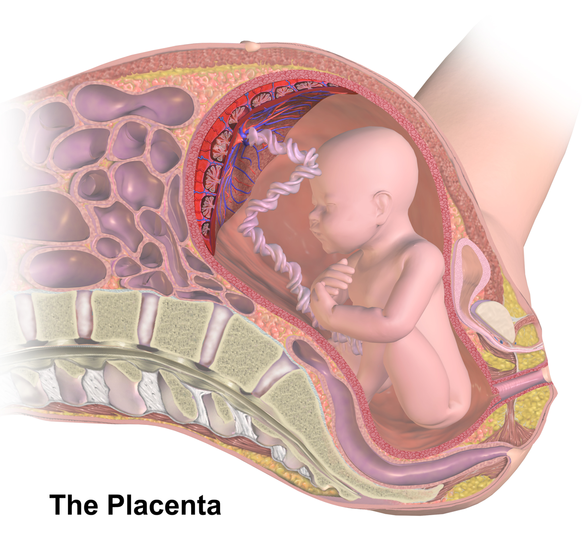
|
Amnion
The amnion (: amnions or amnia) is a membrane that closely covers human and various other embryos when they first form. It fills with amniotic fluid, which causes the amnion to expand and become the amniotic sac that provides a protective environment for the developing embryo. The amnion, along with the chorion, the yolk sac and the allantois protect the embryo. In birds, reptiles and monotremes, the protective sac is enclosed in a shell. In marsupials and placental mammals, it is enclosed in a uterus. The amnion is a feature of the vertebrate clade ''Amniota'', which includes reptiles, birds, and mammals. Amphibians and fish lack the amnion and thus are anamniotes (non-amniotes). The amnion stems from the extra-embryonic somatic mesoderm on the outer side and the extra-embryonic ectoderm or trophoblast on the inner side. Etymology Etymologists have traditionally assumed that the Greek term ἀμνίον (''amnion'') relates to Ancient Greek ἀμνίον : , "little lam ... [...More Info...] [...Related Items...] OR: [Wikipedia] [Google] [Baidu] |
Umbilical Cord
In Placentalia, placental mammals, the umbilical cord (also called the navel string, birth cord or ''funiculus umbilicalis'') is a conduit between the developing embryo or fetus and the placenta. During prenatal development, the umbilical cord is physiologically and genetically part of the fetus and (in humans) normally contains two arteries (the umbilical arteries) and one vein (the umbilical vein), buried within Wharton's jelly. The umbilical vein supplies the fetus with oxygenated, nutrient-rich blood from the placenta. Conversely, the fetal heart pumps low-oxygen, nutrient-depleted blood through the umbilical arteries back to the placenta. Structure and development The umbilical cord develops from and contains remnants of the yolk sac and allantois. It forms by the fifth week of human embryogenesis, development, replacing the yolk sac as the source of nutrients for the embryo. The cord is not directly connected to the mother's circulatory system, but instead joins the pla ... [...More Info...] [...Related Items...] OR: [Wikipedia] [Google] [Baidu] |
Embryo
An embryo ( ) is the initial stage of development for a multicellular organism. In organisms that reproduce sexually, embryonic development is the part of the life cycle that begins just after fertilization of the female egg cell by the male sperm cell. The resulting fusion of these two cells produces a single-celled zygote that undergoes many cell divisions that produce cells known as blastomeres. The blastomeres (4-cell stage) are arranged as a solid ball that when reaching a certain size, called a morula, (16-cell stage) takes in fluid to create a cavity called a blastocoel. The structure is then termed a blastula, or a blastocyst in mammals. The mammalian blastocyst hatches before implantating into the endometrial lining of the womb. Once implanted the embryo will continue its development through the next stages of gastrulation, neurulation, and organogenesis. Gastrulation is the formation of the three germ layers that will form all of the different parts of t ... [...More Info...] [...Related Items...] OR: [Wikipedia] [Google] [Baidu] |
Mesoderm
The mesoderm is the middle layer of the three germ layers that develops during gastrulation in the very early development of the embryo of most animals. The outer layer is the ectoderm, and the inner layer is the endoderm.Langman's Medical Embryology, 11th edition. 2010. The mesoderm forms mesenchyme, mesothelium and coelomocytes. Mesothelium lines coeloms. Mesoderm forms the muscles in a process known as myogenesis, septa (cross-wise partitions) and mesenteries (length-wise partitions); and forms part of the gonads (the rest being the gametes). Myogenesis is specifically a function of mesenchyme. The mesoderm differentiates from the rest of the embryo through intercellular signaling, after which the mesoderm is polarized by an organizing center. The position of the organizing center is in turn determined by the regions in which beta-catenin is protected from degradation by GSK-3. Beta-catenin acts as a co-factor that alters the activity of the transcription facto ... [...More Info...] [...Related Items...] OR: [Wikipedia] [Google] [Baidu] |
Embryonic Development
In developmental biology, animal embryonic development, also known as animal embryogenesis, is the developmental stage of an animal embryo. Embryonic development starts with the fertilization of an egg cell (ovum) by a sperm, sperm cell (spermatozoon). Once fertilized, the ovum becomes a single diploid cell known as a zygote. The zygote undergoes mitosis, mitotic cell division, divisions with no significant growth (a process known as cleavage (embryo), cleavage) and cellular differentiation, leading to development of a multicellular embryo after passing through an organizational checkpoint during mid-embryogenesis. In mammals, the term refers chiefly to the early stages of prenatal development, whereas the terms fetus and fetal development describe later stages. The main stages of animal embryonic development are as follows: * The zygote undergoes a series of cell divisions (called cleavage) to form a structure called a morula. * The morula develops into a structure called a bla ... [...More Info...] [...Related Items...] OR: [Wikipedia] [Google] [Baidu] |
Trophoblast
The trophoblast (from Greek language, Greek : to feed; and : germinator) is the outer layer of cells of the blastocyst. Trophoblasts are present four days after Human fertilization, fertilization in humans. They provide nutrients to the embryo and develop into a large part of the placenta. They form during the first stage of pregnancy and are the first cells to Cellular differentiation, differentiate from the fertilized Ovum, egg to become extraembryonic structures that do not directly contribute to the embryo. After blastulation, the trophoblast is contiguous with the ectoderm of the embryo and is referred to as the trophectoderm. After the first differentiation, the cells in the human embryo lose their Cell potency#Totipotency, totipotency because they can no longer form a trophoblast. They become Cell potency#Pluripotency, pluripotent stem cells. Structure The trophoblast proliferates and differentiates into two cell layers at approximately six days after fertilization for ... [...More Info...] [...Related Items...] OR: [Wikipedia] [Google] [Baidu] |
Vitelline Duct
In the human embryo, the vitelline duct, also known as the vitellointestinal duct, the yolk stalk, the omphaloenteric duct, or the omphalomesenteric duct, is a long narrow tube that joins the yolk sac to the midgut lumen of the developing fetus. It appears at the end of the fourth week, when the yolk sac (also known as the umbilical vesicle) presents the appearance of a small pear-shaped vesicle. Function Obliteration Generally, the duct fully obliterates (narrows and disappears) during the 5–6th week of fertilization age (9th week of gestational age), but a failure of the duct to close is termed a vitelline fistula. This results in discharge of meconium from the navel (umbilicus). About two percent of fetuses exhibit a type of vitelline fistula characterized by persistence of the proximal part of the vitelline duct as a diverticulum protruding from the small intestine, Meckel's diverticulum, which is typically situated within two feet of the ileocecal junction and may b ... [...More Info...] [...Related Items...] OR: [Wikipedia] [Google] [Baidu] |
Allantois
The allantois ( ; : allantoides or allantoises) is one of the extraembryonic membranes arising from the yolk sac. It is a hollow sac-like structure filled with clear fluid that forms part of the developing conceptus in an amniote that helps the embryo exchange gases and handle liquid waste. The other extraembryonoic membranes are the yolk sac, the amnion, and the chorion. In mammals these membranes are known as fetal membranes. The allantois, along with the amnion, chorion, and yolk sac (other extraembryonic membranes), identify humans and other mammals, birds, and reptiles as amniotes. These extraembryonic membranes that form the embryo have aided amniotes in the transition from aquatic to terrestrial environments. Fish and amphibians are '' anamniotes'', lacking the allantois. Function This sac-like structure, whose name is Greek for sausage (from ἀλλαντοειδής ''allantoeidḗs'', in reference to its shape when first formed) is primarily involved in nutrition ... [...More Info...] [...Related Items...] OR: [Wikipedia] [Google] [Baidu] |
Wharton's Jelly
Wharton's jelly (''substantia gelatinea funiculi umbilicalis'') is a gelatinous substance within the umbilical cord, largely made up of mucopolysaccharides (hyaluronic acid and chondroitin sulfate). It acts as a mucous connective tissue containing some fibroblasts and macrophages, and is derived from extra-embryonic mesoderm of the connecting stalk. Umbilical cord occlusion As a mucous connective tissue, it is rich in proteoglycans, and protects and insulates umbilical blood vessels. Wharton's jelly, when exposed to temperature changes, collapses structures within the umbilical cord and thus provides a physiological clamping of the cord, typically three minutes after birth. Stem cells Cells in Wharton's jelly express several stem cell genes, including telomerase. They can be extracted, cultured, and induced to differentiate into mature cell types such as neurons. Wharton's jelly is therefore a potential source of adult stem cells, often collected from cord blood. Etymology It ... [...More Info...] [...Related Items...] OR: [Wikipedia] [Google] [Baidu] |
Placenta
The placenta (: placentas or placentae) is a temporary embryonic and later fetal organ that begins developing from the blastocyst shortly after implantation. It plays critical roles in facilitating nutrient, gas, and waste exchange between the physically separate maternal and fetal circulations, and is an important endocrine organ, producing hormones that regulate both maternal and fetal physiology during pregnancy. The placenta connects to the fetus via the umbilical cord, and on the opposite aspect to the maternal uterus in a species-dependent manner. In humans, a thin layer of maternal decidual ( endometrial) tissue comes away with the placenta when it is expelled from the uterus following birth (sometimes incorrectly referred to as the 'maternal part' of the placenta). Placentas are a defining characteristic of placental mammals, but are also found in marsupials and some non-mammals with varying levels of development. Mammalian placentas probably first evolved abou ... [...More Info...] [...Related Items...] OR: [Wikipedia] [Google] [Baidu] |
Endoderm
Endoderm is the innermost of the three primary germ layers in the very early embryo. The other two layers are the ectoderm (outside layer) and mesoderm (middle layer). Cells migrating inward along the archenteron form the inner layer of the gastrula, which develops into the endoderm. The endoderm consists at first of flattened cells, which subsequently become columnar. It forms the epithelial lining of multiple systems. In plant biology, endoderm corresponds to the innermost part of the cortex ( bark) in young shoots and young roots often consisting of a single cell layer. As the plant becomes older, more endoderm will lignify. Production The following chart shows the tissues produced by the endoderm. The embryonic endoderm develops into the interior linings of two tubes in the body, the digestive and respiratory tube. Liver and pancreas cells are believed to derive from a common precursor. In humans, the endoderm can differentiate into distinguishable organs after 5 w ... [...More Info...] [...Related Items...] OR: [Wikipedia] [Google] [Baidu] |


